Medical expert of the article
New publications
Dacryocystitis
Last reviewed: 05.07.2025

All iLive content is medically reviewed or fact checked to ensure as much factual accuracy as possible.
We have strict sourcing guidelines and only link to reputable media sites, academic research institutions and, whenever possible, medically peer reviewed studies. Note that the numbers in parentheses ([1], [2], etc.) are clickable links to these studies.
If you feel that any of our content is inaccurate, out-of-date, or otherwise questionable, please select it and press Ctrl + Enter.

Acute purulent dacryocystitis, or phlegmon of the lacrimal sac, is a purulent inflammation of the lacrimal sac and the fatty tissue that surrounds it. Purulent dacryocystitis can develop without previous chronic inflammation of the lacrimal ducts when infection penetrates from an inflammatory focus on the nasal mucosa or paranasal sinuses.
Causes of dacryocystitis
Many factors play a role in the etiopathogenesis of dacryocystitis: occupational hazards, sudden changes in ambient air temperature, diseases of the nose and paranasal sinuses, injuries, decreased immunity, virulence of microflora, diabetes mellitus, etc. Blockage of the nasolacrimal duct most often occurs as a result of inflammation of its mucous membrane during rhinitis. Sometimes the cause of obstruction of the nasolacrimal duct is its damage during injury, often surgical (during puncture of the maxillary sinuses, maxillary antrotomy). However, most authors believe that the main cause of dacryocystitis is the presence of pathological processes in the nasal cavity and its paranasal sinuses.
Symptoms of acute dacryocystitis
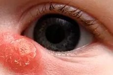
In case of lacrimal sac phlegmon, redness of the skin and dense, sharply painful swelling appear in the area of the inner corner of the eye slit and on the corresponding side of the nose or cheek. The eyelids become edematous, the eye slit narrows or the eye closes completely. The spread of the inflammatory process to the tissue surrounding the sac is accompanied by a violent general reaction of the body (increased temperature, general deterioration, weakness, etc.).
Symptoms of chronic purulent dacryocystitis
Chronic inflammation of the lacrimal sac (chronic dacryocystitis) develops most often as a result of obstruction of the nasolacrimal duct. The retention of tears in the sac leads to the appearance of microorganisms in it, most often staphylococci and pneumococci. Purulent exudate is formed. Patients complain of lacrimation and purulent discharge. The conjunctiva of the eyelids, the semilunar fold and the lacrimal caruncle are reddened. Swelling of the lacrimal sac area is noted, and when pressed, mucopurulent or purulent contents are released from the lacrimal points. Constant lacrimation and purulent discharge from the lacrimal sac into the conjunctival cavity are not only a "discomfort" disease, but also a factor that reduces working capacity. They limit the performance of a number of professions (turners, jewelers, surgical professions, transport drivers, people who work with computers, artists, athletes, etc.).
Chronic dacryocystitis is more common in middle-aged people. Dacryocystitis is more common in women than in men. Lacrimation often increases in the open air, most often in frost and wind, bright light
What's bothering you?
Complications
Dacryocystitis often leads to severe complications and disability. Even the slightest defect in the corneal epithelium, when a speck of dirt gets in, can become an entry point for coccal flora from the stagnant contents of the lacrimal sac. A creeping corneal ulcer develops, leading to persistent visual impairment. Severe complications can also arise if purulent dacryocystitis remains unrecognized before abdominal surgery on the eyeball.
 [ 7 ]
[ 7 ]
What do need to examine?
How to examine?
Who to contact?
Treatment of acute dacryocystitis
At the height of inflammation, antibiotics, sulfonamides, painkillers and antipyretics are prescribed. Gradually, the infiltrate becomes softer, an abscess is formed. The fluctuating abscess is opened and the purulent cavity is drained. The abscess can open on its own, after which the inflammation gradually subsides. Sometimes, at the site of the opened abscess, an unhealed fistula remains, from which pus and tears are released. After acute dacryocystitis, there is a tendency for repeated outbreaks of the phlegmonous inflammatory process. To prevent this, radical surgery is performed in a calm period - dacryocystorhinostomy.
Treatment of chronic dacryocystitis
Currently, chronic dacryocystitis is treated mainly by surgical methods: a radical operation is performed - dacryocystorhinostomy, which restores lacrimal drainage into the nasal cavity. The essence of dacryocystorhinostomy is the creation of an anastomosis between the lacrimal sac and the nasal cavity. The operation is performed with external or intranasal access.
The principle of external surgery was proposed in 1904 by the rhinologist Toti, and was later improved.
Dupuy-Dutant and other authors perform dacryocystorhinostomy under local infiltration anesthesia. A 2.5 cm incision is made in the soft tissues to the bone, retreating 2-3 mm from the attachment point of the internal palpebral ligament towards the nose. The soft tissues are moved apart with a raspatory, the periosteum is cut, it is peeled off together with the lacrimal sac from the bone of the lateral wall of the nose and the lacrimal fossa to the nasolacrimal canal and moved outward. A bone window measuring 1.5 x 2 cm is formed using a mechanical, electric or ultrasonic cutter. The nasal mucosa in the bone "window" and the wall of the lacrimal sac are cut longitudinally, catgut sutures are applied first to the posterior flaps of the nasal mucosa and sac, then to the anterior ones. Before applying the anterior sutures, drainage is inserted into the anastomosis area towards the nasal cavity. The edges of the skin are sutured with silk threads. An aseptic pressure bandage is applied. A gauze tampon is inserted into the nose. The first dressing is done after 2 days. The stitches are removed after 6-7 days.
Endonasal dacryocystorhinostomy according to West with modifications is also performed under local anesthesia.
For correct orientation in the position of the lacrimal sac, the medial wall of the lacrimal sac and the lacrimal bone are pierced with a probe inserted through the inferior lacrimal canaliculus. The end of the probe, which will be visible in the nose, corresponds to the posteroinferior angle of the lacrimal fossa. On the lateral wall of the nose, in front of the middle nasal concha, a flap of the nasal mucosa measuring 1 x 1.5 cm is cut out according to the projection of the lacrimal fossa and removed. At the site of the projection of the lacrimal sac, a bone fragment measuring 1 x 1.5 cm is removed. The wall of the lacrimal sac, protruded by the probe inserted through the lacrimal canaliculus, is dissected in the shape of the letter "c" within the bony window and used for ostectomy. This opens an outlet for the contents of the lacrimal sac into the nasal cavity.
Both methods (external and intranasal) provide a high percentage of recovery (95-98%). They have both indications and limitations.
Intranasal operations on the lacrimal sac are characterized by low trauma, ideal cosmetics, and less disruption of the physiology of the lacrimal drainage system. Simultaneously with the main operation, it is possible to eliminate anatomical and pathological rhinogenic factors. Such operations are successfully performed in any phase of phlegmonous dacryocystitis.
In recent years, endoscopic treatment methods have been developed: endocanalicular laser and intranasal surgery using operating microscopes and monitors.
In case of combined obstruction of the patency of the lacrimal canals and nasolacrimal duct, operations with external and intranasal approaches have been developed - canaliculorhinostomy with the introduction of intubation materials - tubes, threads, etc. - into the lacrimal drainage tract for a long time.
In case of complete destruction or obliteration of the lacrimal ducts, a lacorinostomy is performed - the creation of a new lacrimal duct from the lacrimal lake into the nasal cavity using a silicone or plastic lacoprosthesis, which is inserted for a long period. After epithelialization of the lacostomy walls, the prosthesis is removed.

