Medical expert of the article

New publications
Cystography
Last reviewed: 29.06.2025

All iLive content is medically reviewed or fact checked to ensure as much factual accuracy as possible.
We have strict sourcing guidelines and only link to reputable media sites, academic research institutions and, whenever possible, medically peer reviewed studies. Note that the numbers in parentheses ([1], [2], etc.) are clickable links to these studies.
If you feel that any of our content is inaccurate, out-of-date, or otherwise questionable, please select it and press Ctrl + Enter.
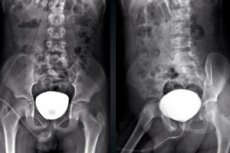
Cystography is a medical procedure used to examine the bladder and urinary tract using X-rays or other imaging techniques. It can be performed to diagnose a variety of medical conditions and diseases of the bladder, urethra, or neighboring organs.
The cystography procedure may include the following steps:
- Contrast agent administration: The patient may be given contrast agent through the urethra or through a catheter. The contrast agent helps to create clear images of the bladder and neighboring organs during X-ray examination.
- X-rays: After the contrast agent is injected, the doctor takes X-rays to visualize the structure of the bladder and its function. These pictures may show the presence of abnormalities, tumors, infections, strictures (narrowings), or other problems.
- Fluoroscopy: At some stages of the procedure, real-time fluoroscopic images may be used to assess the movement of contrast agent in the bladder and urinary tract.
Cystography can be performed in a variety of clinical scenarios, including investigation of lower abdominal pain, pain on urination, presence of blood in the urine (hematuria), evaluation of possible bladder damage after trauma or surgery, and to diagnose urethral reflux in children.
Before performing a cystography, the physician usually discusses the procedure with the patient, explains how it is performed, and discusses the potential risks and benefits. This allows the patient to be informed and prepared for the procedure.
Indications for the procedure
Here are some of the main indications for cystography:
- Urinary outward disease: Cystography can be used to evaluate structural and functional abnormalities of the urinary tract, such as urethral narrowing (stenosis), congenital urinary tract anomalies, or polyps.
- Urinary Incontinence: If a patient suffers from uncontrolled urination, cystography can help identify possible causes such as bladder compression, urethral defects, or urine reflux.
- Suspicion of urolithiasis: Cystography can be used to detect uroliths in the bladder or urinary tract.
- Evaluation after surgical procedures: After bladder or urethral surgery, cystography may be performed to assess efficacy and tissue status.
- Suspicion of tumors: If a tumor is suspected in the bladder, cystography can be used to detect and evaluate it.
- Urinary reflux: Cystography may be performed to diagnose urinary reflux, when urine backs up from the bladder into the urinary tract.
- Trauma assessment: After traumatic injury to the bladder or urethra, cystography can help in assessing the extent of the injury and planning treatment.
Preparation
This procedure may be necessary to detect various pathologies of the urinary system. Preparation for cystography includes the following steps:
- Talking to yourdoctor: Discuss the cystography procedure with your doctor. You will be told about the purpose and benefits of the procedure, as well as possible risks and complications.
- Prepare for possible allergic reactions: If you have previously experienced an allergic reaction to a contrast agent, inform your doctor. In such cases, additional precautions may be necessary.
- Discussing medical conditions: Tell your doctor about any existing medical conditions, allergies, or medications you are taking. This will help your doctor determine if the procedure is appropriate for you and what safety precautions should be taken.
- Fasting: You will usually need to fast before your cystography. Your doctor will give you instructions on how long you should be on an empty stomach before the procedure.
- Pre-procedure tests: You may need to have pre-procedure tests, such as blood or urine tests, to make sure you do not have a urinary infection or other problems.
- Non-pregnancy: If you are a woman who is pregnant or suspect you may be pregnant, inform your doctor, as cystography may not be desirable during pregnancy.
- Preparing for the procedure: You may be asked to undress and put on medical clothing before the procedure. You may also be given a medical gown. Prepare for the fact that the procedure may take several hours.
- Consent: Read and sign the consent for cystography, confirming that you agree to the procedure and understand the possible risks.
The device for carrying out the procedure
The cystography procedure uses x-ray machines or ultrasound machines to visualize the bladder and urinary tract. The specific equipment and techniques used in the procedure may vary depending on the purpose and specifics of the study.
- X-ray machine: X-ray cystography uses an X-ray machine that creates images of internal organs using X-rays. The patient may be injected with a contrast agent through the urethra or catheter, and then x-rays are taken in different projections to visualize the bladder and urethra.
- Ultrasound machine: Ultrasound cystography (transabdominal ultrasound cystography) may use an ultrasound machine to create images of the bladder. In this case, a contrast agent may be injected through the urethra or intravenously. The ultrasound machine is used to visualize the bladder and assess its functionality.
- Fluoroscopy: Fluoroscopic equipment may be used at some stages of X-ray cystography. Fluoroscopy provides real-time images and can be used to assess the movement of contrast agent in the bladder and urinary tract.
Contrast agents
The contrast agents used in the cystography procedure may have different names depending on their composition and properties. Some of the most common contrast agents that may be used in cystography include:
- Urografin: Urografin is one of the commonly used contrast agents to study the bladder and urethra in x-ray examinations.
- Omnipaque (omnipaque): This is another type of contrast agent that can be used in cystography. It is also used to create contrast on x-rays.
- Optiray (optiray): Optiray contrast agent can be used in ultrasound cystography to visualize the bladder.
- Ultracon: Ultracon is another example of a contrast agent used in cystography and other educational medicine procedures.
- Cystographin: The name "Cystographin" may be used to refer to a contrast agent designed specifically for cystography.
It is important to note that the choice of contrast agent and its specific name may depend on the clinical situation, the purpose of the procedure, and the personal preference of the medical staff. The medical professional will usually determine which contrast agent will be used for each specific bladder and urethra examination.
Technique of the cystographs
Here's a general technique for conducting it:
-
Patient Preparation:
- The patient is explained the purpose of the procedure, told what it will involve, and the potential risks and benefits are discussed.
- The doctor may ask about the patient's medical history and possible allergies to contrast agents.
-
Equipment Preparation:
- The X-ray machine or ultrasound machine is prepared for operation.
- Prepare the contrast agent that will be used to inject into the bladder.
-
Patient positioning:
- The patient may be placed on an x-ray or ultrasound table, depending on the type of cystography.
- Patient positioning depends on which areas are to be examined.
-
Bladder preparation:
- In X-ray cystography, the bladder can be filled with contrast agent through the urethra using a catheter. The area around the urethra may be treated with an antiseptic agent beforehand.
- In the case of ultrasound cystography, the contrast agent may be administered intravenously.
-
Image of the bladder:
- After injecting a contrast agent and preparing the patient, x-rays or ultrasound are taken to visualize the bladder.
- During the X-ray procedure, multiple images may be taken in different projections for more detailed examination.
-
Outcome Assessment:
- The resulting images or ultrasound data are analyzed by a specialist.
- The doctor evaluates the condition of the bladder, urethra, and surrounding tissues.
-
Completion of the procedure:
- After cystography is completed, the contrast agent can be removed from the bladder.
- The patient can be advised on further action and treatment depending on the results of the study.
What does cystography show?
This examination can provide information about various aspects of the urinary system and is commonly used for the following purposes:
- Detecting bladder ab normalities: Cystography can help detect abnormalities in the shape, size, or structure of the bladder, such as diverticula (pockets in the bladder wall), stones, or polyps.
- Bladder function assessment: The procedure may allow doctors to assess how the bladder contracts and expels urine. This can be useful in assessing bladder function and identifying abnormalities such as weakness in contractility.
- Urinary tract studies: Cystography can also be used to evaluate the urinary tract, including the ureters and urethra. It can help identify narrowings (strictures) or other abnormalities that may prevent normal urine output.
- Diagnosis of urinary problems: Cystography can help detect problems such as urethral reflux (backflow of urine from the urethra into the bladder), urinary tract infections, and other pathologies.
- Preparation for surgery: Sometimes cystography is performed beforesurgical procedures on the urinary system to provide doctors with additional information about the structure and function of the organs.
Cystography can be useful in identifying and diagnosing various conditions of the urinary system. Doctors use the results of this procedure to develop a treatment plan and determine the best way to manage problems related to the bladder and urinary system.
Types of cystography
There are different types of cystography depending on the way the contrast agent is injected and the direction of its movement:
- Descending (antegrade): In this case, the contrast agent is injected into the bladder through the urethra, i.e. In the direction of urine flow. This type of cystography is used to study the structure and function of the bladder and to evaluate urinary situations.
- Ascending (retrograde): Here, the contrast agent is injected through a catheter placed in the bladder through the urethra. It then rises back up into the ureters and kidneys, allowing the doctor to study the structure and function of these organs. Retrograde cystography is often used to diagnose urethral reflux in children.
- Direct: The direct cystography procedure involves injecting a contrast agent directly into the bladder by puncture through the abdominal wall. This method can be used when there is little control over urination or for a more accurate assessment of the bladder.
- Excretory: This type of cystography combines an examination of the bladder and the urethra. The patient is first injected with contrast agent intravenously and then it is excreted through the urethra into the bladder. Excretory cystography allows you to assess the condition and function of the entire urinary system.
- Intravenous: In this case, the contrast agent is injected into a vein and distributed in the urinary organs, allowing the bladder and urinary tract to be evaluated. This method may be less invasive than retrograde cystography.
Each type of cystography has its own specific indications and is used depending on the specific clinical situation and goals of the study.
Mycotic cystography
It is a diagnostic procedure that is used to examine the bladder and urinary tract using X-rays and a contrast agent. This procedure allows doctors to evaluate the structure and function of the bladder and to detect various anomalies and abnormalities.
The process of micturition cystography involves the following steps:
- You will be asked to strip down to your waist and put on a gown or medical garment.
- You will be placed on the x-ray table in a supine position.
- Medical personnel will insert a flexible catheter tube through the urethra into the bladder. This process can be uncomfortable but is usually done under medically supervised conditions.
- After the catheter is inserted, the bladder will be filled with a special contrast agent through the catheter. This allows the medical staff to view the bladder on x-rays and in real time.
- During the procedure, you may be asked to perform various maneuvers, such as urinating or coughing, to assess urinary tract function.
- Once the procedure is complete, the contrast agent will be removed from the bladder and the catheter will be removed.
Myc cystography can be used to diagnose a variety of conditions and problems including urinary reflux, urinary tract abnormalities, tumors, urethral narrowing, and other bladder and urinary tract pathologies.
It is important to note that the procedure can be uncomfortable, and many patients feel discomfort when the catheter is inserted and the bladder is filled. However, it is an important diagnostic method that can help doctors determine the cause of symptoms and choose the best treatment.
Cystography in children
This is a medical procedure that is performed to examine the bladder and urethra in children. It can be performed for various purposes such as diagnosing abnormalities, assessing bladder functionality, or detecting problems in urination. Here are some important points:
-
Indications: Cystography in children may be recommended in the following cases:
- Suspicion of urinary tract anomalies.
- Recurrent urinary tract infections.
- Suspicion of congenital bladder defects.
- Symptoms related to urination, such as pain, frequent urination, or urinary incontinence.
-
Preparation: Preparing the child for cystography may involve explaining the procedure at a level they can understand and discussing what they can expect. Parents can be present and support the child.
-
Types of cystography:
- Intravenous cystography: A contrast agent is injected intravenously and then the bladder and urinary tract are examined using ultrasound equipment.
- Retrograde cystography: A contrast agent is injected through a catheter placed in the bladder through the urethra. X-rays are then taken for visualization.
-
Safety: The cystography procedure is generally considered safe, but there is a small risk of urinary tract infection and minor discomfort.
-
Results: Cystography results can help your doctor establish a diagnosis and determine a treatment plan, if necessary. They can also be used to monitor the effectiveness of treatment.
Cystography in children is usually performed by specialized specialists such as pediatric urologists or pediatric radiologists. It is important to discuss all aspects of the procedure with the medical staff and follow their recommendations to ensure the child's safety and comfort.
Contraindications to the procedure
Cystography is a diagnostic procedure, and like any other medical test, it can have contraindications in which it may be undesirable or dangerous to perform the procedure. Here are some common contraindications:
- Pregnancy: During pregnancy, X-rays may pose a risk to the developing fetus. Cystography should be avoided in pregnant women unless critically necessary.
- Allergy to contrast agent: If the patient has a known allergy to the contrast agent used during cystography, this may be a contraindication. The physician should be informed of the allergy.
- Acute urinary tract infection: In acute urinary tract infection, cystography may be delayed until the infection is treated, as the procedure may worsen the condition.
- Severe allergic reaction to previous cystography: If the patient has a history of severe allergic reaction to contrast agent or other problems during previous cystography, this may be a contraindication.
- Skin damage to the urethral region: If the patient has open wounds, abrasions, or other skin lesions in the urethral area, cystography may be undesirable or require special precautions.
- Uncooperative patients: Cystography requires the patient's cooperation, as the patient must remain still during the procedure. If the patient is uncooperative (e.g. Due to mental state or age), this may be a contraindication.
- Other Individualized Medical Conditions: Sometimes cystography may not be desirable because of a patient's other individualized medical conditions. The physician must consider the medical history and circumstances of each individual case.
Normal performance
Normal cystography values may vary depending on the purpose and objective of the study. However, the following aspects are usually evaluated when analyzing cystography results:
- Bladdershape and contour: The bladder should have a regular shape and contour with no abnormalities or bulges.
- Bladder filling: The bladder should be completely filled with contrast agent during the procedure. This allows its size and shape to be assessed under normal conditions.
- Contractile function: During cystography, the bladder may contract to expel urine. This function can be evaluated to detect possible abnormalities.
- Urinary tract clearance: The examination also evaluates the clearance of the ureters and urethra. Doctors may pay attention to how the contrast agent moves through the urinary tract.
- No abnormalities or defects: Doctors may look for abnormalities such as diverticula (pockets or pockets in the bladder wall), tumors, strictures (narrowings), or other abnormalities.
Complications after the procedure
Cystography is a relatively safe procedure, but like any medical test, it can have some risks and complications. The following are possible complications after a cystography procedure:
- Infection: Inserting a catheter through the urethra or using other instruments may increase the risk of urinary tract infection. Patients at increased risk of urinary tract infections, such as those with a history of urinary tract infections or a weakened immune system, may be more susceptible to this complication.
- Allergic reaction: In some cases, patients may have an allergic reaction to the contrast agent used during cystography. This may manifest as itching, skin rash, redness or pain.
- Pain and discomfort: After the procedure, patients may experience mild pain or discomfort in the urethral or abdominal area, especially when urinating. These symptoms are usually short-lived and improve with time.
- Microtrauma: Inserting a catheter or other instruments into the bladder can cause microtrauma or irritation of the mucosa. This can lead to temporary symptoms such as minor bleeding or hematuria (blood in the urine).
- Increased urination: Some patients may temporarily experience increased urination after cystography. This may be caused by bladder irritation.
- Peritonitis (rare): In extremely rare cases, the procedure can cause perforation (puncture) of the bladder, which can lead to peritonitis (inflammation of the abdomen). This complication is very rare and can be avoided by following all safety precautions.
Care after the procedure
After a cystography procedure, some care and adherence to recommendations may be necessary to avoid complications and speed recovery. Here are some general guidelines for care after cystography:
- Drink water: Increase your water intake after the procedure to help flush the contrast agent out of the bladder and prevent possible complications such as a urinary tract infection.
- Rest and limitation of physical activity: For the first few hours after cystography, it is recommended to rest and avoid strenuous physical activity.
- Consideration of side effects: You may experience discomfort, slight pain, or frequent urination after the procedure. These symptoms are usually temporary and may improve over time.
- Reduce the risk of infection: Maintain strict genitourinary hygiene. Urinate before and after urinating and avoid bathing in public pools or hot tubs for a few days.
- Monitor your own condition: If you experience serious symptoms such as fever, bleeding, pain, or worsening of the condition, contact your doctor immediately.
- Ongoing medical supervision: After the procedure, you may receive advice from your doctor about the care you need depending on your individual circumstances. Follow the doctor's instructions.
Urography and cystography
They are two different radiologic examinations of the urinary tract that use a contrast agent and x-rays, but they focus on different aspects of the urinary system and have their own characteristics. Here are the main differences between urography and cystography:
-
Study Area:
- Urography: This examination evaluates the structure and function of the kidneys and ureters. Urography includes two main types, intravenous urography (IVU) and excretory urography (EU).
- Cystography: This study evaluates the structure and function of the bladder and urethra.
-
Purpose of the study:
- Urography: The primary purpose of urography is to evaluate the kidneys and their ability to filter blood, and to trace the passage of contrast agent through the ureters and urinary tract.
- Cystography: The main purpose of cystography is to evaluate the bladder, its structure, shape and function, and to detect abnormalities or abnormalities in the urinary tract.
-
Type of contrast agent:
- Urography: Urography uses an intravenous contrast agent that is injected into a vein and distributed into the kidneys and urinary tract.
- Cystography: Cystography uses a contrast agent that is injected directly into the bladder through the urethra or by other means.
-
Specifics of procedure:
- Urography: The urography procedure may involve two steps - first x-rays of the kidneys and ureters (RUT) and then x-rays of the bladder and urethra (UU).
- Cystography: Cystography evaluates the bladder and urethra, and a contrast agent is injected directly into the bladder.
-
Indications:
- Urography: Urography can be used to detect lesions of the kidneys, ureters, bladder, and urethra such as stones, tumors, narrowings, and abnormalities.
- Cystography: Cystography is usually performed to evaluate the bladder, detect urinary reflux and other urinary tract abnormalities.
It is important to realize that both urography and cystography are important methods of diagnosing the urinary system and can be used for a variety of clinical purposes. The choice between these tests depends on the specific situation and medical advice.