Medical expert of the article

New publications
Hip coxitis.
Last reviewed: 12.07.2025

All iLive content is medically reviewed or fact checked to ensure as much factual accuracy as possible.
We have strict sourcing guidelines and only link to reputable media sites, academic research institutions and, whenever possible, medically peer reviewed studies. Note that the numbers in parentheses ([1], [2], etc.) are clickable links to these studies.
If you feel that any of our content is inaccurate, out-of-date, or otherwise questionable, please select it and press Ctrl + Enter.
Inflammation of the hip joint or arthritis can be defined as coxitis of the hip joint, where the term “coxitis” (from Latin coxae – hip) – without specifying the localization of the inflammatory process – is self-sufficient from a medical point of view. [ 1 ]
Epidemiology
The prevalence of coxitis is estimated by experts at 14.2% of all arthritis; the proportion of post-traumatic inflammation of the hip joint does not exceed 5-10% of all cases, and the proportion of reactive coxitis ranges from 0.6 to 2.7 cases per 100 thousand.
According to some data, septic arthritis in children and adolescents is diagnosed in one case per 70 thousand requests for medical care.
Purulent coxitis in elderly adults is detected annually in approximately five people out of 90-100 thousand.
Causes coxitis
The inflammatory process in coxitis has different causes and can affect the synovial membranes and bone structures of the hip joint. And depending on the origin, types or types of the disease are distinguished.
The result of trauma, even a long-standing severe sprain, fracture of the femoral neck or dislocation of the hip joint, is its post-traumatic inflammation - right-sided or left-sided coxitis.
When the joint is affected by Staphylococcus aureus, Streptococcus pneumoniae, and beta-hemolytic cocci (Haemophilus influenzae and Kingella kingae), infectious coxitis develops. Among the viruses involved in this type of disease, specialists most often name the rubella virus (Rubella virus) and the Epstein-Barr virus; hepatitis B, C, and E viruses; parvovirus B19.
In case of hematogenous damage to the joint by Mycobacterium tuberculosis, often caused by the reactivation of previous mycobacterial foci, tuberculous coxitis can develop – in the form of peripheral osteoarticular tuberculosis of the hip joint. [ 2 ]
Septic coxitis, septic arthritis or acute purulent coxitis, which can be streptococcal, staphylococcal, gonococcal, etc., have an infectious etiology. And if there is serous effusion in the cavity of the inflamed joint, serous coxitis is determined.
Reactive coxitis is also associated with infection – reactive arthritis of the hip joint or infectious-allergic coxitis, caused by an increased immune response to previous urogenital or gastrointestinal diseases caused by bacterial infections such as Neisseria gonorrhea, Mycoplasma hominis, Ureaplasma urealyticum, Salmonella enteritenteria, [ 3 ] Yersinia enterocolitica, Campylobacter jejuni. With reactive arthritis, joint inflammation develops several weeks or months after diseases of the genitourinary organs or gastrointestinal tract. [ 4 ]
Read more in the publication - What Causes Reactive Arthritis?
Allergic coxitis, in which joint inflammation occurs as an autoimmune response of the body, is associated with the consumption of certain food proteins.
Transient or transient coxitis (toxic transient inflammation of the synovial membrane of the joint) can be diagnosed in children aged three to ten years after viral infections as a syndrome of acute pain in the hip with stiffness in the hip joint and atraumatic lameness - coxitis syndrome (also called irritable hip syndrome).
In patients with systemic lupus erythematosus (SLE), bilateral coxitis is associated with impaired blood supply to joint tissues and the development of their avascular necrosis.
Read also – Causes of joint pain [ 5 ]
Risk factors
Risk factors for the development of coxitis are:
- hip joint injuries;
- hip dysplasia and slipped capital femoral epiphysis in infants;
- prematurity of children;
- infectious diseases in children and adolescents;
- osteonecrosis;
- osteodystrophy (Paget's disease);
- the presence of autoimmune diseases, primarily rheumatoid arthritis; [ 6 ]
- diabetes;
- overweight.
Pathogenesis
In most cases, the pathogenesis of coxitis is associated with wear and tear and thinning of the cartilage that covers the surfaces of the bone elements of this joint.
When it is affected by tuberculosis, the process may be limited to the synovial membrane (with minimal destruction of the articular surface), but when the inflammation originates in the bone tissue or spreads strongly to it, the surfaces of the joint and the epiphysis are destroyed with subsequent reactive formation of osteophytes.
Viruses can penetrate the synovial membrane of the joints or surrounding tissues, and the immune system perceives them as antigens. In this case, immune cells not only attack the viruses, but also deposit in the joint in the form of so-called immune complexes, causing acute viral inflammation of the hip joint - acute coxitis.
Like reactive arthritis of any joint, reactive coxitis has an immune-mediated mechanism of development associated with the fact that bacteria and viruses entering the bloodstream induce the activity of T-lymphocytes, which spreads to the joint tissues. Studies have revealed the cytotoxic role of human leukocyte antigen B27 (HLA-B27) in the pathogenesis of the reactive form of joint inflammation: this protein of blood leukocytes can change the reaction of the immune system at the cellular level, making it more aggressive.
Symptoms coxitis
Pain in the hip joint, dysfunction of the joint, which leads to stiffness (limited mobility) of the joint, as well as difficulty walking are the main symptoms of coxitis.
In any case, the first signs of inflammation at an early stage of the disease are pain, often minor (except for the acute form). People with arthritis of the hip joint often complain of pain in the morning, getting out of bed. At the same time, for many, the pain subsides 20-30 minutes after getting up.
Stiffness and pain (which may radiate to the knee) lead to complaints of difficulty getting up from a chair, going up and down stairs, bending the torso; inability to squat and abduct the hip.
As the inflammation continues to affect the joint, a Trendelenburg gait (with pelvic tilt) and a so-called antalgic gait - with limping and taking small steps (to reduce pain) - may develop; at a later stage, a fixed limitation of flexion/extension and abduction/adduction of the hip may occur, causing patients to noticeably limp.
With septic coxitis, the skin over the joint is hyperemic and hot, the body temperature rises to fever, there may be general weakness, headache and nausea. And in newborns and infants, the hip joint is usually held in abductive flexion and external rotation.
How hip coxitis manifests itself in children, read in the publication: Hip joint pain in children
Complications and consequences
Coxitis leads to destruction of cartilage with gradual increase in pain. And contracture of periarticular muscles leads to functional or actual shortening of the limb on the side of the affected joint. Scoliosis often develops.
Complications of hip reactive arthritis include ankylosing spondylitis and sacroiliac joint inflammation.[ 7 ]
Reactive coxitis can lead to chronic articular, ophthalmological and cardiac consequences.
In the case of septic coxitis, there is a threat not only of irreversible destruction and dislocation of the joint, but also of death due to the development of sepsis: with treatment, up to 15% of people die, and without treatment - more than 65%.
Diagnostics coxitis
Diagnosis of coxitis begins with a detailed history and physical examination of the patient.
The following tests are taken: general and biochemical blood tests, blood tests for rheumatoid factor, C-reactive protein, antibodies to M. tuberculosis and other bacteria; PCR blood test for viral DNA; serum test for HLA-B27 antigen; general clinical analysis of synovial fluid (obtained by joint aspiration) with subsequent bacterial culture.
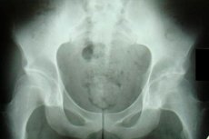
Instrumental diagnostics are used: X-ray and ultrasound of the hip joints, CT and MRI of the hip joint, scintigraphy.
According to experts, differential diagnosis of coxitis can be difficult. For example, it is necessary to differentiate septic arthritis with acute hematogenous osteomyelitis and juvenile idiopathic arthritis, Ewing's sarcoma and Perthes disease.
Who to contact?
Treatment coxitis
In infectious coxitis of bacterial origin, the main drugs are antibiotics: Vancomycin, as well as cephalosporin drugs for injection - Ceftriaxone, Ceftazidime, etc. In tuberculous coxitis, Rifampicin is used, in septic coxitis - Flucloxacillin, Clindamycin, Amoxicillin. More information in the article - Antibiotics for the treatment of arthrosis and arthritis of the joints.
Treatment for other types of hip arthritis is aimed at relieving symptoms and preventing chronic complications. Read more:
- Treatment of joints
- Medicines for joint pain
- Arthritis pills
- Ointment for joint pain: choosing the right one
Physiotherapy treatment, including exercise therapy, is discussed in detail in the publication – Physiotherapy for joint diseases.
Surgical treatment of purulent and serous coxitis involves drainage of the joint. In other cases – with advanced disease that does not respond to conservative measures – a complete replacement (prosthesis) of the hip joint may be required. [ 8 ], [ 9 ]
Prevention
The best way to prevent infectious coxitis is to avoid bacterial and viral infections by observing personal hygiene rules, preferring protected sex, and strengthening the immune system.
Losing extra pounds reduces the mechanical load on the hip and other joints of the lower extremities, which slows down the wear of articular cartilage.
Forecast
The dependence of the prognosis of coxitis of the hip joint on its etiology is obvious. Gonococcal coxitis can be completely cured, while with septic inflammation caused by Staphylococcus aureus, after treatment with antibiotics, the functions of the hip joint are restored in 46-50% of cases, and the remaining patients - due to the functional disorder of the joint - become disabled.