Medical expert of the article
New publications
Choledocholithiasis: causes, symptoms, diagnosis, treatment
Last reviewed: 05.07.2025

All iLive content is medically reviewed or fact checked to ensure as much factual accuracy as possible.
We have strict sourcing guidelines and only link to reputable media sites, academic research institutions and, whenever possible, medically peer reviewed studies. Note that the numbers in parentheses ([1], [2], etc.) are clickable links to these studies.
If you feel that any of our content is inaccurate, out-of-date, or otherwise questionable, please select it and press Ctrl + Enter.

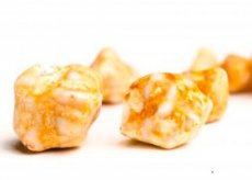
Choledocholithiasis is the formation or presence of stones in the biliary tract. Choledocholithiasis may cause attacks of biliary colic, biliary obstruction, gallstone pancreatitis, or biliary tract infection ( cholangitis ).
Diagnosis of choledocholithiasis usually requires verification by magnetic resonance cholangiopancreatography or ERCP. Timely endoscopic or surgical decompression is indicated.
What causes choledocholithiasis?
Primary stones (usually pigment stones) may form in the biliary tract. Secondary stones (usually cholesterol stones) form in the gallbladder and then migrate to the biliary tract. Forgotten stones are stones that were not detected during cholecystectomy. Recurrent stones form in the ducts more than 3 years after surgery. In developed countries, more than 85% of common bile duct stones are secondary; these patients were also diagnosed with cholelithiasis. At the same time, in 10% of patients, gallstone symptoms are associated with common bile duct stones. After cholecystectomy, brown pigment stones may form due to bile stasis (eg, postoperative strictures) and infection. There is a direct correlation between the formation of ductal pigment stones and the increase in time after cholecystectomy.
Causes of biliary obstruction (except stones and tumors):
- Damage to the ducts during surgical interventions (most common)
- Scarring due to chronic pancreatitis
- Obstruction of the duct due to external compression by a common bile duct cyst (choledochocele) or pancreatic (rare) pseudocyst
- Extrahepatic or intrahepatic stricture as a result of primary sclerosing cholangitis
- AIDS-induced cholangiopathy or cholangitis; direct cholangiography may show features similar to primary sclerosing cholangitis or papillary stenosis; infectious etiology is possible, most likely cytomegalovirus infection, Cryptosporidium, or Microsporidia
- Clonorchis sinensis can cause obstructive jaundice with intrahepatic duct inflammation, proximal stasis, stone formation and cholangitis (in Southeast Asia)
- Migration of Ascaris lumbricoides into the common bile duct (rare)
Symptoms of choledocholithiasis
Biliary tract stones may migrate into the duodenum without symptoms. Biliary colic develops when their movement is impaired and they become partially obstructed. More complete obstruction causes dilation of the common bile duct, jaundice, and eventually bacterial infection (cholangitis). Stones blocking the ampulla of Vater may cause gallstone pancreatitis. In some patients (usually the elderly), biliary obstruction by stones may develop without prior symptoms.
Acute cholangitis due to obstructive lesions of the biliary tract is initiated by the flora of the duodenum. Although most (85%) cases result from biliary tract stones, biliary obstruction may be caused by tumors or other causes. The flora consists mainly of gram-negative organisms (eg, Escherichia coli, Klebsiella Enterobacter); less commonly, gram-positive organisms (eg, Enterococcus) and mixed anaerobic flora (eg, Bacteroides Clostridia). Symptoms include abdominal pain, jaundice, fever, and chills (Charcot's triad). Palpation reveals abdominal tenderness and an enlarged and tender liver (abscesses often form). Confusion and hypotension are manifestations of advanced disease, and the mortality rate is approximately 50%.
Where does it hurt?
Diagnosis of choledocholithiasis
Common bile duct stones should be suspected in patients with jaundice and biliary colic. Liver function tests and instrumental examination should be performed. Increased levels of bilirubin, alkaline phosphatase, ALT, and gamma-glutamyl transferase, characteristic of extrahepatic obstruction, are of diagnostic value, especially in patients with signs of acute cholecystitis.
Ultrasound can verify stones in the gallbladder and sometimes in the common bile duct. The common bile duct is dilated (> 6 mm in diameter if the gallbladder was not removed; > 10 mm after cholecystectomy). If there is no dilation of the common bile duct (e.g. on the first day), the stones have probably migrated. If doubt remains, more informative magnetic resonance cholangiopancreatography (MRCP) should be performed to diagnose residual stones. ERCP is performed if MRCP is uninformative; this study can be both therapeutic and diagnostic. CT is less informative than ultrasound.
If acute cholangitis is suspected, a complete blood count and blood culture should also be performed. Leukocytosis is characteristic, and an increase in aminotransferases to 1000 IU/L suggests acute liver necrosis, primarily due to microabscess formation. The choice of antibiotic should be guided by the results of blood culture.
 [ 10 ]
What do need to examine?
How to examine?
Who to contact?
Treatment of choledocholithiasis
If biliary obstruction is detected, ERCP with stone removal and sphincterotomy should be performed. Laparoscopic cholecystectomy, which is not entirely suitable in cases where intraoperative cholangiography is required or for common bile duct examination in general, can be performed strictly individually after ERCP and sphincterotomy. Open cholecystectomy with common bile duct examination carries a higher mortality rate and a more severe postoperative course. For patients with a high surgical risk of cholecystectomy, such as the elderly, sphincterotomy is the only alternative.
Acute cholangitis is a disease that requires emergency care, active complex therapy and urgent removal of stones by endoscopic or surgical means. Antibiotics are prescribed as in acute cholecystitis. More preferable alternative drugs are imipenem and ciprofloxacin; metronidazole is prescribed to very severe patients to affect anaerobic infection.

