Medical expert of the article

New publications
Cerebellum
Last reviewed: 07.07.2025

All iLive content is medically reviewed or fact checked to ensure as much factual accuracy as possible.
We have strict sourcing guidelines and only link to reputable media sites, academic research institutions and, whenever possible, medically peer reviewed studies. Note that the numbers in parentheses ([1], [2], etc.) are clickable links to these studies.
If you feel that any of our content is inaccurate, out-of-date, or otherwise questionable, please select it and press Ctrl + Enter.
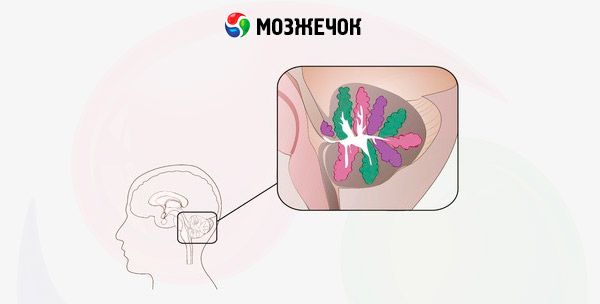
The cerebellum (cerebellum; small brain) is located posterior (dorsal) to the pons and the upper (dorsal) part of the medulla oblongata. It lies in the posterior cranial fossa. The occipital lobes of the cerebral hemispheres hang over the cerebellum, separated from the cerebellum by the transverse fissure of the cerebrum (fissura transversa cerebralis).
The cerebellum has an upper and lower surface, the boundary between which is the posterior edge of the cerebellum, where a deep horizontal fissure (fissura horizontalis) passes. It begins at the point where the middle peduncles enter the cerebellum. The upper and lower surfaces of the cerebellum are convex. On the lower surface there is a wide depression - the cerebellar valley (vallecula cerebelli). The dorsal surface of the medulla oblongata is adjacent to this depression. The cerebellum has two hemispheres (hiispheria cerebelli) and an unpaired middle part - the cerebellar vermis (vermis cerebelli, a phylogenetically older part). The upper and lower surfaces of the hemispheres and the vermis are cut by numerous transverse parallel fissures of the cerebellum (fissura cerebelli), between which are long and narrow sheets (gyri) of the cerebellum (folia cerebelli). Groups of gyri, separated by deeper grooves, form lobules of the cerebellum (lobuli cerebelli). The cerebellar grooves run uninterrupted through the hemispheres and through the vermis. Each lobule of the vermis corresponds to two (right and left) lobules of the hemispheres. A more isolated and phylogenetically older lobule of each hemisphere is the flocculus. It is adjacent to the ventral surface of the middle cerebellar peduncle. With the help of the long peduncle of the flocculus (pedunculus flocculi), the flocculus is connected to the cerebellar vermis, to its nodulus. The cerebellum is connected to the adjacent parts of the brain by three pairs of peduncles. The inferior cerebellar peduncles (pedunculi cerebellares cauddles, s. inferiores; rope-shaped bodies) go downwards and connect the cerebellum with the medulla oblongata. The middle cerebellar peduncles (pedilnculi cerebellares medii) are the thickest, they go forward and pass into the pons. The superior cerebellar peduncles (peduncuii cerebellares rostrales, s. siiperiores) connect the cerebellum with the midbrain. The cerebellar peduncles contain fibers of the conducting pathways that connect the cerebellum with other parts of the brain and the spinal cord.
The cerebellar hemispheres and the vermis consist of the internally located brain body (corpus medullare), white matter, and a thin plate of gray matter covering the white matter on the periphery - the cerebellar cortex (cortex cerebelli).
The cerebellar cortex has three cellular layers. The most superficial is the molecular layer, below it is a layer of pear-shaped neurons (ganglionic layer), and even deeper is the granular layer.
The molecular layer is formed mainly by basket and stellate neurons. Basket neurons are located in the lower part of the molecular layer. These cells are 10 to 20 µm in size, have an irregular shape, and long processes. The dendrites of basket neurons branch mainly across the convolutions of the cerebellum. The axons of basket neurons also go across the convolutions above the piriform neurons. Collaterals extend from the axons downwards to the bodies of piriform neurons, braiding them, forming basket-like figures. Basket neurons inhibit the functions of piriform cells with their impulses. Stellate cells have dendrites of varying lengths and an axon that forms synapses on the dendrites of piriform cells.
The granular layer is formed by numerous small neurons - granule cells. The processes of the granule cells form numerous synapses (synaptic tangles) on other cells of this layer, as well as the endings of fibers ("mossy") ending in the cerebellum and transmitting excitatory impulses.
The layer of piriform neurons is formed by large cells (Purkinje cells) arranged in a single row. The axons of the piriform cells emerge from the cerebellar cortex and terminate on the cells of its nuclei.
Afferent nerve impulses arriving in the cerebellum have an excitatory effect on the piriform neurons. These impulses are transmitted along the fibers of the spinocerebellar and vestibulocerebellar tracts. The nerve fibers pass through the granular layer to the piriform cells, spread along their dendrites ("climbing" fibers) and end in synapses on the bodies of the piriform neurons. Afferent impulses arriving in the cerebellum from the vestibular (statovestibular) receptors of the inner ear, from the proprioceptors of the skeletal muscles are analyzed and compared with impulses coming from the cortex of the cerebral hemispheres. In the thickness of the cerebellar sheets, the white matter has the appearance of thin white stripes (laminae albae).
The white matter of the cerebellum contains paired cerebellar nuclei (nuclei cerebelli). The most significant of these is the dentate nucleus (nucleus dentatus). On a horizontal section of the cerebellum, this nucleus has the shape of a thin curved gray strip, with its convex part facing laterally and backwards. In the medial direction, the gray strip is not closed; this place is called the gate of the dentate nucleus (hilum nuclei dentati). Inside the dentate nucleus, in the white matter of the cerebellar hemisphere, are the cork-shaped nucleus (nucleus iboliformis) and the spherical nucleus (nucleus globosus). Here, in the white matter of the vermis, is the most medial nucleus - the tent nucleus (nucleus fastigii).
The white matter of the worm, bordered by the bark and divided along the periphery by numerous deep and shallow grooves, on the sagittal section has a bizarre pattern reminiscent of a tree branch, hence its name “tree of life” (arbor vitae cerebelli).
 [
[ What's bothering you?
What do need to examine?
How to examine?