Medical expert of the article

New publications
A bartholin gland abscess
Last reviewed: 05.07.2025

All iLive content is medically reviewed or fact checked to ensure as much factual accuracy as possible.
We have strict sourcing guidelines and only link to reputable media sites, academic research institutions and, whenever possible, medically peer reviewed studies. Note that the numbers in parentheses ([1], [2], etc.) are clickable links to these studies.
If you feel that any of our content is inaccurate, out-of-date, or otherwise questionable, please select it and press Ctrl + Enter.
A distinction is made between true and false abscess of the Bartholin gland.
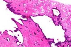
The large vestibular gland (Bartholin's gland) is a paired formation. It belongs to the complex tubular glands, has a round shape and the size of a large pea. The glandular acini are lined with a single-row cylindrical epithelium that secretes mucus.
The main excretory duct of the large vestibular gland is formed from the fusion of several ducts. It opens into the vestibule of the vagina in front and outside the hymen. The duct is lined with transitional epithelium and is 1.5-2 cm long.
What causes a Bartholin gland abscess?
A true abscess of the Bartholin gland is characterized by the involvement of the process and acute purulent melting of the tissue of the entire gland and the surrounding tissue. True bartholinitis is caused by pyogenic cocci, most often gonococci, which have a tropism for the columnar epithelium lining the gland. In gonorrhea of the lower genital tract, the ducts of the Bartholin glands are involved in the process in 20-30% of cases, often indirectly due to the presence of abundant purulent discharge in the vagina.
A false abscess (primary - as a result of infection and abscessing of a retention cyst that has formed for the first time as a result of blockage of the duct, or secondary - suppuration of a long-existing Bartholin's gland cyst) is most often caused by associative flora (staphylococci, streptococci, colibacillary flora, anaerobes, fungi).
Nowadays, false abscess formation is more common. Certain anatomical prerequisites contribute to the formation of a "false" glandular abscess, namely, a significant narrowing of the duct along its length. In the place where small ducts flow into the main one, a kind of ampulla containing a secret is formed in the gland; then the main duct narrows, and at the exit to the outside it is already a pinpoint opening. The presence of inflammatory edema in the area of its external opening in vulvitis, colpitis, as well as inflammation of the mucous membrane of the duct itself (canaliculitis), contributes to its rapid closure, retention and infection of the discharge of the abundantly secreting gland, which leads to the formation of a false abscess (primary) or cyst.
Symptoms of Bartholin's Gland Abscess
The clinical picture of the disease does not depend on the type of abscess (true or false) and has the following symptoms:
- The process is often one-sided.
- When the excretory duct becomes infected (canaliculitis), redness is detected around its external opening - the so-called "gonorrheal spot"; when palpating the gland, scanty purulent discharge appears; infiltration and soreness in the area of the duct projection are also detected.
- When the infection spreads directly to the gland or gland cyst, swelling of the middle and lower third of the labia majora appears and rapidly increases, spreading to the skin of the labia majora, the mucous membrane of the labia minora and the mucous membrane of the entrance to the vagina, which is explained by the looseness of the subcutaneous tissue in this area; hyperemia of the corresponding areas subsequently appears.
- Inflammatory infiltration of the gland area and adjacent tissues (cellulose) appears, subsequently a clear fluctuation zone begins to be determined in the infiltrate, more often along the lower pole. It should be noted that with a true abscess of the Bartholin gland (when the tissue of the gland itself melts, and not the accumulation of pus in the cyst cavity), the general and local inflammatory reaction is expressed more clearly: sharp pain and swelling of the surrounding tissues are noted; unlike a false abscess, the skin over the true abscess is immobile, signs of concomitant inguinal lymphadenitis are determined.
- An abscess of the Bartholin gland is characterized by pronounced painfulness of the formation. A sharp increase in pain is observed in a sitting position, when walking, defecating, in connection with which patients often take a forced position (lying down). The use of analgesics gives only a short-term effect.
In the suppuration and abscess formation stage, there is a hectic temperature and other signs of intoxication - weakness, loss of appetite, sleep disturbance. The "sleepless night" sign, characteristic of surgical pathology, indicates suppuration and the need to open the abscess.
Unlike acute, chronic purulent bartholinitis is characterized by a recurrent course with periods of remission and exacerbation. Palpation reveals a cystic formation of uneven, mostly dense consistency in the lower third of the labia majora, fused with the underlying tissues, slightly painful, the size of a plum. The abscess periodically opens through the outlet duct of the gland on the inner surface of the labia or in the vestibule of the vagina (it empties into the rectum extremely rarely). Therefore, such patients often have deformation of the labia, vagina or perineum as a result of repeated scarring of the passages during spontaneous and (or) surgical opening of the abscess. In some cases, a functioning fistula tract is detected on the skin or mucous membrane of the labia, in the vagina or in the perineum (the result of repeated spontaneous or artificial (marsupialization of the gland) opening of the abscess.
In the remission stage, patients are bothered by dyspareunia and leucorrhoea, caused, among other things, by the presence of concomitant chronic vulvovaginitis.
In case of exacerbation of the process due to activation of infection and/or disruption of outflow (the perforation hole often closes), all the signs of acute inflammation described above appear.
Diagnosis of Bartholin's gland abscess
Diagnosis of a Bartholin gland abscess is simple and consists of inspection and palpation. Additional research methods are usually not required.
The area of the opening of the excretory duct is carefully examined, paying attention to the nature of the discharge, the presence of spots, swelling (edema), hyperemia around the opening, and asymmetry. To do this, spread the labia with the thumb and index finger of the left hand. Then palpate the gland, determining signs of inflammation (edema, hyperemia), the location and size of the inflammatory formation, its consistency (dense or uneven consistency with areas of fluctuation), and soreness. An abscess of the Bartholin's gland is characterized by the presence of pronounced asymmetry - the genital slit has a sickle shape, its convex side faces the healthy side. Sometimes the tumor completely or partially covers the genital slit.
The condition of the regional (inguinal) lymph nodes is assessed; if the process becomes complicated, signs of inguinal lymphadenitis appear on the corresponding side.
In specific (gonorrheal) bartholinitis, one should remember about metastatic lesions, and in particular about gonorrheal arthritis.
 [ 7 ], [ 8 ], [ 9 ], [ 10 ], [ 11 ], [ 12 ], [ 13 ]
Differential diagnosis of Bartholin's gland abscess
As a rule, recognizing a Bartholin gland abscess is not difficult. However, there may be some purulent diseases, the ignoring of the symptoms of which leads to diagnostic errors. First of all, these include furunculosis of the skin of the labia majora.
A furuncle is an acute purulent inflammation of the hair follicle and surrounding tissues (sebaceous gland and connective tissue). It is often caused by Staphylococcus aureus and occurs in people with metabolic disorders and decreased immunity (diabetes, vitamin deficiencies, chronic infections). Upon examination, an inflammatory cone-shaped infiltrate is determined on the labia majora, with a collection of pus with a black dot (necrosis) at the top under the epidermis. Furunculosis of this area is accompanied by significant swelling of the surrounding tissues. In advanced cases with large furuncles, patients have signs of purulent intoxication (weakness, fever), lymphangitis and regional lymphadenitis, and in the most severe cases - acute thrombophlebitis.
Carbuncle is an acute purulent-necrotic inflammation of several hair follicles and sebaceous glands with the formation of general and extensive necrosis of the skin and subcutaneous tissue. The patient is bothered by severe, "tearing" pain, a high temperature is noted, other symptoms of intoxication are sharply expressed (weakness, loss of appetite, nausea, headache). During examination, an infiltrate is determined in the area of the labia majora, the skin above it is purple in color, with many thinnings, from which thick greenish-gray pus is released (the "sieve" symptom). Often the holes merge, forming a large defect in the skin. The disease is often complicated by lymphangitis and regional lymphadenitis.
Suppurating cyst of the Gartner's duct. Typical localization of the cyst is the upper or middle third of the lateral vaginal wall, very rarely - the lower sections; in this case, the cyst is always located above the lower third of the labia majora. The cyst has the shape of an elongated oval, the upper pole "goes" deep into the paravaginal, and sometimes into the paravesical tissue. Infection of the contents (yellow mucinous fluid) is rare.
Complications of bone tuberculosis (in particular, tuberculosis of the pubic arch). In this disease, "flows" can spread to the pararectal and paravaginal tissue and labia, simulating an abscess of the Bartholin gland. A thorough collection of anamnesis, as well as an X-ray examination (X-ray or CT of the lungs and pelvic bones) helps to recognize this disease.
Bartholin's gland cancer. Palpation in the corresponding area reveals a dense, lumpy, painless formation fused with the underlying tissues. The discharge is hemorrhagic, serous, or purulent. Ulcers appear late. Cytological examination of the exudate, puncture, or biopsy confirms the diagnosis of the tumor.
What do need to examine?
What tests are needed?
Treatment of Bartholin's gland abscess
Conservative treatment is acceptable and successful only in the initial stages of the disease (infiltrative stage) with at least partial drainage from the gland preserved. In such cases, therapy for acute purulent inflammation is prescribed.
In case of abscess formation, the only adequate method of treatment is surgical opening of the abscess. Delayed surgical intervention leads to complications - lymphangitis, lymphadenitis, spontaneous opening of the abscess into the vagina or rectum and transition of the acute disease into a chronic purulent-infiltrative process.
It should be noted that attempts to widen the outlet of the main duct of the gland to improve the outflow of purulent secretion are always unsuccessful. Puncture of the abscess, aspiration of its contents and washing with antiseptic solutions, as a rule, gives a short-term effect associated with the evacuation of pus; the puncture opening then immediately closes and does not provide a constant outflow from the purulent cavity.
An adequate aid is a wide opening of the abscess along the lower pole in the fluctuation zone from the side of the mucous membrane of the labia. After complete emptying (as a rule, there is one purulent cavity), the cavity is sanitized with antiseptic solutions (they are introduced with a syringe through a tube until a "clean" solution is obtained). The condition of the patients immediately improves, pain decreases, and symptoms of purulent intoxication disappear. To ensure natural outflow after opening the abscess, patients need to walk. On the first day, it is advisable to additionally wash the abscess cavity 2-3 times, then it is enough to perform the manipulation once a day.
It is not recommended to leave tubes (except for APD) in the abscess cavity, or to insert turundas, especially gauze ones, since this does not provide drainage, but only prevents the outflow; in addition, these objects, being foreign bodies, absorb purulent secretions.
It is also illogical to use locally (turundas, pads, tampons) ointments, especially those containing components that promote enhanced regeneration, since the rapid epithelialization of the wound that occurs in this case is the cause of impaired outflow, and the risk of relapse increases.
In parallel with the surgical component, naturally, drug treatment of acute purulent inflammation is also carried out, including the fight against microbes, swelling, etc.
Further treatment includes resorption treatment, physiotherapy, and general strengthening treatment.
If there was a false abscess of the Bartholin gland and after the treatment a cyst of the Bartholin gland is determined, in the “cold” period (after 2-3 months) a planned surgical intervention is carried out, in which, to prevent relapse, the entire capsule of the cyst is necessarily removed.
The operation of marsupialization of the gland (opening the cyst cavity and suturing its walls to the vaginal mucosa), as palliative and ineffective, is not currently used.
In cases of chronic purulent bartholinitis, only surgical treatment is effective - extirpation of the gland, removal of cicatricial and purulent-necrotic tissue, excision of fistula tracts. The operation is performed during the remission period after preliminary preparation (as with other forms of chronic purulent inflammation, prescribing antibiotics during the remission period is pointless, local sanitation, the use of immunocorrectors, eubiotics, tissue metabolites are necessary).