Medical expert of the article

New publications
Atheroma on the face
Last reviewed: 04.07.2025

All iLive content is medically reviewed or fact checked to ensure as much factual accuracy as possible.
We have strict sourcing guidelines and only link to reputable media sites, academic research institutions and, whenever possible, medically peer reviewed studies. Note that the numbers in parentheses ([1], [2], etc.) are clickable links to these studies.
If you feel that any of our content is inaccurate, out-of-date, or otherwise questionable, please select it and press Ctrl + Enter.
Atheroma is a benign retention neoplasm that develops in the sebaceous gland. Accordingly, its favorite localization is those areas of the body that are rich in glandulae sebacea (alveolar glands), most often these are the so-called seborrheic zones, which include the facial part of the head - forehead, cheeks, superciliary area, nasolabial triangle, wings of the nose, chin, ears (lobes and area behind the ears).
 [ 1 ]
[ 1 ]
Epidemiology
Atheroma on the face is formed as a result of accumulation of sebum in the sebaceous duct and its subsequent obturation (blockage). A benign cyst can be congenital and is defined as an anomaly of intrauterine development, such cysts are diagnosed extremely rarely, more often in the facial area secondary, retention cysts are determined, which develop in patients aged from 16-17 to 55-60 years, regardless of gender and social status.
Causes facial atheromas
Before understanding and justifying the cause of atheroma on the face, it is necessary to remember how the sebaceous glands are structured and work.
Glandulae sebacea differ in their action from other glandular tissue, such as sweat glands. They not only produce a specific secretion, but also activate the destruction and division of cells of the secretory fluid during this process, i.e. the mechanism of such secretion is fully related to the holocrine type. The period of production, destruction and replacement of sebaceous secretion lasts from 3 to 4 weeks, this provides a reliable protective effect for the entire skin of the body, which protects more than 900,000 sebaceous glands. Glandulae sebacea (sebaceous glands) serve as reliable protection for the skin, providing it with bactericidal treatment due to the composition of the secretory fluid, and also control thermal insulation and retain moisture in the deep layers of the dermis.
The glandulae sebacea are most densely represented in the head area, especially in its hairy part, in the face area. The causes of atheroma on the face are due to a violation of the production of dendrite in three types of sebaceous glands:
- Large sebaceous glands - the scalp, the middle part of the face - the nose, cheeks, chin. The areas where the glands are located are from 450 to 900 per square centimeter of skin.
- The second-order glands are located in the area of long vellus hair (lanugo hair in infants and vellus hair in adults) all over the face and body.
- Small sebaceous glands are located in the follicles of long hair in the upper layers of the dermis.
In addition, sebaceous glands are divided into two types:
- Glands that have a duct that opens onto the surface of the skin (free).
- Glands in which the excretory duct opens directly into the hair follicle.
Accordingly, cysts of free sebaceous glands may depend on gender. Thus, in women, the excretory ducts of the glands are localized throughout the entire face area, in men only in those places where there is no growth of long hair or within the red border of the lips. Follicular cysts do not know gender preferences and are formed with the same frequency in women and men.
[ 7 ]
Risk factors
Since a sebaceous cyst is formed as a result of the accumulation of dendrite (secretory fluid) and subsequent blockage of the duct, the causes of atheroma on the face can be due to regulatory factors that control the work of glandulae sebacea:
- Neurohumoral regulation due to the balance of hormones, mainly sex hormones. Dendrite hypersecretion is most often associated with hormonal dysfunctions (the period of puberty or fading - menopause).
- Congenital facial atheromas in infants are caused by the influence of maternal hormones (pituitary hormones and progesterone).
- Regulation of the sebaceous glands by the autonomic peripheral or central nervous system can be disrupted, as a result of which benign neoplasms, including atheromas, often form.
- Metabolic disorder.
- Diseases associated with the anterior pituitary gland.
- Diseases of the adrenal cortex.
- Viral encephalitis, which leads to disruption of the autonomic centers.
- Diseases associated with decreased activity of the immune system and the development of seborrheic dermatitis.
- Diseases associated with dysfunction of the digestive tract.
Dermatologists note that hypersecretion of the facial sebaceous glands is more often and earlier observed in girls during puberty, later the production of dendrite in women decreases faster than in men, women's skin "dries out" faster with all the signs of increasing dryness. In this sense, men's skin is more protected by the produced dendrite, which is associated with an increased level of testosterone, but this factor also provokes the formation of sebaceous gland cysts.
In addition, the causes of atheroma on the face can be purely age-related, when the work of the glands becomes less intense. Dystrophy of the sebaceous glands can be associated with congenital pathology, a hereditary factor, or autoimmune diseases, such as scleroderma. The causes that provoke factors of cystic neoplasms, as a rule, are important in terms of further preventive actions that are recommended after the main therapeutic stage. Since atheroma is a benign neoplasm, its etiological pathways are important, but do not play a significant role in the choice of treatment, which in 99.9% is surgical, that is, the cyst is removed completely, regardless of symptoms and localization.
Symptoms facial atheromas
A sebaceous gland cyst may not show any visible signs for a long time. Atheroma forms slowly, the process of accumulation of secretion inside the sebaceous duct takes from six months to 1 year or more. The secretion inside the excretory duct consists of cholesterol, lipid elements, dead epithelial cells, mucus, horny scales. The size of the cyst varies from very tiny, barely noticeable formations to large ones - up to 5-7 centimeters in diameter.
Symptoms of atheroma on the face most often manifest visually, when a person notices an unusual compaction in one or another area of the facial zone. Signs of a cyst in a clinical sense can be as follows:
- Tumor-like neoplasm.
- The cyst has a dense structure that can be determined by palpation.
- The atheroma has a round shape, quite clearly defined, limited to the face.
- The skin surrounding the cyst is not changed in color or structure.
- A simple atheroma is not accompanied by pain.
- The skin over the atheroma is mobile, but is not capable of gathering into a fold characteristic of other neoplasms.
- Atheroma is prone to inflammation and suppuration, these processes cause pain, an increase in local temperature in the cyst area. Fluctuation is possible, determined by palpation. The skin around the cyst is hyperemic.
- A purulent atheroma has the typical appearance of a developing abscess – a swollen formation with a white center.
Symptoms of atheroma on the face may vary depending on the location of the neoplasm. The localization of atheroma on the facial part of the head is as follows:
- Earlobe cyst.
- Atheroma of the eyebrow area.
- Quite rarely – atheroma of the forehead area.
- Atheroma in the area of the wings of the nose, including the cheek area (nasolabial fold).
- Very rare – atheroma of the eyelid.
- Sebaceous duct cyst of the chin.
- Very rare – atheroma of the lips.
It should be taken into account that a suppurating atheroma is prone to spontaneous opening and breakthrough of pus on the skin surface, but much more dangerous in terms of consequences are cases when the purulent contents of the cyst break through into the subcutaneous tissue and as a result form phlegmon. Phlegmon, in turn, has characteristic symptoms - a sharp increase in body temperature to 39-40 degrees, increasing swelling in the suppuration zone, hyperemic skin area, necrosis of soft tissues in the area of the purulent process. Such a complication in the facial area is very dangerous and is fraught with the development of a systemic inflammatory reaction, up to sepsis.
Atheroma of the lacrimal caruncle
The lacrimal apparatus is considered an accessory part of the eye structure, its main task is to protect the eyes from external factors and preserve the cornea, conjunctiva, maintaining a normal level of moisture in them. Lacrimal secretion is drained outward or into the nasal cavity with the help of the lacrimal gland, small glands, lacrimal ducts
The lacrimal organs produce and drain lacrimal fluid into the nasal cavity; they consist of the lacrimal gland, additional small lacrimal glands and specific pathways – rivus lacrimalis (lacrimal streams), lacus lacrimalis (lacrimal lake), canalicu us lacrimalis (lacrimal canals). It is in the lacrimal lake area that the caruncula lacrimalis is localized – the lacrimal caruncle – the visible part of the eye surface, covered with conjunctiva, slightly convex and protruding at the inner corner. Atheroma of the lacrimal caruncle is not common and only occurs in those rare patients whose caruncula lacrimalis is covered with the finest hairs. This area of the eye is considered non-functional and belongs to the category of residual rudimentary organs that were passed on to humans “by inheritance” from possible distant ancestors. A similar part of the eye is well developed in reptiles and snakes in the form of the so-called “third eyelid”, which is completely unnecessary in the human body, most likely for this reason an organ that has atrophied in the process of evolution and is not functioning.
Any neoplasms in the lacrimal glands of the human eye are considered very rare, if they are determined, then in 75-80% they are benign and not capable of malignancy. Cysts of the lacrimal caruncle are often diagnosed as epithelioma, fibroma, lipodermoid or atheroma, for differentiation of the diagnosis a histological examination of the secretion inside the formation is required. All these neoplasms do not pose a danger to health and are not capable of having a significant impact on visual acuity. However, atheroma of the lacrimal caruncle can be accompanied by the following symptoms:
- Burning sensation in the eye.
- A sensation of a foreign body in the area of the lacrimal caruncle.
- Absence of increased lacrimation.
- Absence of pain.
- There may be an increase in size and redness of the lacrimal caruncle.
The causes of benign neoplasms in this area are not fully understood, but in most cases they are associated with eyelashes and foreign bodies getting into the eye, as well as with microtrauma of the eye and subsequent infection. Congenital pathologies of the lacrimal apparatus are diagnosed less often, which include acute dacryocystitis or atresia of the lacrimal points and canals.
Treatment of a benign cyst of the lacrimal caruncle is always performed surgically. The operation is performed under local anesthesia for patients aged 7 years and older; general anesthesia is indicated for children. The sooner the neoplasm is removed, the lower the risk of its inflammation, suppuration and complications in terms of infection of other eye structures.
[ 14 ]
Atheroma on the cheek
A sebaceous cyst on the cheek is not uncommon; this area is very rich in large glandulae sebacea, due to which the skin in this area looks the most prominent and often causes a lot of trouble from an aesthetic and cosmetic point of view.
The reasons why an atheroma forms on the cheek can be varied:
- Disruption of the digestive tract.
- Hormonal imbalance, especially during puberty and menopause.
- Acne, blackheads, comedones, which the patient seeks to cure (squeeze out) on his own.
- Failure to comply with facial skin care rules.
- Specific skin type – oily or combination skin.
- Seborrhea. The cheeks are typical seborrheic areas.
- Congenital anomalies of the sebaceous glands (rare).
- Infectious skin diseases.
- Systemic autoimmune processes, including scleroderma.
- Facial injuries.
- Operations on the facial area, scars, cicatrices (atheroma develops due to a disruption in the normal process of removing sebum).
Symptoms of atheroma on the cheek are typical for all cysts of this kind:
- Painless stage of cyst formation.
- A distinct, visible raised formation on the cheek.
- The cyst is firm to the touch.
- The skin over the atheroma is not changed in color.
- The cyst has an oval shape and can reach quite large sizes due to the well-developed subcutaneous tissue and the specific structure of the skin in this area.
Treatment of sebaceous cysts on the face is considered more complicated, as the operation requires caution and delicacy. The most unpleasant complication after removing atheroma on the cheek is a scar, the size of which depends on the size of the neoplasm and the depth of its occurrence. Atheroma is always excised totally, together with the capsule, otherwise it is impossible to avoid relapses and repeated operations. On the other hand, such an operation is inevitably accompanied by a dissection of the skin, even when using a radio wave or laser method, therefore, the procedure cannot do without a scar. It is for this reason that atheroma should be removed as early as possible, before it increases in size and becomes inflamed, this is the only way to achieve a virtually invisible suture and not disturb the overall aesthetics and beauty of the face.
Atheroma on the forehead
A sebaceous gland cyst "chooses" a specific place for formation, it needs either a hair follicle, where the excretory duct glandulae sebacea enters, or an area rich in many alveolar glands. Atheroma on the forehead most often develops in the hair growth zone, that is, closer to the actual scalp, such a neoplasm is considered benign, retention, formed as a result of the accumulation of sebum and blockage of the duct outlet.
Atheroma on the forehead can be provoked by the following factors:
- Disruption of the sebaceous glands as a result of age-related hormonal changes (adolescence, menopause, old age).
- Improper care of the forehead skin, blockage of the excretory ducts of the glands, skin pores with cosmetics.
- Endocrine pathologies (diseases of the ovaries, adrenal glands).
- Taking medications (glucocorticosteroids).
- Digestive disorders, gastrointestinal diseases.
- Chronic acne.
- Demodicosis is a microscopic mite that parasitizes hair follicles and sebaceous glands.
- Hypotrophic scars after injury, post-acne.
Atheroma on the forehead can be similar to lipoma, fibroma, epithelioma in its clinical manifestations, therefore it requires precise differentiation. In addition, a specific neoplasm related to venereal diseases can develop in the forehead area - syphilitic gumma, which is also a painless, dense subcutaneous node not fused with the skin.
Treatment of sebaceous gland cysts is always surgical, atheroma can be removed at any stage of its development, and differential diagnostics are carried out in parallel, when tissue is collected for histology during enucleation. Removal of atheroma on the forehead can be carried out in various ways, their choice depends on the size and condition of the neoplasm. Small cysts are well removed with a laser, purulent atheromas of the forehead are first opened, processed, drained, total excision of the capsule and its contents is possible only after neutralization of the symptoms of inflammation. One of the most effective and safe methods is considered to be the radio wave method, in which there is practically no scar left on the skin. It should be noted that proposals to remove atheroma on the face without sutures and incisions are incorrect. Without a minimal incision of the skin, it is impossible to remove the cyst, since complete extraction of its capsule is required, otherwise the atheroma will recur, accordingly, the operations will have to be repeated more than once. The radio wave method involves cutting the skin within 1.5-2 millimeters, evaporating the contents of the neoplasm, its capsule and coagulating the tissue. From an aesthetic point of view, this method is the most gentle, thus, the forehead atheroma can be removed forever.
Atheroma on the eyebrows
Eyebrow hairs are of the bristly type, they grow much slower than their "brothers" on the head and other parts of the body, in addition, they are more vulnerable to the effects of external factors and more resistant to internal changes in the body, for example, hormonal changes. That is why the main reason why an atheroma on the eyebrow can form is considered to be a violation of hygiene rules or simply contamination of the sebaceous gland duct with both household elements (dirt, dust) and cosmetics. An atheroma on the eyebrow is often called a trichodermal cyst, since it is related to the hair follicle, where it is actually located.
Symptoms of atheroma in the eyebrow area:
- Painless lump on the eyebrow.
- Dense elastic structure of the cyst.
- Atheroma on the eyebrow rarely reaches large sizes; more often it is determined within the boundaries of 0.3 to 1 centimeter.
- The cyst is mobile and has an outlet in the middle.
- Atheroma in the eyebrow area often becomes purulent and opens on its own, with purulent contents leaking out.
- Once opened, a sebaceous cyst of the eyebrow is prone to recurrence and cannot disappear without surgical treatment.
Atheroma in any part of the body is subject to surgical removal, in the eyebrow area its enucleation is not difficult, since this area is considered safe enough for cosmetic procedures. Cyst removal belongs to the category of minor surgery and is performed on an outpatient basis, a minimal incision and the subsequent postoperative scar are almost invisible, as they are hidden by the hard hairs of the eyebrow. During the operation, the isolated tissues are sent for histological examination to differentiate atheroma from fibroma, lipoma, hygroma and other benign formations of the skin and subcutaneous tissue.
[ 17 ]
Atheroma on the lip
The sebaceous glands in which atheroma is formed are divided into two types - glands located in the hair follicle and free, separate glands. Atheroma on the lip is associated with the second type - free sebaceous glands, which are localized in the mucous membranes of the eyelids, nipples, including in the lip area. The excretory ducts of such glands go directly to the surface of the skin, protecting it with secreted sebum, providing a normal level of moisture and elasticity.
Reasons why a sebaceous gland cyst (atheroma) may develop on the lip:
- Genetic predisposition to blockage of excretory ducts of glands.
- Disorders of the digestive tract.
- Infectious lesion of the skin around the lips.
- Malformations of free sebaceous glands – asteatosis, heterotopia, Fordyce disease.
- Hyperkeratosis (excessive thickening of the upper layer of the dermis) due to exposure to sunlight, as a result of mechanical trauma, due to vitamin deficiency.
- Contamination of the gland's excretory duct with cosmetics, including lipstick.
- Independent attempts to remove acne, comedones (squeezing).
Clinical signs of atheroma on the lip:
- In Fordyce disease, there are small atheromatous rashes in the form of small pale nodules in the area of the mucous membrane of the lip.
- When a retention cyst of the lip forms, it is a painless small lump (usually on the lower lip) that rises above the border.
Dermatologists and cosmetologists often call atheroma on the lip a mucocele, although such a neoplasm does not belong to the sebaceous gland, it is a cyst of the salivary gland, which is also removed surgically.
A retention neoplasm on the lip is considered benign, but it must be operated on as early as possible to avoid inflammation and suppuration of the cyst. Atheroma is subject to total excision with a scalpel, laser or radio wave method.
Atheroma of the eye
A sebaceous gland cyst in the eye area is associated with a blockage of the excretory duct. Most often, an eye atheroma is initially mistaken for a stye or a fatty tumor (lipoma), but the cyst is an independent disease that requires specific treatment.
The eyelids have so-called free glandulae sebacea, which come out directly onto the skin. These glands are located along the entire length of the upper eyelid plate and in the cartilaginous tissue of the lower eyelid. Atheroma of the eye is most often diagnosed on the upper eyelids, since there are almost 2 times more sebaceous glands there than in the lower ones (up to 40 glandulae sebacea). The secreted fatty secretion moves with the lacrimal fluid to the medial corner of the eye in the lacrimal lake and can accumulate there during the night, which is especially noticeable in the morning, after sleep.
An atheroma of the eye is rarely large, rather it resembles a small white nodule, painless and dense to the touch. Such a cyst often suppurates, often opens on its own and recurs again over a long period of time.
Atheroma in the eye area should be differentiated from the following neoplasms:
- Lipoma of the eye, which, unlike lipomas in other parts of the body, is prone to developing into liposarcoma, a malignant neoplasm.
- Papilloma of the eye.
- Chalazion (inflammation and blockage of the meibomian gland).
- Seborrheic keratosis.
- Benign nevus of the eyelid.
- Adenoma of the eyelid.
- Syringoma.
- Fibropapilloma.
- Senile wart.
Eye atheroma is treated surgically, the method is chosen depending on the initial examination and the condition of the cyst. Inflamed, suppurating atheroma is treated symptomatically, then removed, simple cysts of small sizes are operated under local anesthesia in patients over 10 years old, operations under general anesthesia are indicated for younger children. The cyst is excised totally to avoid relapse, in this sense it must be removed as early as possible, without waiting for inflammation. Atheroma tissues are necessarily sent for histology to exclude malignant processes in the eye area.
[ 20 ]
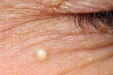
Atheroma of the eyelid
Eyelid diseases in ophthalmology are conventionally divided into inflammatory, infectious, benign tumor and malignant tumor pathologies. Atheroma of the eyelid is considered a benign neoplasm, not capable of malignancy, but one that requires timely treatment in the form of surgical removal. Atheroma is a cyst formed as a result of the accumulation of sebum and blockage of the excretory duct of the alveolar free gland. Such a neoplasm is differentiated from tumors with similar symptoms:
- Keratoacanthoma (epithelial neoplasm).
- Hemangioma.
- Wart.
- Papilloma.
- Nevus.
- Lipoma.
- Chalazion (meibomian cyst).
- Fibroma.
- External undeveloped barley of the eyelid.
- Internal stye of the eyelid.
- Blepharitis (simple, ulcerative, angular).
- Moll's cysts.
- Zeiss cysts.
- Contagious molluscum of viral etiology.
- Dermoid cyst of the eyelid.
- Seborrheic keratosis.
- Xanthelasma is a collection of lipid elements in the medial area of the eyelids.
- Follicular conjunctivitis.
- Hemangioma.
Atheroma of the eyelid is prone to inflammation, including purulent inflammation, which significantly complicates its treatment. It is much easier and safer to remove a small, simple cyst, which is enucleated completely - together with the capsule and contents in an outpatient setting. Inflamed atheromas often recur even after surgery due to the fact that access to the cavity is difficult, in addition, the boundaries of the neoplasm are erased and precise excision of the cyst is almost impossible. In this regard, a purulent cyst is treated, they wait for the symptoms to subside and a period of remission, after which a complete excision of the atheroma of the eyelid is performed. The tissue recovery period usually lasts no more than one and a half months, the suture is so microscopic that it is completely invisible and is not considered a cosmetic defect.
[ 21 ]
Atheroma of the lower eyelid
The fat layers of the upper and lower eyelids differ from each other. The largest accumulation of fat is noted at the septum of the eye, the upper eyelid contains two layers, the lower one is more saturated - it has three sections of the fat layer. Accordingly, there are more sebaceous glands below, which determines the reasons why atheroma of the lower eyelid is diagnosed 1.5 times more often than a similar cyst above.
Atheroma of the lower eyelid is a small dense neoplasm in the form of a tumor, painless and barely visible. The cyst does not affect vision until it grows to an impressive size, it takes a long time to form, but when inflamed, it quickly grows sometimes to 2-3 centimeters, covering the eyeball.
Differential diagnosis of lower eyelid atheroma is carried out with the following eye diseases:
- Xanthoma (xanthelasma) is a yellowish tumor that protrudes above the surface of the eyelid.
- Lipoma is a typical fatty tumor.
- Fibropapilloma.
- Hygroma.
- Senile wart.
- Meibomian gland cyst.
- Benign nevus of the eyelid.
Eyelid atheroma is treated only surgically. Adult patients do not require inpatient treatment, the procedure is performed on an outpatient basis under local anesthesia. Children under 7 years of age are hospitalized, since the cyst is removed under general anesthesia. The operation is a fairly simple procedure, complications are possible only in the form of recurrence of the atheroma due to its incomplete excision.
Atheroma on the nose
The largest sebaceous glands are located in the nasal area, especially in the skin of the wings of the nose and in the nasolabial triangle. The skin around the nose is quite thin, the tip of the nose and wings are denser and more textured, have enlarged pores. Since atheroma tends to form in the sebaceous glands, this is the determining factor in its localization in this area. Most often, nasal atheroma is diagnosed in the vestibulum nasi - the inner side of the wings, a place rich in small hairs and glandulae sebacea (alveolar glands). The outer part of the nose is also prone to the formation of lipomas, among which atheroma occupies a leading position.
Atheroma on the nose is similar in visual signs to the following skin neoplasms and diseases:
- Internal boils of the nose.
- Inflamed acne vulgaris.
- Lipomas.
- Fibromas.
- Phlegmonous acne.
- Dermoid cyst of the base of the nose.
- Papilloma.
The causes that provoke a sebaceous gland cyst in the nasal area may be the following:
- Oily skin type.
- Failure to observe hygiene and rules for facial skin care.
- Diseases of the gastrointestinal tract.
- Endocrine pathologies.
- Hypersecretion of sebaceous glands caused by hormonal disorders.
- Chronic acne, comedones.
- Seborrhea of the skin (the nose is one of the seborrheic zones).
Atheroma of the nose looks like a seal, clearly outlined, painless and slowly increasing. The cyst can become inflamed and transform into an abscess. After its opening, the atheroma increases again until its total excision by surgery. Independent removal or resorption of the cyst is impossible due to its structure, the capsule consists of epithelial cells, the contents - of cholesterol crystals, keratinized particles and sebum.
[ 24 ], [ 25 ], [ 26 ], [ 27 ]
How is atheroma in the nasal area treated?
There are several ways to remove a sebaceous cyst:
- Total enucleation of atheroma – the capsule, its contents, and often nearby tissues affected by the inflammatory process are removed. The operation is performed using a scalpel.
- Laser removal of cysts is possible only for small-sized neoplasms (up to 2-3 centimeters), in the absence of symptoms of inflammation or suppuration.
- Radio wave methods of evaporation of the capsule, contents and parallel coagulation of tissues and vessels.
All options for removing sebaceous gland cysts are considered effective if the atheroma has not become purulent, the operation does not take more than 30 minutes, the recovery period lasts no more than a month, when small scars after surgical manipulations are completely absorbed.
Diagnostics facial atheromas
Diagnosis of atheroma is not difficult, as a rule, the cyst is determined by inspection and palpation. A more accurate, specific picture is given by the result of histological examination, when tissue sampling is performed during removal.
Diagnosis of atheroma on the face does not require specific methods, most often it is enough to collect anamnesis, examine and palpate. An exception may be identified cysts in the eye and nose area, then CT - computed tomography, ultrasound, radiography in several projections are prescribed to clarify the diagnosis. A more accurate result, one way or another, is given by histology, which confirms the benign or other nature of the neoplasm on the face.
Differential diagnosis
Specific diagnostics of atheroma on the face consists precisely in differentiation, during which the cyst must be separated from similar tumors of the skin and subcutaneous tissue by external signs. These may be the following diseases:
- Molluscum contagiosum – contagious mollusk. Small seals in the form of nodules, painless, dense, with a small depression in the middle.
- Eyelid hail or meibomian gland cyst (chalazion).
- Lipoma is a typical fatty tumor, which is a classic benign fatty tumor.
- Fibroma.
- Blepharitis (eyelids).
- Milia are whiteheads.
- Hernia of the nasal root.
- Dermatomyofibroma.
- Keloid scar.
- Elastoma.
- Fibrous papule.
- Xanthogranuloma.
- Papilloma.
- Warts (seborrheic, senile).
- Nevus.
- Adenoma.
- Xanthoma.
- Dermoid cyst.
- Syringoma (blockage of sweat glands).
Who to contact?
Treatment facial atheromas
Treatment of sebaceous gland cysts in 100% of cases is surgery. It is necessary to immediately determine and learn the fact that due to its structure, atheroma cannot resolve on its own or with the help of conservative therapy, especially folk methods. A short-term reduction of the cyst is possible due to a breakthrough of the contents, it is good if this happens externally - on the skin, it is worse if the dendrite seeps into the subcutaneous tissue, this is fraught with an abscess, phlegmon. In the facial area, this is not only unacceptable, but also dangerous in terms of general blood poisoning, sepsis.
Treatment of atheroma on the face is carried out surgically at any stage of the process, with the exception of the period of inflammation and suppuration. Small cysts are removed with a laser without consequences for beauty, small stitches dissolve within a month and become almost invisible. Large atheromas are removed with a scalpel, in such cases, dissection of the skin is inevitable, accordingly, the scar can be quite large. Therefore, waiting for the cyst to increase in size is inappropriate, as well as relying on its “magical” spontaneous disappearance. The sooner the atheroma is cut out, the lower the risk of getting a cosmetic defect.
The operations are performed under local anesthesia, the procedure does not take much time, and recovery after surgery is not required.
Purulent atheromas require longer treatment. The abscess is opened, the wound is drained, antibacterial therapy is prescribed, 14-21 days after the symptoms of inflammation subside, the atheroma is excised completely to avoid relapses. The prognosis for treating atheroma is 100% favorable, such neoplasms are not prone to malignancy and never transform into a malignant process.
Removal of atheroma on the face
There are several generally accepted methods for removing atheroma on the face. Of course, every patient, regardless of gender, strives to keep the face intact and safe, that is, to avoid the appearance of unwanted scars. In this regard, removing atheroma on the face is really more specific, in contrast to operations on other parts of the body. However, excision of a cyst in the face is not difficult, the procedure lasts no more than 30 minutes, given the achievements of medicine and new technologies, atheroma can be called one of the safest and most favorable in terms of prognosis of diseases.
Removal of atheroma on the face, options:
- Surgical method using a scalpel. The atheroma is removed along with the membrane through a tiny incision, after which cosmetic stitches are applied.
- Laser removal of atheroma in the facial area is indicated for small neoplasms that do not have signs of inflammation. This method is considered effective, painless, and there are practically no scars left after the laser, which is very important for manipulations on the face.
- Radio wave method of "evaporation" of atheroma is one of the most popular methods that guarantees a relapse-free result. Contactless technology allows to do without sutures, without complications with the most precise, targeted introduction into the area of cyst formation. Radio wave removal of atheroma in the eye area, nasolabial triangle and cheeks is especially effective.
The choice of method depends on the condition of the atheroma - its size, the presence of signs of inflammation, its location, as well as the age of the patient. Removal of benign cysts is considered quite simple and is not accompanied by postoperative complications, so timely neutralization of atheroma can currently be considered a simpler procedure than even a facelift or other manipulations from the category of contour plastics.
Prevention
The main rule that helps prevent the development of various neoplasms on the face is considered to be regular skin care, including professional cleaning in beauty salons. Prevention of atheroma on the face can also include the following recommendations:
- Cleansing of skin pores with carefully selected products.
- Using steam baths and gently removing excess oil from the skin.
- Maintaining a healthy diet, including foods rich in fiber, vitamins and microelements. Limiting the consumption of spicy, sweet, fatty foods.
- Regular visits to a cosmetologist and following all of his or her advice on caring for problem areas of the face.
- It is mandatory to remove makeup every day before going to bed.
- Limit sun exposure (direct sunlight), use protective cosmetics with UV protectors.
- Taking vitamins A, E, C, complexes containing zinc, copper, iron, which help maintain the turgor and elasticity of facial skin.
- Avoid any attempts to remove pimples, acne, and comedones on the face on your own.
- Use only high-quality, certified cosmetics and skin care products.
- Timely actions to prevent the appearance of lipomas and cysts before the expected period of hormonal changes (puberty, menopause) - rational nutrition, use of special antiseptic agents (lotions, gels, scrubs, creams).
- Mandatory skin protection during winter to prevent dehydration, dryness and ultraviolet radiation.
Atheroma on the face is not a malignant neoplasm and never degenerates into an oncological process. However, in order to avoid purely cosmetic defects and the psychological discomfort associated with them, you should carefully care for your facial skin and promptly contact a cosmetologist if any atypical seals appear on it.