Medical expert of the article

New publications
Angiomyolipoma of the left and right kidney
Last reviewed: 04.07.2025

All iLive content is medically reviewed or fact checked to ensure as much factual accuracy as possible.
We have strict sourcing guidelines and only link to reputable media sites, academic research institutions and, whenever possible, medically peer reviewed studies. Note that the numbers in parentheses ([1], [2], etc.) are clickable links to these studies.
If you feel that any of our content is inaccurate, out-of-date, or otherwise questionable, please select it and press Ctrl + Enter.
Among benign tumors, a specific neoplasm such as angiomyolipoma stands out, which can be accidentally detected during visualization of abdominal organs. Although, reaching a certain size, this tumor can cause a number of symptoms and complications.
The most common location of angiomyolipoma is the kidneys, [ 1 ] the second is the liver; occasionally, such a tumor forms in the spleen, retroperitoneal space, lungs, soft tissues, ovaries, and fallopian tubes.
Epidemiology
Renal angiomyolipoma is the most common benign tumor of this organ; it develops sporadically in 0.2-0.6% of people after 40 years of age, and women predominate among patients. [ 2 ]
According to clinical statistics, in 80% of cases, single formations occur.
Angiomyolipoma is less common in children than in adults. According to research, 75% of children with tuberous sclerosis at the age of ten years are found to have renal angiomyolipoma, and in more than 50% of cases its size increases. [ 3 ], [ 4 ]
Causes angiomyolipomas
The exact causes of angiomyolipoma are unknown, and it can occur in middle-aged adults sporadically. But the version of the genetic origin of these tumors has strong evidence. Thus, most often, renal angiomyolipoma is associated with a hereditary genetic disease - tuberous sclerosis, which is caused by a mutation in one of two tumor suppressor genes: TSC1 (on the long arm of chromosome 9q34) or TSC2 (on the short arm of chromosome 16p13) and in which - due to excessive proliferation of cells - benign neoplasias of various localizations are formed, including multiple renal angiomyolipomas.
Renal parenchymal angiomyolipoma with cystic changes is detected in almost a third of patients with such a progressive systemic disease as pulmonary lymphangioleiomyomatosis (LAM), as well as in its fairly common combination with tuberous sclerosis. [ 5 ]
In children, there is a connection between the occurrence of multiple angiomyolipomas and the presence of congenital encephalotrigeminal angiomatosis (Sturge-Weber syndrome), caused by random mutations of the GNAQ gene (encoding guanine nucleotide-binding protein G), or with neurofibromatosis type I, which is the result of a mutation of the neurofibromin 1 (NF-1) protein gene.
Risk factors
Experts believe that the most serious risk factor for the development of this neoplasm is tuberous sclerosis: bilateral angiomyolipomas – angiomyolipoma of the left kidney and, at the same time, angiomyolipoma of the right kidney – are detected in 50-80% of patients.
Among the general factors that increase the risk of developing these tumors are genetic mutations that are inherited or occur spontaneously during intrauterine development, as well as the presence of certain systemic syndromes.
Pathogenesis
Angiomyolipomas are defined as mesenchymal tumors, that is, they are neoplasms of mesenchymal stem cells - adult pluro or multipotent stem cells of adipose, muscle and vascular tissues.
It is assumed that the pathogenesis of tumor formation is due to the increased ability of these cells to proliferate, self-renew by dividing and differentiating into cells of various tissues (including adipose, muscle and connective). And the morphological feature of classical angiomyolipoma is the presence in its composition of adipose tissue cells (adipocytes), smooth muscle cells and blood vessels (with thickened walls and abnormal lumens).
In addition, this tumor is considered a perivascular epithelioid cell neoplasm, which, as many researchers claim, originates from epithelioid cells (epithelioid histiocytes) adjacent to the walls of blood vessels. Being derivatives of activated macrophages, these cells are similar to epithelial cells, but have a modified cytoskeleton, denser cell membranes and an increased ability to aggregate and adhere. [ 6 ]
Symptoms angiomyolipomas
If the tumor is larger than 30-40 mm in diameter, the main symptoms – in cases where the tumor is located in the kidneys – are sudden pain (in the abdomen, side or back); nausea and vomiting; fever, hypotension/hypertension, anemia. Sometimes larger angiomyolipomas can bleed spontaneously, leading to hematuria.
But the clear first signs always manifest themselves as abdominal pain of varying intensity.
An extremely rare tumor, angiomyolipoma of the liver, is asymptomatic in most patients and is detected by chance; it may present itself as minor pain in the right hypochondrium and abdominal discomfort. However, intratumoral hemorrhage with bleeding into the abdominal cavity is also possible. [ 7 ]
Angiomyolipoma of the adrenal gland is also a very rare neoplasm (with a prevalence of 0.5-5%). As a rule, it does not manifest itself in any way, but when large, it causes abdominal pain. [ 8 ], [ 9 ]
Angiomyolipoma of the spleen may be isolated, but is more often associated with renal angiomyolipoma and tuberous sclerosis; the formation grows slowly and asymptomatically. With angiomyolipoma of the mediastinum against the background of tuberous sclerosis, dyspnea, chest pain, nausea and vomiting develop, pleural effusions are noted.
Angiomyolipoma of soft tissues behaves differently; it can form, for example, in the anterior abdominal wall (in the form of a dense node), in the skin of any localization, in deep muscle tissue.
Angiomyolipoma and pregnancy. If a pregnant woman has a renal angiomyolipoma that is not detected in time, its hemorrhagic rupture, causing bleeding, can have fatal consequences for both the mother and the fetus. [ 10 ], [ 11 ]
Forms
In the absence of a unified classification, the types or kinds of angiomyolipomas are distinguished not so much by localization as by their histology and visualized characteristics.
Renal angiomyolipomas are divided into sporadic (isolated) and hereditary (associated with tuberous sclerosis). Based on histological features, isolated angiomyolipomas are divided into three-phase (typical) and single-phase (atypical).
In addition, using computer and magnetic resonance imaging methods, the three-phase type is divided into tumors with a high content of fatty tissue and, accordingly, with a low content; specialists also take into account how fat cells are distributed in the tumor mass.
Tumors with a low content of adipocytes and a predominance of smooth muscle cells are called epithelioid or atypical, classifying them as potentially malignant neoplasms.
Complications and consequences
The main consequences and complications of angiomyolipoma include:
- spontaneous retroperitoneal bleeding that can lead to shock;
- dilation of tumor blood vessels (aneurysm);
- destruction of the renal parenchyma, leading to decreased renal function and end-stage renal failure (with the risk of requiring dialysis or nephrectomy).
Epithelioid angiomyolipoma – in rare cases – can transform into a malignant tumor with metastases to the lymph nodes. [ 12 ]
Diagnostics angiomyolipomas
Visualization, that is, instrumental diagnostics, plays a key role in the detection of renal angiomyolipomas. [ 13 ]
And analysis (microscopic examination) of a tumor tissue sample obtained by percutaneous renal biopsy [ 14 ] is necessary to differentiate benign mesenchymal tumor with a small content of adipocytes from renal cell carcinoma. In case of epithelioid angiomyolipoma, immunohistochemical analysis of a tumor tissue sample may additionally be required. [ 15 ]
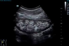
Classic angiomyolipoma with a sufficient amount of fatty tissue on ultrasound shows a hyperechoic mass, and formations smaller than 30 mm can give acoustic shadowing. The echogenicity is lower, the less fat cells in the neoplasm, so experts note the insufficiency of the diagnostic reliability of ultrasonography (especially for small tumors).
The density of adipocytes in the renal tumor is clearly demonstrated by angiomyolipoma on CT – in the form of dark (hypodense) foci. [ 16 ]
If the tumor contains too few fat cells, angiomyolipoma is examined on MRI, which allows visualization of fat areas in the mass and their location by comparing T1-weighted images with and without frequency-selective fat suppression.[ 17 ]
Differential diagnosis
Differential diagnostics is extremely important. If the tumor has a low number of adipocytes, i.e. it is closer to an epithelioid formation, angiomyolipoma or kidney cancer (renal cell carcinoma, sarcoma, etc.) should be differentiated.
Differentiation is also made with retroperitoneal liposarcoma with renal involvement, oncocytoma, Williams tumor, and adrenal myelolipoma.
Treatment angiomyolipomas
For each patient, treatment of angiomyolipoma is carried out taking into account the tumor size and clinical picture. Its goal is to protect the renal parenchyma while maintaining their functions. [ 18 ]
Methods vary from active surveillance (from the moment the tumor is detected) to drug treatment and surgery. Monitoring of small renal angiomyolipomas (less than 40 mm in size) includes annual ultrasound of the kidneys, and if the tumor grows (usually by 5% per year) – CT of the kidneys.
The criteria for eligibility for treatment, according to the recommendations of the European Association of Urology (EAU), are large tumors and the presence of symptoms.
Although pharmacological therapy for angiomyolipomas is still under investigation, the main drugs used for renal angiomyolipomas associated with tuberous sclerosis and/or lymphangioleiomyomatosis are mTOR inhibitors of the protein rapamycin (mTOR), which stop cell proliferation: Rapamycin (Sirolimus), Everolimus (Afinitor), Temsirolimus, Zotarolimus.
The use of these drugs is long-term, so one should keep in mind the high risk of their side effects, including a decrease in the number of leukocytes and platelets and an increase in the level of cholesterol in the blood, arterial hypertension, abdominal pain and intestinal disorders, an increase in the frequency of infections (bacterial, viral, fungal). [ 19 ]
For large neoplasms in the kidneys, surgical treatment is performed:
- selective vascular embolization; [ 20 ]
- radiofrequency or cryoablation of the tumor; [ 21 ]
- partial or total nephrectomy.
For patients with decreased kidney function, therapeutic nutrition is additionally recommended – diet No. 7.
Prevention
Preventive measures include early detection of angiomyolipomas, and arterial embolization is used as a preventive treatment for symptomatic or large tumors (due to the risk of bleeding).
Forecast
If angiomyolipomas do not enlarge rapidly and do not have dilated blood vessels, the prognosis is favorable. Otherwise, severe renal failure develops, requiring removal of the kidney or constant dialysis.