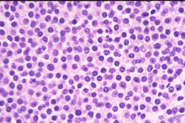
Plasmacytoma
Last reviewed: 23.04.2024

All iLive content is medically reviewed or fact checked to ensure as much factual accuracy as possible.
We have strict sourcing guidelines and only link to reputable media sites, academic research institutions and, whenever possible, medically peer reviewed studies. Note that the numbers in parentheses ([1], [2], etc.) are clickable links to these studies.
If you feel that any of our content is inaccurate, out-of-date, or otherwise questionable, please select it and press Ctrl + Enter.

Such a disease as a plasmacytoma causes quite a lot of interest in the field of immunological research, since it is distinguished by the production of a huge number of immunoglobulins with a homogeneous structure.
Plasmacytoma refers to malignant tumors consisting of plasma cells growing in soft tissues or within the axial skeleton.
Causes of the plasmacytomas
Doctors still have not figured out exactly what causes the B-lymphocytes to mutate into myeloma cells.
 [11],
[11],
Risk factors
The main risk factors for this disease were identified:
- Elderly men and middle-aged men - plamocytoma begins to develop when the amount of male hormone testosterone in the body decreases.
- People under the age of 40 account for only 1% of patients with this disease, so it can be argued that the disease more often affects after 50 years.
- Heredity - about 15% of patients with plasmacytoma have grown up in families where relatives of cases of mutations of B lymphocytes have been registered.
- People with excess weight - with obesity, there is a decrease in metabolism, which can lead to the development of this disease.
- Irradiation with radioactive substances.
Pathogenesis
Plasmacytoma can occur in any part of the body. A single plasmacytoma of the bone arises from plasma cells located in the bone marrow, while the extramedullary plasmacytoma is believed to arise from plasma cells located on the mucous membranes. Both variants of the disease are different groups of neoplasms in terms of location, tumor progression and overall survival. Some authors consider single plasmacytic bone as marginal cellular lymphoma with extensive plasmacytic differentiation.
Cytogenetic studies show periodic losses in chromosome 13, chromosome arm 1p and chromosome arm 14Q, as well as areas in the chromosomal arms of 19p, 9q and 1Q. Interleukin 6 (IL-6) is still considered the main risk factor for the progression of disorders caused by plasma cells.
Some hematologists consider single plasmacytic bone as an intermediate stage in evolution from monoclonal gammopathy of unclear etiology to multiple myeloma.
Symptoms of the plasmacytomas
With the plasmacytoma or myeloma, the kidneys, joints and immunity of the patient most severely suffer. The main symptoms depend on the stage of the disease. It is noteworthy that in 10% of cases the patient does not notice any unusual symptoms, since the paraprotein is not produced by cells.
With a small number of malignant cells, the plasmacytoma does not manifest itself. But when the critical level of these cells is reached, the synthesis of paraprotein occurs with the development of the following clinical symptoms:
- Lomit joints - there are pain in the bones.
- The tendons are sick, the pathological protein is deposited in them, which violates the basic functions of the organs and irritates their receptors.
- Pain in the region of the heart
- Frequent fractures of bones.
- Reduced immunity - the defenses of the body are inhibited, as the bone marrow produces too few leukocytes.
- A large amount of calcium from the destroyed bone tissue enters the blood.
- Violation of the kidneys.
- Anemia.
- DIC-syndrome with the development of hypocoagulation.
Forms
There are three separate disease groups defined by the International Working Group on myeloma: a single bone plasmacytoma (SPB), an extraosteal, or extramedullary plasmacytoma (EP) and a multifocal form of multiple myeloma that is either primary or recurrent.
For simplicity, single plasmacytomas can be divided into 2 groups depending on the location:
- Plasmacytoma of the bone system.
- Extramedullary plasmacytoma.
The most common of these is a single plasmacytoma of the bone. It accounts for about 3-5% of all malignant tumors caused by plasma cells. Occurs in the form of lytic lesions within the axial skeleton. Extramedullary plasmacytoma is most common in the upper respiratory tract (85%), but can be localized in any soft tissue. Approximately half of the cases are paraproteinemia.
The solitary plasmacytoma
A solitary plasmacytoma is a tumor that consists of plasma cells. This disease of bone tissue is local, which is its main difference from plasmacytoma. In some patients, solitary myeloma first develops, which can then be transformed into a multiple.
With a solitary plasmacytoma, bone is affected in one area. When conducting laboratory examinations, the patient is diagnosed with impaired renal function, hypercalcemia.
In some cases, the disease is completely invisible, even without changing the main clinical indicators. The prognosis for the patient is more favorable than for multiple myeloma.
 [19], [20], [21], [22], [23], [24]
Extramedullary plasmacytoma
Extramedullary plasmacytoma is a serious disease in which plasma cells are transformed into malignant tumors with rapid spread throughout the body. As a rule, such a tumor develops in the bones, although in some cases it can be localized in other tissues of the body. If the tumor affects only the plasma cells, then an isolated plasmacytoma is diagnosed. With multiple plasmacytomas, one can speak of multiple myeloma.
Plasmocytoma of the spine
Plasmocytoma of the spine is characterized by the following symptoms:
- Strong pain in the spine. In this case, the pain can grow gradually, simultaneously with the increase in the tumor. In some cases, pain is localized in one place, in others - in the hands or feet. Such pain does not go away after taking OTC analgesics.
- The patient changes the sensitivity of the skin of the legs or hands. Often there is complete numbness, a tingling sensation, hyper- or hypoesthesia, an increase in body temperature, a fever, or vice versa a feeling of cold.
- It is difficult for the patient to move around. Gait changes, and walking problems may occur.
- Difficulty urinating and emptying the intestines.
- Anemia, frequent fatigue, weakness in the whole body.
Plasmocytoma of bone
When maturing B-lymphocytes in patients on the plasmacytoma bone under the influence of certain factors, a failure occurs - instead of plasmocytes, a myeloma cell is formed. It is characterized by malignant properties. The mutated cell begins to clone itself, which increases the number of myeloma cells. When these cells begin to accumulate, the plasmacytoma of the bone develops.
A myeloma cell forms in the bone marrow and begins to germinate from it. In bone tissue it is actively divided. Once these cells enter bone tissue, they begin to activate osteoclasts that destroy it and create voids inside the bones.
The disease proceeds slowly. In some cases, it may take about twenty years from the moment of B-lymphocyte mutation before the diagnosis of the disease.
Plasmocytoma of the lungs
Plasmacytoma of the lung is a relatively rare disease. Most often it affects men aged 50 to 70 years. Usually, atypical plasma cells germinate in the large bronchi. When diagnosing, you can see clearly limited, rounded greyish-yellow uniform nodules.
When the lung plasmacytoma is not affected by the bone marrow. Metastases are spread by the hematogenous way. Sometimes neighboring lymph nodes are involved in the process. Most often, the disease is asymptomatic, but in rare cases, such symptoms are possible:
- Frequent cough with sputum.
- Painful sensations in the chest.
- Increase in body temperature to low-grade figures.
During blood tests, no changes are detected. Treatment consists in conducting an operation with the removal of pathological foci.
Diagnostics of the plasmacytomas
Diagnosis of the plasmacytoma is carried out using the following methods:
- There is an anamnesis - the expert asks the patient about the nature of the pain, when they appeared, what other symptoms he can isolate.
- The doctor examines the patient - at this stage it is possible to identify the main signs of the plasmacytoma (the pulse is increasing, the skin is pale, multiple hematomas, tumor compaction on the muscles and bones).
- Conducting a general blood test - with myeloma, the indicators will be as follows:
- ESR - not lower than 60 mm per hour.
- Decrease in the number of erythrocytes, reticulocytes, leukocytes, platelets, monocytes and neutrophils in blood serum.
- Reduced hemoglobin level (less than 100 g / l).
- Several plasmatic cells can be detected.
- Carrying out a biochemical blood test - when a plasmacytoma is found:
- Increase in the amount of total protein (hyperproteinemia).
- Decrease in albumin (hypoalbuminemia).
- Increase of uric acid.
- Increase in the level of calcium in the blood (hypercalcemia).
- Increase in creatinine and urea.
- Conducting a myelogram - in the process the structure of cells that are in the bone marrow is studied. In the sternum, a puncture is made with the help of a special tool, from which a small amount of bone marrow is extracted. With myeloma, the indicators will be as follows:
- High level of plasma cells.
- A large number of cytoplasm was found in the cells.
- Normal hematogenesis is depressed.
- There are immature atypical cells.

- The study of laboratory markers of the plasmacytoma - blood from the vein is mandatory in the early morning. Sometimes you can use urine. When the plasmacytoma in the blood will be found paraproteins.
- Conducting a general analysis of urine - the determination of the physico-chemical characteristics of the patient's urine.
- Carrying out a radiography of bones - with the help of this method it is possible to find out the places of their defeat, and also to make the final diagnosis.
- Conducting spiral computed tomography - do a whole series of X-ray images, thanks to which you can see: where exactly the bone and spinal deformity has been destroyed and where the soft tissues have tumors.
Diagnostic criteria for a single bone plasmacytoma
The criteria for determining a single plasmacytoma of the bone are different. Some hematologists include patients with more than one lesion and an elevated myelomal protein and exclude patients who have progressed to a disease within 2 years or if they have a pathological protein after radiation therapy. Based on the results of magnetic resonance imaging (MRI), flow cytometry and polymerase chain reaction (PCR), the following diagnostic criteria are currently used:
- The destruction of bone tissue in one place under the influence of clones of plasma cells.
- Infiltration of bone marrow by plasma cells is not more than 5% of the total number of nucleated cells.
- Absence of osteolytic lesions of bones or other tissues.
- Absence of anemia, hypercalcemia or renal dysfunction.
- Low levels of serum or urinary monoclonal protein concentration
Diagnostic criteria for extramedullary plasmacytoma
- Determination of monoclonal plasma cells by tissue biopsy.
- Infiltration of bone marrow by plasma cells is not more than 5% of the total number of nucleated cells.
- Absence of osteolytic lesions of bones or other tissues.
- Absence of hypercalcemia or renal insufficiency.
- Low concentration of whey protein M, if present.
Differential diagnosis
Skeletal forms of the disease often progress to multiple myeloma for 2-4 years. Because of their cellular similarity, plasmacytomas should be differentiated from multiple myeloma. For SPB and extramedullary plasmacytoma, the presence of only one lesion localization (either in bone or in soft tissues), normal bone marrow structure (<5% of plasma cells), absence or low level of paraproteins is characteristic.
Who to contact?
Treatment of the plasmacytomas
Plasmacytoma or myeloma is treated with several methods:
- Operation of stem cell transplantation or bone marrow transplantation.
- Conducting chemotherapy.
- Radiation therapy.
- The operation to remove the bone that was damaged.
Chemotherapy is used for multiple plasmacytoma. As a rule, treatment is carried out with the help of only one drug (monochemotherapy). But in some cases a complex of several drugs may be needed.
Chemotherapy is a fairly effective method of treating myeloma. In 40% of patients there is complete remission, in 50% - partial remission. Unfortunately, many patients with the passage of time relapse disease.
To eliminate the main symptoms, plasmacytomas are prescribed various painkillers, as well as procedures:
- Magnetoturbotron - treatment is carried out using a low-frequency magnetic field.
- Electrosleep - treatment is carried out with the help of low-frequency impulse currents.
With myeloma, it is also necessary to treat associated diseases: kidney failure and calcium metabolism.
Treatment of a single bone plasmacytoma
Most oncologists use approximately 40 Gy for spinal injury and 45 Gy for other bone lesions. For lesions larger than 5 cm, the power of 50 Gy should be considered.
As reported in the Liebross et al. Study, there is no link between the dose of radiation and the disappearance of monoclonal protein.
Surgical treatment is contraindicated in the absence of structural imbalance or neurological disorders. Chemotherapy can be considered as the preferred method of therapy for patients who do not respond to radiation therapy.
Treatment of extramedullary plasmacytoma
Treatment of extramedullary plasmacytoma is based on the radiosensitivity of the tumor.
Combination therapy (surgical intervention and radiation therapy) is a common treatment, depending on the resectability of the lesion. Combined treatment can provide the best results.
The optimal dose of irradiation for local lesions is 40-50 Gy (depending on the size of the tumor) and is carried out for 4-6 weeks.
Because of the high rate of lymph node involvement, these zones should also be included in the radiation field.
Chemotherapy can be considered for patients with a refractory form of the disease or with recurrence of the plasmacytoma.
Forecast
Complete recovery with plasmacytoma is almost impossible. Only with single tumors and timely treatment can we talk about a complete cure. The following methods are used: removal of bone that has been damaged; transplantation of bone tissue; stem cell transplantation.
If the patient complies with certain conditions, then a prolonged remission may occur:
- With myeloma, no serious comorbidity was diagnosed.
- The patient shows high sensitivity to cytostatic drugs.
- There were no serious side effects in treatment.
With properly selected treatment from chemotherapy and steroid drugs remission can last two to four years. In rare cases, patients can live after diagnosis and treatment for ten years.
On average, with chemotherapy, 90% of patients live more than two years. If treatment is not to conduct a life span does not exceed two years.
 [40]
[40]

