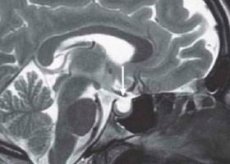
Syndrome "Empty Turkish saddle"
Last reviewed: 23.04.2024

All iLive content is medically reviewed or fact checked to ensure as much factual accuracy as possible.
We have strict sourcing guidelines and only link to reputable media sites, academic research institutions and, whenever possible, medically peer reviewed studies. Note that the numbers in parentheses ([1], [2], etc.) are clickable links to these studies.
If you feel that any of our content is inaccurate, out-of-date, or otherwise questionable, please select it and press Ctrl + Enter.

The phrase "empty Turkish saddle" (PTS) entered medical practice in 1951. After anatomical work, it was proposed by S. Busch, who studied autopsy material of 788 people who died from diseases not associated with pituitary pathology. In 40 cases (34 women), a combination of the almost complete absence of the Turkish saddle diaphragm with splaying of the pituitary in the form of a thin layer of tissue at its bottom was found. The saddle turned out to be empty. Similar pathology was previously described by other anatomists, but Busch was the first to relate a partially empty Turkish saddle to a diaphragm deficiency. His observations were confirmed by later studies. In the literature, this phrase means various nosological forms, a common feature of which is the expansion of the subarachnoid space into the intracellular region. The Turkish saddle at the same time, as a rule, increased.
Causes of the empty Turkish saddle syndrome
The cause and pathogenesis of the empty Turkish saddle are not completely clear. An empty Turkish saddle that develops after radiation or surgical treatment is secondary, and arising without prior intervention in the pituitary gland is primary. The clinical manifestations of the secondary empty Turkish saddle are due to the underlying disease and the complications of the applied therapy. This chapter is devoted to the problem of the primary empty Turkish saddle. It is believed that the development of an “empty Turkish saddle” requires the insufficiency of its diaphragm, i.e., a thickened protrusion of the dura mater forming the roof of the Turkish saddle and closing the exit from it. The diaphragm separates the cavity of the saddle from the subarachnoid space, excluding only the hole through which the pituitary stem passes. The attachment of the diaphragm, its thickness and the nature of the hole in it are subject to significant anatomical variations.
The line of its attachment to the back of the saddle and its tubercle may be reduced, the total surface is uniformly thinned, and the opening is expanded due to the almost complete reduction of the diaphragm, which remains in the form of a thin (2 mm) rim around the periphery. The resulting failure in this case leads to the spread of the subarachnoid space in the intracellular region and to the ability of the CSF to directly affect the pituitary gland, which can lead to a decrease in its volume.
All variants of the congenital pathology of the structure of the diaphragm determine its absolute or relative insufficiency, which is a necessary prerequisite for the development of the empty Turkish saddle syndrome. Other factors only predispose to the following changes:
- an increase in pressure in the suprasellar subarachnoid space, which, through an inferior diaphragm, enhances the effect on the pituitary gland (with intracranial hypertension, hypertension, hydrocephalus, intracranial tumors);
- a decrease in the size of the pituitary gland and a violation of the volumetric ratio between it and the Turkish saddle, in violation of the blood supply and infarction of the pituitary gland or adenoma (in diabetes, head injuries, meningitis, sinus thrombosis) as a result of physiological involution of the pituitary gland (during pregnancy, during this period the pituitary gland can double, moreover, in multiparous women, it becomes even larger, since after birth it does not return to its original volume after menopause, when the volume of the pituitary gland decreases, - such an involution can be observed in patients with primary hypofunction of the peripheral endocrine glands, in which there is an increase in the secretion of tropic hormones and hyperplasia of the pituitary gland, and the start of replacement therapy leads to involution of the pituitary and the development of an empty Turkish saddle; a similar mechanism is described after taking oral contraceptives);
- to one of the rare options for the development of an empty Turkish saddle - the rupture of an intracellular tank containing a liquid.
Thus, an empty Turkish saddle is a polyetiological syndrome, the main cause of which is the inferior diaphragm of the Turkish saddle.
 [1],
[1],
Symptoms of the empty Turkish saddle syndrome
An empty Turkish saddle is often asymptomatic and is accidentally detected during X-ray examination. The “empty Turkish saddle” is found mainly in women (80%), more often after 40 years of age, multipathing. About 75% of patients are obese. Clinical signs varied. Headache occurs in 70% of patients, which is the reason for the initial radiography of the skull, which in 39% of cases shows a modified Turkish saddle and leads to further more detailed examination. Headache varies widely in localization and extent - from mild, intermittent, to unbearable, almost constant.
Possible reduction of visual acuity, generalized narrowing of its peripheral fields, bitemporal hemianopsia. Swelling of the nipple of the optic nerve is rarely observed, but its descriptions in the literature are found.
Rhinorrhea is a rare complication associated with rupture of the bottom of the Turkish saddle under the influence of pulsation of cerebrospinal fluid. The emerging connection between the suprasellular subarachnoid space and the sphenoid sinus increases the risk of meningitis. The appearance of rhinorrhea requires surgical intervention, for example, the Turkish saddle tamponade with a muscle.
Endocrine disorders with an empty Turkish saddle are manifested in a change in the tropic functions of the pituitary gland. Studies using sensitive radio-immune methods and stimulation samples revealed a high percentage of hormone secretion dysfunction (subclinical forms). So, K. Brismer et al. Found that in 8 of 13 patients the somatotropic hormone secretion response to insulin hypoglycemia was reduced, and in the study of the pituitary-adrenal cortex axis, the secretion of cortisol after intravenous administration in 2 of 16 ACTH patients changed inadequately; reaction to metyrapone was normal in all patients. Unlike these data, Faglia et al. (1973) observed inadequate release of corticotropin on various stimuli (hypoglycemia, lysine-vasopressin) in all examined patients. The reserves of TSH and GT were also studied using TRG and RG, respectively. Samples showed a number of changes. The nature of these violations is still unclear.
There are more and more works describing the hypersecretion of tropic hormones in combination with an empty Turkish saddle. The first of these was information about a patient with acromegaly and an elevated level of somatotropic hormone. JN Dominique et al. Reported an empty turkish saddle in 10% of patients with acromegaly. Usually these patients also have pituitary adenoma. The primary empty Turkish saddle develops as a result of necrosis and involution by adenomas, and adenomatous residues continue to hypersecret the somatotropic hormone.
Most often, an increase in prolactin is observed in the “empty Turkish saddle” syndrome. Reported his growth in 12-17% of patients. As with GH hypersecretion, hyperprolactinemia and empty Turkish saddle are often associated with the presence of adenomas. Analysis of the observations shows that 73% of patients with an empty Turkish saddle and hyperprolactinemia have had adenomas during the operation.
There is a description of the primary “empty Turkish saddle” in patients with ACTH hypersecretion. These are more often cases of disease, Itsenko-Cushing disease with pituitary microadenoma. However, it is known about a patient with Addison's disease, in whom prolonged stimulation of corticotrophs due to adrenal insufficiency led to ACTH-secreting adenoma and an empty Turkish saddle. Of interest is the description of 2 patients with an empty Turkish saddle and ACTH hypersecretion at normal cortisol levels. The authors put forward an assumption about the production of ACTH-peptide with low biological activity and the subsequent infarction of hyperplastic corticotrophs with the formation of an empty Turkish saddle. A number of authors cite examples of isolated ACTH deficiency and an empty Turkish saddle, a combination of an empty Turkish saddle and adrenal carcinoma.
Thus, endocrine dysfunction in the empty Turkish saddle syndrome is extremely diverse. There are both hyper- and hyposecretion of tropic hormones. Violations range from subclinical forms detected by stimulation samples to pronounced panhypopituitarism. The variability of changes in endocrine function corresponds to the breadth of aetiological factors and the pathogenesis of the formation of a primary empty Turkish saddle.
Diagnostics of the empty Turkish saddle syndrome
The diagnosis of the empty Turkish saddle syndrome is usually established during the examination to identify a pituitary tumor. It should be emphasized that the presence of neuro x-ray data, indicating an increase and destruction of the Turkish saddle, does not necessarily indicate a pituitary tumor. The incidence of primary intrasellar pituitary tumors and empty Turkish saddle syndrome was similar in these cases, 36 and 33%, respectively.
The most reliable for diagnosing an empty Turkish saddle is pneumoencephalography and computed tomography, especially in combination with the introduction of contrast media intravenously or directly into the cerebrospinal fluid. However, already on conventional X-ray and tomograms, it is possible to reveal signs characteristic of the empty Turkish saddle syndrome. These are localization of changes below the diaphragm of the Turkish saddle, symmetrical arrangement of its bottom in frontal projection, “closed” saddle shape, an increase mainly in vertical size, no signs of thinning and erosion of the cortical layer, a double-contour bottom on the sagittal image, with the lower one of the lines thick and clear, and the top one is blurred.
Thus, the presence of an “empty Turkish saddle” with its characteristic increase should be assumed in patients with minimal clinical symptoms and unchanged endocrine function. In these cases, there is no need for pneumoencephalography; the patient should simply be monitored. It should be emphasized that the empty Turkish saddle, accompanied by an increase in its size, is often observed with an erroneous diagnosis of pituitary adenoma. However, the presence of an “empty Turkish saddle” does not exclude a pituitary tumor. At the same time, differential diagnosis is aimed at determining the overproduction of hormones.
Of radiological methods for diagnosis, the most informative is a combination of pneumoencephalography and polytomographic studies.
What do need to examine?
How to examine?
Who to contact?
Treatment of the empty Turkish saddle syndrome
Special treatment for the empty Turkish saddle is not carried out. Although the combination with an empty Turkish saddle does not affect the treatment plan for a tumor, it is important for a neurosurgeon to know about its coexistence, since in these cases the risk of developing postoperative meningitis increases.
Prevention
Prevention of empty Turkish saddle includes the prevention of injuries, inflammatory diseases, including intrauterine, as well as thrombosis and tumors of the brain and pituitary.
Forecast
Empty Turkish saddle syndrome has a different prognosis. It depends on the nature and course of concomitant diseases of the brain and pituitary gland.

