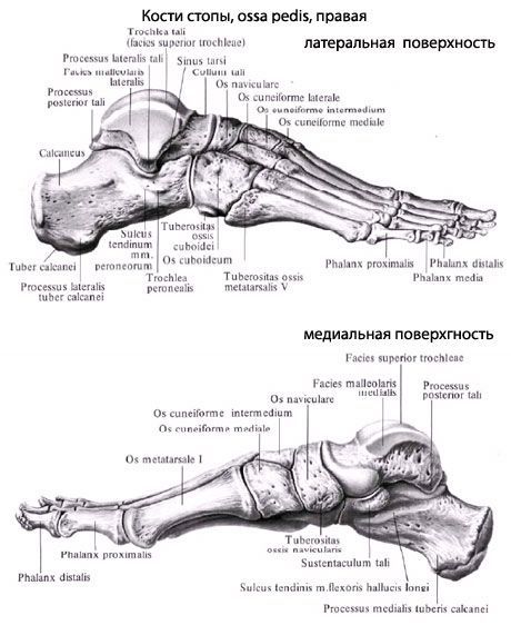
Heel bone (heel)
Last reviewed: 23.04.2024

All iLive content is medically reviewed or fact checked to ensure as much factual accuracy as possible.
We have strict sourcing guidelines and only link to reputable media sites, academic research institutions and, whenever possible, medically peer reviewed studies. Note that the numbers in parentheses ([1], [2], etc.) are clickable links to these studies.
If you feel that any of our content is inaccurate, out-of-date, or otherwise questionable, please select it and press Ctrl + Enter.
Calcaneus calcaneus is the largest bone of the foot. It is located under the talus bone and extends from under it. Behind the body of the calcaneus is a downward inclined tuber calcanei. On the upper side of the calcaneus, three articular surfaces are distinguished: the anterior, middle and posterior ram joint surfaces (faciei articulares talaris anterior, media et posterior). These surfaces correspond to the heel joints of the talus. Between the middle and posterior articular surfaces, the calcaneus groove (sulcus calcanei) is visible, which together with the same groove on the talus bone forms the sinus tarsi (sinus tarsi). The entrance to this bosom is located on the rear of the foot from its lateral side. From the anterior margin of the calcaneus, from the medial side, a short and thick process departs - the support of the talent bone (sustentdculum tali). On the lateral surface of the calcaneus there is a furrow of the tendon of the long fibular muscle (sulcus tendinis m.peronei longi). On the distal (anterior) end of the calcaneus, for articulation with the cuboid bone, there is a cuboid articular surface (facies articularis cuboidea).

Where does it hurt?
What do need to examine?
How to examine?


 [
[