
Patellar dislocation
Last reviewed: 07.06.2024

All iLive content is medically reviewed or fact checked to ensure as much factual accuracy as possible.
We have strict sourcing guidelines and only link to reputable media sites, academic research institutions and, whenever possible, medically peer reviewed studies. Note that the numbers in parentheses ([1], [2], etc.) are clickable links to these studies.
If you feel that any of our content is inaccurate, out-of-date, or otherwise questionable, please select it and press Ctrl + Enter.
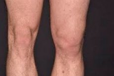
Patellar dislocation (patellar dislocation or patellar subluxation), is a medical condition in which the patella (the bony part located on the front of the knee) moves out of its normal position and moves to the side or around the joint of the knee. This condition can be caused by a variety of factors, and is most commonly associated with trauma or anatomical features of the knee structure. [1]
Symptoms of patella withdrawal may include:
- Pain: Sharp pain in the knee area at the time of dislocation and during movement.
- Swelling: Swelling and edema around the knee due to soft tissue and joint damage.
- Inability to move: The patient may have difficulty moving the leg and knee due to pain and discomfort.
- Muscle Spasms: Muscle spasms around the knee can occur in an attempt to hold the patella in place.
- Visual change: In case of a complete dislocation of the patella, its position may be visibly altered and even visible from the outside.
- Crunching or clicking: In patella dislocation, a clicking sound may be heard when the patella moves from its normal position.
Treatment for patellar luxation depends on the severity of the condition and often includes the following measures:
- Manual repositioning: The doctor can manually return the patella to its normal position.
- Immobilization: A cast, bandage or splint may be required to stabilize and protect the knee.
- Physical Therapy: Physical therapy exercises and rehabilitation can help restore strength and stability to the knee.
- Surgical treatment: In some cases, especially relapses or severe cases, surgery may be required to restore the structure and stability of the knee joint.
Treatment and prognosis for patella dislocation can vary depending on individual circumstances, and it is recommended to see a doctor for diagnosis and appropriate treatment.
Causes of the patella dislocation
This can happen for a variety of reasons, including:
- Trauma: One of the most common causes of patella dislocation is knee injury. This can be due to a bump, fall, accident, or sports injuries that can cause dislocation of the patella.
- Muscle and Ligament Weakness: Underdevelopment or weakness of the surrounding muscles and ligaments around the knee can contribute to patellar dislocation.
- Genetic factors: Some people may have more mobile or less stable joints, which can increase the risk of patellar luxation.
- Joint wear and tear: Osteoarthritis, in which the cartilage tissue in the joint wears down, can increase the likelihood of patellar dislocation.
- Congenital Anomalies: In some cases, abnormalities in the joint structure of the knee from birth can contribute to patellar dislocation.
- Increased stress on the knee: For example, athletes who jump or run with frequent stress on the knee may have an increased risk of patellar dislocation.
- Dysfunction of the tibialis anterior muscle (quadriceps): Problems with the function of this muscle can increase the risk of patellar luxation.
Symptoms of the patella dislocation
This is a serious injury that can come with a variety of signs and symptoms. Here are some of them:
- Acute pain: Usually when the patella is dislocated, there is a sharp and intense pain in the knee area. The pain may worsen with movement or attempts to use the leg.
- Swelling: The site of a dislocation can swell quickly due to fluid and blood pooling in the area of injury.
- Limited movement: A dislocated patella can make the leg stiff and restricted in movement. The injured person may not be able to bend or straighten the leg at the knee joint.
- Instability: The knee joint may feel unstable, and the injured person may feel as though their leg is unsupported.
- Bruising and redness: Bruising and redness may occur at the site of injury due to damage to blood vessels.
- Sensitivity and numbness: In some cases, there may be sensitivity or numbness in the area of injury.
Stages
Patellar dislocation (patellar dislocation) can be classified according to its severity. There are the following degrees of patellar dislocation:
-
Grade I (mild):
- In this degree of dislocation, the patella exits the patellofemoral sulcus but immediately returns to its place without intervention.
- Typically, patients may feel pain or discomfort in the knee, but usually this type of dislocation is easily corrected on its own or with chiropractic intervention.
-
Grade II (moderate):
- In this degree, the patella exits the patellofemoral sulcus and remains everted, but can be returned to position without the use of surgery.
- Patients feel more severe pain and discomfort than with Grade I, and medical attention may be needed to correct the position of the patella.
-
Grade III (severe):
- In this degree, the patella comes out of the patellofemoral sulcus and becomes stuck on the outside. Returning the patella back into place can be painful and may require medical attention.
- Pain and discomfort increase and surgery may be necessary to restore the patella to its normal position.
-
Grade IV (permanent dislocation):
- In this degree, the patella remains permanently dislocated and cannot be put back in place without surgical intervention.
- This is the most severe form of patellar dislocation and requires surgical treatment to restore the structure and function of the knee.
Forms
There are several different types of patellar dislocations, including the following:
- Traumatic patella dislocation: This type of dislocation is caused by trauma or injury to the knee. For example, a sudden movement or injury can cause the patella to dislocate. A traumatic dislocation may be accompanied by pain, swelling, and other symptoms.
- Habitual patellar dislocation: Habitual dislocation means that the patella goes out of its normal position after an injury, but can also easily return to normal. This may be due to lability or instability of the joint that requires attention and treatment.
- Recurrent patellar dislocation: This type of dislocation is characterized by repeated instances of patellar dislocation even after recovery. It can be caused by structural abnormalities, muscle weakness, or other factors that make the knee joint less stable.
- Congenital dislocation of the patella: Congenital dislocation is caused by abnormalities in the structure of the joint or bones of the leg that result in the patella not being in its normal position from the beginning of life. This condition may require surgery to correct.
- Medial patellar dislocation: Medial dislocation means that the patella is displaced inward from its normal position when viewed from the frontal plane (looking at the front of the knee). This type of dislocation can be caused by anatomical features and requires special attention and treatment.
Complications and consequences
A patellar dislocation can lead to a variety of complications and consequences, especially if it does not receive proper treatment and care. Uncontrolled or recurrent patella dislocation can have serious consequences on the health and function of the knee joint. Here are some of the possible complications and consequences:
- Soft tissue damage: A patella dislocation can be accompanied by damage to the soft tissues around the knee, such as ligaments, tendons and the joint bag. This can cause pain, swelling, inflammation and restriction of movement.
- Synovitis: Synovitis, an inflammation of the joint lining, can develop as a result of damage to the joint bag. This can lead to pain, swelling and restricted movement.
- Chronic instability: Recurrent patellar dislocations can lead to chronic instability of the knee joint, making it difficult to function normally and increasing the risk of further damage.
- Osteoarthritis: Constant injury and instability can accelerate the development of osteoarthritis in the knee joint. This condition is characterized by cartilage destruction and pain in the joint.
- Surgery: In some cases, especially severe dislocations and instability, surgical treatment such as ligament reconstruction or correction of anatomical anomalies may be required. Surgical intervention may involve risks and may require rehabilitation.
- Loss of function: In uncontrolled cases of patellar dislocation, especially in the absence of effective treatment and rehabilitation, the knee joint can lose function, which can limit movement and the patient's ability to perform daily tasks.
- Psychological aspects: Persistent pain and limitations due to patellar dislocation can affect the patient's psychological state, causing depression, anxiety and limiting quality of life.
Diagnostics of the patella dislocation
Diagnosis of patellar dislocation usually involves physical examination, clinical tests, and instrumental studies. Here are some diagnostic methods that may be used:
- Physical Exam: The doctor will perform a thorough physical examination of the knee, including evaluation of symptoms, motion, and joint stability. He or she may also try to activate the displaced patella back into the joint (manipulation).
- Clinical Tests: The physician may perform specific clinical tests such as the Lachman'a test and the McMurray test to assess joint stability and determine if there is a patellar output.
- Radiography: Radiography can be performed to evaluate bone structure and determine if there are any abnormalities in the position of the patella. It also helps to rule out other conditions that may mimic patellar luxation.
- Magnetic resonance imaging (MRI): MRI can be used to visualize the soft tissues, ligaments, and cartilage in the knee joint in greater detail. This helps to identify damage associated with patellar output.
- Ultrasound: Ultrasound can be used to visualize joint structures and detect ligament and soft tissue injuries.
- Arthroscopy: Some cases may require arthroscopy, a minimally invasive procedure that uses an endoscope to visualize the joint and perform surgical procedures.
Who to contact?
Treatment of the patella dislocation
Treatment for patella dislocation may depend on the severity of the injury and the presence of complications. Treatment usually involves the following methods and steps:
-
Assessment and diagnosis:
- If a patella dislocation is suspected, the patient should immediately see a physician or go to the nearest medical facility to have the injury diagnosed and evaluated.
- The doctor performs a physical exam, and may order x-rays or other educational studies to confirm the diagnosis and determine the extent of the injury.
-
Reduction (recovery):
- The primary step is to restore the proper position of the patella in the knee joint, which is called reduction.
- Reduction is usually performed by an experienced physician who carefully and gently performs maneuvers to put the joint back in place.
-
Immobilization:
- After a successful reduction, it may be necessary to wear a special bandage, plaster cast, or other fixation to prevent re-dislocation and provide stability to the joint.
- The length of time immobilization is worn may depend on the severity of the injury and the doctor's recommendations.
-
Medication treatment:
- The patient may be prescribed anti-inflammatory drugs or painkillers to relieve pain and inflammation.
- Antibiotic therapy may be required if infection or other complications are present.
-
Physical therapy and rehabilitation:
- After fixation and immobilization, physiotherapy and rehabilitation begins. Physiotherapy exercises help to restore strength, flexibility and functionality of the joint.
- Rehabilitation may include massage, balance exercises, muscle strengthening, and other methods appropriate for each individual case.
-
Surgical intervention (if necessary):
- In cases of severe damage, complications or recurrences of patella dislocation, surgery may be required to repair the knee joint.
- Surgical treatment may include ligament reconstruction, repair of damaged structures, or other procedures.
Immobilization, plaster and taping
Immobilization, plaster and taping can be used in the treatment of patellar dislocation, depending on the characteristics and extent of the injury. Here is how they can be applied:
-
Immobilization:
- Immobilization involves restricting movement of the joint to prevent additional damage and promote healing.
- For this purpose, various types of bandages or orthotics can be used to fix the patella in the correct position and provide support. These bandages can be soft or rigid, depending on the extent of the injury and the doctor's recommendations.
- Immobilization may be used in the initial phase of treatment and then decreased as the patient recovers.
-
Gypsum:
- Plaster immobilization may be recommended in cases of severe dislocation or to eliminate the risk of additional injury and provide stable fixation of the patella.
- The plaster bandage usually stays in place for a certain period of time, which can vary depending on the nature of the injury.
- After the cast is removed, physical therapy may be required to restore motor function.
-
Taping:
- Taping (sticking) can be an option for treating patella dislocation, especially in cases where support and stabilization is needed but a cast is not required.
- A physical therapist or medical professional may use medical tape (adhesive tape such as Kinesio tape) to create support and stabilize a joint.
- Taping can provide support without complete immobilization, allowing the patient to be more involved in the recovery process.
Operation
Surgery to repair a dislocated patella can be performed using different methods and techniques depending on the specific circumstances and surgical practice. The following are common steps and techniques for performing patella repair surgery:
-
Patient Preparation:
- The patient undergoes a preoperative evaluation, including a physical examination and discussion of medical history.
- Additional diagnostic tests, such as X-rays, MRI or CT scans, may be needed to more accurately evaluate the knee joint.
-
Anesthesia: Before the surgery begins, the patient is given general or local anesthesia to ensure a pain-free operation.
-
Access to the knee joint:
- The surgeon creates access to the knee joint, usually by making a small incision in the skin above the knee. The incision may be made on the front or side of the knee, depending on the specific technique.
-
Ligament Restoration:
- If the surgery involves repairing damaged ligaments, the surgeon may use the patient's own tissues (most commonly tendons or tendon grafts) or artificial materials.
- Ligament repair restores stability to the knee joint and prevents patella dislocation.
-
Assessment and Testing:
- Once the ligaments have been repaired, the surgeon evaluates and tests the stability of the knee joint to ensure that the patella is no longer out of position.
-
Wound closure:
- After the surgery is complete, the surgeon closes the wound using stitches or special adhesives.
-
Post-operative care:
- After surgery, the patient is sent to the intensive care unit for observation and recovery from anesthesia.
- Postoperative care includes controlling pain, inflammation, and infection, as well as beginning physical therapy and rehabilitation to restore knee function.
-
Physical therapy and rehabilitation:
- Physical therapy and rehabilitation play an important role in the recovery process after surgery. The patient is prescribed exercises to restore strength and mobility of the knee, as well as to strengthen the surrounding muscles and tendons.
Rehabilitation
Rehabilitation after a patella dislocation plays an important role in restoring joint function and strengthening the surrounding muscles and ligaments. Here are some general guidelines and exercises that can help in the recovery process after a patella dislocation:
- Start with a physical therapist: It is recommended that you be evaluated by a physical therapist or rehabilitation therapist before beginning any exercises. They can design an individualized recovery program, taking into account the nature and extent of the injury.
- Move gradually: Don't rush your recovery. Start with light and slow movements to avoid the risk of re-injury.
- Work on mobility: Exercises to restore mobility in the patella include smooth and gentle movements such as flexibility and stretching. For example, smoothly bending and extending the leg at the knee joint.
- Muscle Strengthening: Exercises to strengthen the thigh and calf muscles will help improve patellar stability and prevent recurrent dislocation. Examples include raising your leg while lying or sitting.
- Balance and coordination: Balance and coordination training can help restore joint stability and reduce the risk of falls. Single leg exercises and the use of balance platforms can be helpful.
- Load control: It is important to avoid putting excessive strain on the patella and not to overload it. Listen to your body and stop if you feel pain or discomfort.
- Therapeutic exercises: The physiotherapist may prescribe specific exercises to restore the function of the patella. These may be exercises using elastic bands, elastic bandages or special exercise machines.
- Medical supervision: Regular consultations with the doctor and physiotherapist will help to monitor progress in recovery and make necessary adjustments to the rehabilitation program.
- Working with the level of pain and swelling: If pain and swelling are present, cold and heat therapy should be used as recommended by your doctor.
- Adherence to recommendations: It is important to follow your healthcare professional's recommendations and instructions to ensure optimal recovery.
List of authoritative books and studies related to the study of patellar dislocation
-
"Knee Ligament Injuries: Extraarticular Surgical Techniques" (Author: Guy Lavoie, Genre-Jacques Yves), 2006.
- This book focuses on surgical techniques for treating a variety of knee injuries and trauma, including ligamentous injuries that may be associated with patellar dislocations.
-
"The Knee: A Comprehensive Review" (by John A. Feagin Jr., Robert D. Warren), 2010.
- This book is an overview of the anatomy and function of the knee joint, as well as various conditions and injuries, including traumatic patellar dislocations.
-
"Anterior Knee Pain and Patellar Instability" (Author: Vicente Sanchis-Alfonso), 2011.
- This study investigates the problem of anterior knee pain and patellar instability, which may be associated with dislocations.
-
"Patellofemoral Pain, Instability, and Arthritis: Clinical Presentation, Imaging, and Treatment" (by Jack Farr, Robert Nirschl), 2010.
- This book discusses various aspects of patellar pain and instability and may contain information on patellar dislocations.
Literature
Kotelnikov, G. P. Traumatology / edited by Kotelnikov G. P.., Mironov S. P. - Moscow: GEOTAR-Media, 2018.