
Chronic odontogenic osteomyelitis
Last reviewed: 07.06.2024

All iLive content is medically reviewed or fact checked to ensure as much factual accuracy as possible.
We have strict sourcing guidelines and only link to reputable media sites, academic research institutions and, whenever possible, medically peer reviewed studies. Note that the numbers in parentheses ([1], [2], etc.) are clickable links to these studies.
If you feel that any of our content is inaccurate, out-of-date, or otherwise questionable, please select it and press Ctrl + Enter.
The consequence of complicated acute osteomyelitis can become chronic odontogenic osteomyelitis - a severe dental pathology that runs with purulent inflammatory reaction and accumulation of purulent masses in the cavities of bone tissue. Affects the bone, bone marrow, as well as the surrounding soft tissues against the background of previous sensitization of the body. The disease has different variants of course, its diagnostic and therapeutic features. [1]
Epidemiology
In childhood, chronic odontogenic osteomyelitis is caused predominantly by obligate-anaerobic and facultative-anaerobic microorganisms. The composition of purulent microflora depends on the age of the patient. Thus, the older the patient, the greater the number of associations and strict anaerobes can be discussed.
It has been found out that in odontogenic osteomyelitis the microflora is often represented by an average of five or six varieties of aerobic and anaerobic microorganisms, or more.
Chronic odontogenic osteomyelitis is not an uncommon condition in the practice of dental surgeons. It occurs as often as jaw periostitis or chronic periodontitis. Among all cases of osteomyelitis to the share of odontogenic pathological process accounts for about 30%. The disease is more often found in young and middle-aged people (the average age of the diseased is 25-35 years). Men are somewhat more often ill than women. In most cases, the lower jaw is affected.
Causes of the chronic odontogenic osteomyelitis.
The primary cause of chronic odontogenic osteomyelitis is actually acute osteomyelitis, which has not been treated, or it was treated incorrectly or incompletely. In turn, acute pathology can develop as a result of multiple causes, which are closely related to the entry of pathogens into the bone tissue through the circulatory system. "The culprits" more often become bacteria, less often - viruses and fungal infection.
Infection of the bone occurs due to the following factors:
- Dental trauma, carious teeth, other dental pathologies, including periodontitis, periostitis, granuloma, etc.;
- Sepsis, bacteremia;
- Any acute and chronic infectious diseases in the body;
- Lack of oral hygiene, or insufficiently careful observance of hygiene rules;
- Facial boils;
- Purulent otitis media, tonsillitis;
- Scarlet fever;
- Umbilical inflammatory reactions (purulent-septic complications);
- Diphtheria.
In childhood, the causes are often specific, as they are related to the anatomical and functional features of the child's body. Thus, among the most common "pediatric" causes are the following:
- Active bone growth;
- The change of baby teeth and the formation of permanent molars;
- Alteration of the maxillofacial structure;
- Thinning dental plates and wide tubular spaces;
- An extensive capillary network;
- Imperfect immune system, excessive susceptibility to pathologic pathogens.
Odontogenic osteomyelitis occurs when pathogens enter from diseased teeth or other dental infectious foci. [2]
Risk factors
- Physiologic and anatomical features of the jaw structure:
- Active growth of the bone system;
- Changes in the replacement of deciduous teeth;
- Enlarged Haversian canals;
- Susceptible trabeculae of bone;
- Infection-susceptible myeloid bone marrow;
- Extensive blood and lymphatic network.
- Weak nonspecific defenses, weakened by fatigue, stress, hypothermia, infectious diseases (ARVI, adenovirus, etc.), injuries, other pathological conditions.
- Immunopathologies, both congenital and acquired, associated with diabetes mellitus, hemopathologies, etc.
- General immunological disorders, prolonged existing odontogenic pathology, unfavorable changes in the tissues and vessels of the bone marrow.
Pathogenesis
To date, the following pathogenetic versions of the development of chronic odontogenic osteomyelitis are known:
- Infectious-embolic version of Bobrov-Lexer: inflammatory bone reaction develops due to embolic transportation of the infectious agent with its blockage in the end segments of capillary vessels, or when they are thrombosed. Disorder of blood flow and improper bone trophism leads to bone necrosis, and subsequent infection entails the development of purulent inflammation.
- Dr. S. Derijanov's version of allergic conditioning: bone deadening occurs due to toxic effects of re-formed autoimmune bodies, as a response to repeated penetration of "foreign" protein.
- The inflammatory reaction extends beyond the periodontal boundaries, and the primary source and area of entry of infectious agents becomes the previous pathology of soft tissue or hard tissue dental structures, as well as the periodontium.
- The processes of regeneration in periosteum and bone in acute osteomyelitis are absent or insufficiently manifested, which leads to the predominance of bone destruction and the formation of the following destructive foci.
Symptoms of the chronic odontogenic osteomyelitis.
From the moment the infection enters the bone tissue to the appearance of the first pathological manifestations can take a long time. At first, the patient begins to experience discomfort when chewing food, then - and in a calm state. Periostitis begins to develop. With the increase in inflammatory phenomena, the clinical picture expands:
- Pain syndrome increases, there is an irradiation to the ear, temple;
- Oral tissues swell, gums become painful;
- The teeth on the inflamed side become pathologically mobile;
- Difficulty chewing and swallowing food;
- In mandibular odontogenic osteomyelitis, sometimes the chin area is numb;
- There's bad breath;
- Speech impediments;
- The regional lymph nodes are enlarged;
- Changes the roundness of the face.
With the development of a purulent abscess, the temperature rises, a fistulous canal is formed, through which purulent masses flow outward.
After the acute period (about 2 weeks), the pathology passes into the subacute stage: the purulent mass comes out through the fistula, swelling subsides, pain subsides, but problems with chewing remain, the teeth are still loose (may also fall out). Then formed directly chronic course of odontogenic osteomyelitis. The clinical picture becomes more sluggish, for several weeks there is tissue rejection. After some time, necrotized tissues together with pus come out through the fistulous canal, or the development of an extensive abscess is noted. [3]
First of all, in the exacerbation of chronic odontogenic osteomyelitis, there are signs of general intoxication:
- Elevated temperature;
- General weakness, malaise, chills;
- Dyspepsia;
- The patient is passive, the skin is pale, the general condition is moderate to severe.
On external examination, facial asymmetry due to collateral soft tissue edema is noteworthy. There is a muft-like infiltrate, the teeth on the affected side are mobile, there is edema of the gingiva and transitional fold of the mucosa. The tissues are hyperemic, the gingiva is painful on palpation.
Regional lymph nodes are enlarged and painful. The patient cannot open the mouth, or opens it with difficulty and incompletely. There is a putrid odor from the oral cavity. [4]
Chronic odontogenic osteomyelitis in children
Features of the course of odontogenic osteomyelitis in childhood:
- Chronicity of the process in children is much less frequent than in adult patients;
- More often develop complications such as lymphadenitis, phlegmons, abscesses;
- If the pathological process spreads to the rudiments of teeth, partial adentia may occur;
- Pathology in the frontal teeth is not as severe as in the molars;
- Pediatric odontogenic osteomyelitis is characterized by a particularly intense start, rapid development of the inflammatory response and faster recovery (provided competent radical treatment);
- There is virtually no sequestrum capsule formation.
Stages
The course of chronic odontogenic osteomyelitis goes through three stages:
- At the first stage, the acute symptomatology subsides, temperature indicators stabilize to normal, signs of intoxication are also leveled. Some time after the start of the inflammatory reaction, some relief is observed: the pain syndrome ceases to bother, patients practically return to their previous way of life. Such a "lull" can last for several weeks. At the same time, cavity spaces are formed in the bone, purulent mass from the fistula holes almost does not come out. On external examination, swelling is present only to a small extent.
- At the second stage, recurrent inflammation develops like an acute form of odontogenic osteomyelitis, but the temperature does not exceed +38°C, pain is not severe, and signs of intoxication may not be present at all. The fistula hole becomes blocked. The purulent mass spreads to the bone and soft tissue structures. It is possible to develop complications in the form of phlegmon or abscess. Their formation causes the appearance of severe pain syndrome and fever: the condition normalizes only after the repeated breakthrough of pus outside.
- The third stage is characterized by deformation of the affected bone structures against the background of recurrence of chronic odontogenic osteomyelitis. Externally, curvature and changes in the size of the bone and the face as a whole are noticeable.
Forms
Depending on the clinical and radiologic picture, the following forms of chronic odontogenic osteomyelitis are distinguished:
- Destructive;
- Productive;
- Destructive-productive form.
Common to all forms of chronic osteomyelitis is a prolonged course and periodic relapses, so the disease requires long-term therapy and medical supervision.
Any of the forms of the disease can be considered as an unstable state, which under the influence of a provoking factor (a strong drop in immunity as a result of viral infection, stress, hypothermia, etc.) will again manifest itself as a relapse.
- Destructive variant of chronic odontogenic osteomyelitis involves a large proportion of bone tissue. In the area of mucosa or skin, fistulous canals with protruding granulation appear. X-rays show bone lysis with the formation of sequestra.
- Destructive-productive variant is usually preceded by acute osteomyelitis and there is a secondary immunodeficiency state. Destruction and restoration of bone tissue occur in equilibrium. Bone substance is fused diffusely (small sparse foci and small sequestration). The sequestration capsule is not defined.
- The productive variant is otherwise known as hyperplastic: it develops in children and young adults during the active period of facial bone development (approximately 12-18 years of age). Such osteomyelitis is characterized by a particularly long course and frequent relapses (about 7 times a year). Pathogenetic indicators of this form of odontogenic lesions: virulent microorganisms and a weak immune response of the body. Secondary foci of infection are usually represented by infected teeth and embryos of dead teeth. The radiograph reveals pronounced layering of periosteal bone tissue with a slight trabecular pattern and small focal sclerosis.
Depending on the localization of the pathological process, odontogenic mandibular or maxillary osteomyelitis is distinguished.
- Chronic odontogenic osteomyelitis of the mandible predominantly spreads to the alveolar bone lobe, sometimes to the mandibular body and branch. Due to anatomical and structural features, the pathology has a severe course, multiple small and large sequestrations are formed (within 6-8 weeks). In many patients, as a result of destructive changes, pathologic fractures occur, caused even by a minor contusion of the jaw.
- Chronic odontogenic osteomyelitis of the maxilla is characterized by a more rapid development and relatively easy course, in contrast to mandibular lesions. The formation of sequestrations occurs within 3-4 weeks. Diffuse pathology is characterized by destructive changes in the anterior wall of the maxillary sinus, and sometimes the process spreads to the lower part of the eye cavity.
Complications and consequences
In many cases, provided that the patient is timely referred to specialists of maxillofacial surgery and competently designed therapeutic measures patients fully recover.
If the patient seeks medical attention late or receives inadequate or incorrect treatment, there is an increased likelihood of adverse effects and complications, such as:
- Recurrence (redevelopment) of chronic odontogenic osteomyelitis);
- Jaw and facial deformities;
- Pathologic fractures (occur when a small mechanical impact occurs that would not break a healthy bone);
- Phlegmons and abscesses of the facial tissues;
- Vascular thrombosis, occlusion of the cavernous sinus;
- Inflammation of the mediastinum.
Some of the most common complications include:
- Sepsis - the result of an active purulent inflammatory process - a particularly complex and dangerous pathology;
- Spread of purulent infection in the maxillofacial space, the formation of abscesses and phlegmons;
- Development of inflammatory processes in the sinuses;
- Phlebitis of the facial venous vessels;
- Lymphadenitis;
- Inflammatory lesions of the temporomandibular joint, muscle contractures;
- Traumatic fractures.
The greatest number of complications occur in pediatric and elderly patients. [5]
Diagnostics of the chronic odontogenic osteomyelitis.
Diagnostic measures in suspected chronic odontogenic osteomyelitis begin with the collection of anamnesis and examination of the patient, and continue with radiography.
Collecting anamnesis allows you to find out whether a person has had acute osteomyelitis (possibly without seeking medical attention, or with non-compliance with basic therapeutic recommendations). In either case, a complete follow-up examination of the patient is carried out. [6]
The symptomatology of chronic odontogenic osteomyelitis is usually broad, so it is almost impossible to make a diagnosis based on the clinical picture alone. The patient in many cases is able to open the mouth normally, but sometimes the opening is incomplete, which is due to inflammatory changes in the masticatory muscles.
Lymph nodes are normal or slightly enlarged and palpatorily painful.
Examination of the oral cavity reveals inflammatory swelling, redness of the mucous tissues, a diseased tooth or a pathologically altered cavity of a previously extracted tooth. On the mucosal or skin side, there are fistulous canals through which the formed sequestrations are probed.
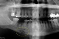
Instrumental diagnostics is represented mainly by radiography, magnetic resonance or computed tomography. Sequestrations are present on the radiograph: it is optimal to perform an orthopantomogram or X-ray in the forward and lateral projections to detect the disease. In the productive course of the disease, sequestration is not determined, but the volume of tissue mineralization increases, which is due to the periosteal reaction. Externally, facial asymmetry and increased bone volume are detected.
Laboratory tests are prescribed as part of general diagnostic measures. Blood analysis shows inflammatory signs, urinalysis - no changes. [7]
Differential diagnosis
|
Diseases requiring differential diagnosis |
Basis for differential diagnosis |
Diagnostic measures and evaluation criteria |
|
Subcutaneous granuloma (odontogenic) |
Sluggish odontogenic inflammatory process in the subcutaneous tissue of the face. The primary infectious focus is a diseased tooth, at the level of which a rounded painless infiltrate up to 15 mm in diameter is formed. The skin over it acquires a bluish-black color, on the side of the oral cavity there is a thrust, it can be felt in the submucous layer, starting from the corresponding dental cavity and up to the infiltrate. Periodically there is suppuration of the infiltrate and its independent opening with the formation of a fistula: the amount of purulent discharge is small. The space of the granuloma is filled with sluggish granulations. |
X-ray examination is performed - panoramic, dental, in lateral mandibular projection. Microscopy reveals granulations of different stages of maturity. |
|
Jaw actinomycosis |
Secondary pathology is associated with the spread of a specific infection from a soft tissue infiltrate near the jaw. The structure of the infiltrate is dense, multiple fistulous channels are possible, from which a crumb-like purulent mass is released. The primary form of actinomycosis has many similarities with hyperplastic osteomyelitis. |
Microscopic examination of the excreted mass, skin tests with actinolysate, determination of the reaction of immunocompetent cells to actinolysate are performed. |
|
Tuberculosis of the jawbones |
Typical are a slow course, sharp pain, marked enlargement and painful lymph nodes. Other facial bones may be involved, and characteristic "retracted" scars are formed in the area of the inflammatory reaction. |
Fluorography (X-ray or CT scan), Mantoux test (in children), exudate culture, specific skin tests are prescribed. |
|
Jaw syphilis |
The pathology develops due to gummosis melting of bone structures at the tertiary stage of syphilis. The nasal bones, the central zones of the maxillary palatine processes, and the alveolar process of the maxilla are most often affected. The formation of softening areas and ossifying periostitis (depending on the form of the disease) is typical. |
Serologic diagnostic methods are used. |
|
Benign tumor processes (suppuration of odontogenic cyst, osteoclastoma, eosinophilic granuloma, osteoidosteoma). |
Benign tumors often grow painlessly, there are no acute inflammatory signs. Periodic decrease and increase in the volume of the neoplasm is not characteristic of such pathologies. |
X-ray (panoramic, dental, lateral mandibular projection), computed tomography are performed. The outcome of histologic analysis is decisive. |
|
Ewing's sarcoma |
Pathology has many symptoms similar to chronic osteomyelitis. Ewing's sarcoma is accompanied by fever, leukocytosis, local bone pain, swelling. Tumor progression is slow at first, then sharply accelerated. The formation of sequestrations is not typical. |
X-rays, computerized or magnetic resonance imaging, biopsy are used. The diagnosis is established on the basis of the outcome of histologic analysis. |
Treatment of the chronic odontogenic osteomyelitis.
The therapeutic procedures include the following steps:
- Surgical treatment:
- Extraction of a focal tooth;
- Periostomy;
- Osteoperforation;
- Opening of the peri-mandibular purulent inflammatory focus.
- Conservative therapy:
- Antibiotic therapy with macrolides that inhibit the growth of 100% of Bacteroides and Fusobacterium strains, III generation cephalosporins, inhibitor-protected penicillins;
- Vancomycin and carbapenems become reserve drugs in difficult situations;
- Taking desensitizing drugs and immunocorrectors;
- Vascular and anti-inflammatory therapy;
- Infusion and vitamin therapy.
The criteria for effective treatment are the absence of pain in the affected area, the absence of inflammatory signs and fistula.
Possible medication prescriptions:
- Cefazolin 500-1000 mg, Cefuroxime 750-1500 mg with Metronidazole 0.5% 100 ml;
- Ketoprofen 100 mg per 2 mL, or orally 150 mg (prolonged version is 100 mg), Ibuprofen 100 mg per 5 mL, or orally 600 mg;
- Hemostatic Etamsilat 12.5% 2 ml intravenously or intramuscularly.
Upon completion of treatment, the patient is registered and observed by a specialist of maxillofacial surgery (visits - twice a year). A follow-up radiography or panoramic tomography is mandatory, and if indicated, dental prosthetics are performed. [8]
Prevention
To prevent the development of chronic odontogenic osteomyelitis is quite possible - for example, if you listen to the advice of doctors and follow the following recommendations:
- Observe thorough oral hygiene, timely sanitize dental infectious foci - in particular, caries, pulpitis and periodontitis;
- Timely visit the dentist, do not ignore the first manifestations of the disease;
- To monitor the health of the entire body;
- Strictly follow all doctor's orders, do not self-medicate.
In general, prevention consists of eliminating factors that could lead to the development of odontogenic osteomyelitis, as well as from the rationality of treatment of this disease from its acute stage. It is important to localize the purulent inflammatory process as soon as possible, prevent bone tissue necrosis and further sequestration: the patient at the first signs of pathology should be hospitalized in a surgical inpatient department.
Forecast
Unfortunately, the disease is often complicated by pathologic fractures, ankyloses of the maxilla, formation of false joints and scar contractures of the masticatory muscles. In the productive type of pathology, renal and cardiac amyloidosis may develop.
To improve the prognosis, it is important to seek medical help in a timely manner, sanitize infectious foci in the body, strengthen immunity, carefully fulfill all the prescriptions of the doctor.
Provided timely diagnosis of the correct management of the patient chronic odontogenic osteomyelitis in most cases ends with recovery. Unfavorable course with ascending spread of purulent-infectious reaction can cause the development of meningitis, encephalitis, brain abscess. With descending spread there is a danger of developing a pulmonary abscess, mediastinitis, sepsis. Such complications significantly increase the risk of death.
Literature
Dmitrieva, L. A. Therapeutic stomatology: national guide / edited by L. A. Dmitrieva, Y. M. Maksimovskiy. - 2nd ed. Moscow: GEOTAR-Media, 2021.