
Mucoceles
Last reviewed: 07.06.2024

All iLive content is medically reviewed or fact checked to ensure as much factual accuracy as possible.
We have strict sourcing guidelines and only link to reputable media sites, academic research institutions and, whenever possible, medically peer reviewed studies. Note that the numbers in parentheses ([1], [2], etc.) are clickable links to these studies.
If you feel that any of our content is inaccurate, out-of-date, or otherwise questionable, please select it and press Ctrl + Enter.
A mucocele is a cyst or bubble that forms due to a buildup of mucin in the sebaceous or salivary glands. It can develop in different parts of the body, but mucocele is most commonly found in the salivary gland area of the mouth.
The main characteristics of mucocele:
- Origin: Mucoceles usually form due to blockage of the sebaceous or salivary glands, resulting in the accumulation of mucin in the glandular ducts.
- Symptoms: Symptoms of a mucocele can include swelling or increased volume in the area of the mass, soreness, redness, and even infection around the mass. In the case of oral mucocele, patients may experience discomfort when chewing and speaking.
- Localization: Mucoceles can occur in a variety of locations, including the lips, tongue, inside of the cheek, or hyoid glands.
- Treatment: Treatment of a mucocele usually involves removing the mass to restore normal function of the gland or glands. This procedure may be performed by a medical professional such as a surgeon, dentist, or oral surgeon.
A mucocele is usually a benign condition, but it can cause discomfort and soreness. Therefore, if you suspect a mucocele or other masses, it is important to see a medical professional for diagnosis and treatment.
Causes of the mucocele
For different locations and types of glandular ducts, there are different causes for the development of mucocele:
- Salivary gland mucocele: This type of mucocele develops due to blockage of the salivary gland ducts. This can occur due to a variety of causes including trauma, infection, inflammation, or other abnormalities in the glandular ducts. Damage or inflammation of the gland can lead to blockage and accumulation of mucin.
- Mucoceles of the tonsils: Mucoceles of the tonsils can develop if the ducts of the tonsils become blocked, for example due to infection or other factors.
- Appendiceal mucocele: In case of appendiceal mucocele, the cause is blockage of the lumen of the appendix, resulting in accumulation of mucin in its cavity.
- Lattice labyrinth mucocele: In rare cases, a lattice labyrinth mucocele may result from blockage of the glandular ducts of the inner ear.
- Trauma: Trauma or mechanical injury can lead to blockage of the glandular ducts and the development of mucocele.
- Genetic Factors: In some cases, mucocele may have a genetic predisposition where a person is more prone to blocked glandular ducts.
Symptoms of the mucocele
Symptoms of a mucocele can vary depending on the location and extent of the cyst. A mucocele usually develops as a result of blockage of the glandular ducts and accumulation of mucin (mucus) inside the cyst. Here are some of the common symptoms of mucocele:
- Tumor or volume increase: The most common symptom of a mucocele is the appearance of a tumor or volume increase in the area where the cyst has developed. The size and shape of the cyst can vary.
- Pain or discomfort: A mucocele may cause pain or discomfort in or around the area of the tumor. The pain may be more severe if the cyst has become inflamed or infected.
- Feeling of heaviness: Patients may feel a sense of heaviness or pressure at the site where the mucocele is located.
- Deformity of the area: There may be deformity of the area due to an enlarged tumor.
- Change in appearance: In cases of mucocele in an area of the mouth, such as the lip or tongue, patients may notice a change in the appearance or shape of the lip or tongue.
- Changes in function: Depending on the location of the mucocele, it can affect the functionality of the area involved. For example, a mucocele in the salivary gland area can lead to difficulty in chewing and speaking.
Mucoceles symptoms usually worsen with mucin accumulation and may temporarily improve after drainage of the cyst or removal of its contents.
Forms
Depending on the location and the glandular gland in which the blockage has occurred, mucocele can have different types. Here are a few types of mucocele:
Salivary gland mucocele
It is a mass that results from blockage or damage to the glandular ducts of the salivary glands in the mouth. Salivary glands produce saliva, which helps in moisturizing and digesting food. When the glandular duct of a salivary gland becomes blocked or damaged, mucin (a viscous fluid found in saliva) can begin to build up inside the gland, resulting in a mucocele.
Here are the main characteristics of salivary gland mucocele:
- Localization: Salivary gland mucocele most commonly develops in the hyoid (submandibular) salivary glands or tonsils. However, it can also occur in other salivary glands of the oral cavity.
- Symptoms: Symptoms of mucocele include swelling or increased volume in the salivary gland area, soreness, discomfort when chewing and speaking, and sometimes draining mucin from the gland into the mouth.
- Treatment: Treatment of salivary gland mucocele usually involves surgical removal of the mass. The procedure can be performed by an oral surgeon or dentist. After mucocele removal, patients usually experience relief from pain and discomfort.
A salivary gland mucocele is a benign condition and is usually not associated with serious complications. However, it can cause discomfort and sometimes recurs after removal, especially if the entire glandular duct system is not removed. Therefore, it is important to see a medical professional for diagnosis and treatment if you suspect a salivary gland mucocele.
Mucoceles of the appendix
This is a condition in which the appendix, a small blind branch, undergoes changes in its structure due to the accumulation of mucin and other secretions in its cavity. This can occur because the lumen of the appendix is blocked, causing secretions to accumulate, increasing the pressure inside the appendix and causing it to dilate.
The symptoms of appendiceal mucocele can be similar to those of appendicitis, making their diagnosis and distinction important tasks:
- Pain: Symptoms usually begin with pain that starts in the abdominal area and then centers in the right lower quadrant of the abdomen where the appendix is located.
- Loss of appetite: Loss of appetite can be another symptom of appendix mucocele.
- Nausea and vomiting: Some patients may experience nausea and vomiting.
- Fever: In some cases, signs of inflammation, including an elevated body temperature, may occur.
- Abdominal muscle rigidity: The abdomen may become painful on palpation and the abdominal muscles may be tight.
Appendiceal mucocele usually does not cause as sharp and severe pain as appendicitis and most often has a less acute course. However, it can cause discomfort and, if left untreated, can lead to complications.
Treatment for appendix mucocele usually involves surgical removal of the appendix (appendectomy) to prevent complications and relieve symptoms. Diagnosis and treatment of this condition should be done by doctors in a medical facility.
Mucoceles of the maxillary sinus.
This is a medical condition in which there is a buildup of fluid (mucocele fluid) in the maxillary sinus (antrum), which is located in the area of the upper face and adjacent to the upper jaw. This fluid usually consists of mucus and is the result of a blockage of the maxillary sinus exit canal, which can occur due to various reasons.
Here are some of the main characteristics of maxillary sinus mucocele:
-
Symptoms: The main symptoms of maxillary sinus mucocele may include:
- A swelling or lump in the upper face area, often around the upper lip or under the eye.
- Soreness or discomfort in the area of the tumor.
- Possible symptoms of thickened mucus in the nasal or oral area.
- Itching or burning in the upper lip area.
- Causes: The main cause of maxillary sinus mucocele is blockage of the exit canal of the maxillary sinus. This can occur due to various factors such as inflammation, infection, trauma or anatomical features.
- Treatment: Treatment of maxillary sinus mucocele usually involves surgical removal of the mucocele cyst and restoration of normal drainage of the maxillary sinus. The procedure may be performed surgically or using an endoscope, depending on the specific situation.
If you suspect a maxillary sinus mucocele or are experiencing symptoms, it is important to see a doctor to get professional advice and prescribe appropriate treatment. Do not attempt to treat a mucocele on your own as this can lead to complications.
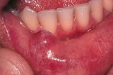
Mucoceles on the lip
A mucocele is a cystic mass, usually filled with mucus, that can occur on the mucous membrane of the mouth or lip. This mass is usually painless, but it can cause discomfort and impair quality of life. A mucocele on the lip usually has the following characteristics:
- Appearance: A mucocele on the lip looks like a small, clear or translucent vesicle or cystic mass that may contain viscous mucus.
- Localization: It usually appears on the inner side of the lip, closer to the oral mucosa.
- Symptoms: Patients may experience discomfort when a mucocele spreads the mucosa of the lip. In some cases, the mucocele may rupture, causing mucus to be released into the mouth.
If a mucocele appears on the lip, it is recommended that you see a doctor or dentist for evaluation and diagnosis. Usually, treatment of a mucocele involves its removal. This can be performed using local anesthesia. After removal of a mucocele, recovery is usually quick.
It is important to avoid trying to squeeze or pierce the mucocele yourself, as this can lead to infection or damage to the lip mucosa.
Nasal sinus mucocele
It is a medical condition in which there is a buildup of mucus or mucosal fluid in the nasal sinuses. The sinuses are air cavities inside the head that connect to the nasal cavity. A mucocele can occur due to a blockage in one of the exit ducts of the nasal sinus, leading to a buildup of mucus and an increase in the size of the sinus.
Symptoms of a nasal sinus mucocele may include:
- Nasal congestion.
- Nasal discharge, which may be mucous or contain gnosserous (mucus and pus) inclusions.
- Pain in the nose or face area.
- Headache.
- Deterioration of the sense of smell.
- External changes in the nasal area if the mucocele becomes prominent due to an increase in the size of the sinus.
Treatment for sinus mucocele usually involves surgery to remove accumulated mucus and restore normal sinus drainage. This may be performed endoscopically using minimally invasive techniques. After surgery, medications may be prescribed to reduce inflammation and relieve pain.
If you suspect a sinus mucocele or are experiencing the symptoms listed above, it is important to see your doctor to get an accurate diagnostic evaluation and prescribe appropriate treatment.
Mucoceles of the lattice labyrinth.
This is a rare condition that occurs when mucin (mucus) builds up in the lattice labyrinth, which is located in the inner ear. The labyrinth is a system of fluid channels and chambers responsible for balance and coordination of body movements.
Symptoms of a mucocele of the lattice labyrinth may include:
- Dizziness: Patients with lattice labyrinth mucocele often experience dizziness or a feeling of unsteadiness. This can occur due to impaired lattice labyrinth function and balance.
- Nausea and vomiting: Similar to dizziness, nausea and vomiting can be symptoms of a mucocele of the lattice labyrinth.
- Hearing loss: Patients may also experience hearing loss or other ear-related symptoms such as tinnitus (tinnitus).
- Nystagmus: Nystagmus is an involuntary rhythmic eye movement that can occur with mucocele of the lattice labyrinth.
Diagnosis and treatment of a mucocele of the lattice labyrinth requires specialized medical intervention. Treatment usually involves removing accumulated mucus or mucin from the lattice labyrinth and, if necessary, restoring balance and function to the inner ear. These treatments are performed by otorhinolaryngologists (ENT physicians) or neurootorhinolaryngologists specializing in ear, throat and nose diseases and neurootorhinolaryngology.
Mucoceles of the maxillary sinus (sinus mucoceles)
It is a medical condition in which there is a buildup of fluid (mucocele fluid) in the maxillary sinus, which is one of the sinuses of the nose. The maxillary sinuses are located on either side of the nose and are connected to it through openings. A maxillary sinus mucocele is usually associated with blocked drainage channels and mucus buildup in the sinus.
Here are some of the main characteristics of maxillary sinus mucocele:
- Symptoms: The main symptoms of maxillary sinus mucocele can be:
- Nasal congestion.
- Nasal discharge, which may be clear or cloudy and may contain mucus.
- Pain or pressure in the facial area or above the eye, especially on the side of the affected maxillary sinus.
- Headache.
- Deterioration of the sense of smell.
- Causes: The main cause of maxillary sinus mucocele is a blockage of the drainage channels that normally carry mucus out of the sinus. This can occur due to inflammation, infection, trauma, or anatomical features.
- Treatment: Treatment of maxillary sinus mucocele usually involves surgical removal of the mucocele cyst and restoration of normal drainage of the maxillary sinus. This may be performed using an endoscope or surgical procedure, depending on the specific situation.
If you suspect a maxillary sinus mucocele or are experiencing symptoms, it is important to see an otorhinolaryngologist for diagnosis and appropriate treatment. Do not attempt to treat a mucocele on your own, as this can lead to complications.
Mucoceles of the temporal bone
It is a medical condition in which there is a buildup of mucus or mucosal fluid in the temporal bone of the skull. The temporal bone is part of the skull, and there are air cavities within it that can contain mucus. A mucocele in the temporal bone can occur due to blockage of the exit ducts, which leads to mucus accumulation and an increase in the size of the air cavities.
Symptoms of temporal bone mucocele may include:
- Headache, often unilateral and localized in the temple area.
- Pressure and discomfort in the temple area.
- A sensation of congestion in the ear or the appearance of tinnitus.
- In some cases, changes in hearing.
- External changes such as an increase in the size of the temple, although these may not be noticeable.
Treatment for temporal bone mucocele usually involves surgery to remove accumulated mucus and restore normal drainage to the temporal bone. This may be performed using endoscopic techniques or through a small incision on the scalp in the temple area. The surgeon may also remove the blocking factors that led to the formation of the mucocele.
If you suspect a temporal bone mucocele or are experiencing the above symptoms, it is important to see your doctor for diagnosis and to determine the best method of treatment. This condition can be successfully treated with a good prognosis after surgery.
Complications and consequences
Mucoceles, if left untreated, can cause a variety of complications and problems, including:
- Infection: Mucoceles can become a source of infection, especially if the contents of the cyst become infected. This can cause an increase in symptoms such as pain, swelling and fever.
- Long-term symptoms: Without treatment, mucocele can last for many months or even years, causing discomfort and pressure in the face and nose area.
- Deterioration of the sense of smell: Mucoceles can put pressure on adjacent areas of the face, which can lead to a worsening of the sense of smell.
- Damage to surrounding tissues: Uncontrolled mucocele growth can cause compression and damage to surrounding tissues, including bones and nerves in the facial area.
- Recurrence: Even after removal of the mucocele, there is a risk of cyst reoccurrence, especially if the cause of the cyst, such as an anatomical feature or chronic inflammation, has not been addressed.
Diagnostics of the mucocele
Diagnosing a mucocele usually involves a physical examination and instrumental tests to confirm the presence of a cyst and determine its location. Here are some methods that can be used to diagnose a mucocele:
- Physical Exam: The doctor may begin the diagnosis by visually examining and evaluating the area where the tumor or volume increase is located. This can help the doctor determine the size, shape, and consistency of the tumor.
- Ultrasound: Ultrasound can be performed to visualize internal structures and confirm the presence of a cyst. This method can be used, for example, to diagnose mucocele of the salivary glands.
- Computed tomography (CT) scan: A CT scan can provide a more detailed image of the area where the mucocele is located and help your doctor determine its size and location.
- Magnetic resonance imaging (MRI): MRI may be useful for diagnosing mucocele in some areas, especially if more detailed soft tissue imaging is needed.
- Puncture or aspiration: Sometimes, to confirm the diagnosis of a mucocele and examine its contents, a puncture or aspiration may be performed, in which a medical professional extracts a sample of mucin from the cyst using a needle.
- Biopsy: In some cases, a biopsy may be necessary to rule out other pathologic processes. This may be necessary if other types of tumors are suspected.
Differential diagnosis
Differential diagnosis of mucocele can be important to rule out other conditions and determine the exact medical condition. Below are some conditions that may have symptoms similar to mucocele and require a differential diagnosis:
- Nasal polyps: Nasal polyps are masses that can occur in the nose or maxillary sinuses. They can cause nasal congestion and other symptoms similar to mucocele.
- Inflammation of the maxillary sinus (maxillary sinusitis): A maxillary sinus infection is an inflammation of the maxillary sinus, which can cause similar symptoms such as pain in the eye or cheek area, swelling and nasal congestion.
- Nasalcyst: Nasal cysts may be similar in appearance to mucocele, but their contents may be different.
- Dental infections: Sometimes infections of the teeth or gums can cause pain and swelling in the upper jaw area, which can be similar to the symptoms of mucocele.
- Malignant neoplasms: Although rare, some malignant tumors in the maxillary sinus area can have symptoms that resemble mucocele.
Treatment of the mucocele
Treatment for mucoceles depends on the location and size of the mass, as well as the symptoms it causes. Usually, a mucocele is treated surgically to remove the accumulated mucus and restore normal drainage. There are two main methods of surgical treatment for mucocele:
- Mucocellectomy: This is a procedure in which the mucocele is removed and then normal drainage is restored. The procedure can be done using endoscopy or done through a small incision in the skin, depending on the location of the mucocele.
- Marsupialization: This method involves creating a new drainage hole in the mucocele, allowing mucus to escape. This can be done without removing the entire mass.
Treatment is usually performed under local or general anesthesia and is usually effective. After the procedure, medications may be prescribed to reduce inflammation and pain and to prevent infection.
After treatment, it is important to monitor the healing process and follow the doctor's recommendations. The healing time may vary depending on the complexity of the case and the surgical technique. It is important to follow your doctor's instructions carefully and have routine check-ups to monitor your condition.
Forecast
The prognosis for a mucocele is usually favorable after its removal or treatment. A mucocele is usually not a serious condition and is usually treated without complications. Highlights:
- Mucoceles Removal: The main method of treating a mucocele is to remove it through a small surgical procedure. This procedure is usually quick and without complications, provided it is performed by a qualified doctor or dentist.
- Rehabilitation: After removal of a mucocele, there is usually no need for prolonged rehabilitation. Most patients can return to their normal life and diet the same day or within a few days after the procedure.
- Recurrence: Although a mucocele is successfully removed, recurrence can sometimes occur, especially if the underlying cause remains, such as blocked salivary ducts. In such cases, additional treatment or surgery may be required.
- Additional measures: It is important to follow up by eliminating or managing the cause of the mucocele (e.g., blocking salivary ducts) to reduce the risk of recurrence.
A list of some books and studies related to the study of mucocele
-
Book: "Salivary Gland Pathology: Diagnosis and Management"
- Authors: Eric R. Carlson, David L. Mandel et al.
- Year of release: 2012
-
Book: "Salivary Gland Disorders and Diseases: Diagnosis and Management"
- Author: Robert L. Witt
- Year of release: 2016
-
Book: "Oral and Maxillofacial Pathology."
- Authors: Angela C. Chi, Brad W. Neville
- Year of release: 2015
-
Study: "Mucoceles: Clinical Features, Diagnosis, and Management"
- Published in the journal Oral Surgery, Oral Medicine, Oral Pathology, and Oral Radiology
- Year of publication: 2017
-
Study: "Management of pediatric mucoceles with a simplified approach: a clinical study"
- Published in Journal of Indian Society of Pedodontics and Preventive Dentistry
- Year of publication: 2013
-
The book: "Mucocele and Ranula."
- Author: Jaime D. Alvarado
- Year of release: 2019
Literature
Chissov, V. I. Oncology / Ed. By V. I. Chissov, M. I. Davydov - Moscow: GEOTAR-Media, 2008. I. Chissov, M. I. Davydov - Moscow: GEOTAR-Media, 2008.