
Onychocryptosis of the nails
Last reviewed: 07.06.2024

All iLive content is medically reviewed or fact checked to ensure as much factual accuracy as possible.
We have strict sourcing guidelines and only link to reputable media sites, academic research institutions and, whenever possible, medically peer reviewed studies. Note that the numbers in parentheses ([1], [2], etc.) are clickable links to these studies.
If you feel that any of our content is inaccurate, out-of-date, or otherwise questionable, please select it and press Ctrl + Enter.
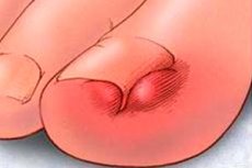
A disorder in which the nail grows into the lateral nail shaft is called "onychocryptosis". The problem most often occurs in the area of the big toe, although the affection of the fingers and toes is not excluded. Onychocryptosis is accompanied by an inflammatory reaction, as a consequence - pain syndrome, redness, swelling. The ingrowth occurs on one side, very rarely - on both sides. In most cases, the pathology has to be treated surgically: if this is not done, the process can become complicated, suppurate, spread to neighboring tissues. With timely treatment, the outcome is usually favorable. [1]
Historical facts
Onychocryptosis of toenails is a long-known problem, described as early as by Hippocrates in the 5th century BC, the medieval scientist Avicenna, the Byzantine doctor Paul of Aegina (7th century), and the Arab physician Abu-al-Qasima (Albukasis). In ancient times, healers treated the disease by removing the lateral nail shaft, the edge of the nail plate, and by cauterizing the granulations of the nail fold.
Somewhat later, the French physician Ambroise Paré (16th century) suggested treating onychocryptosis by radical removal of hypertrophic tissues with further cauterization of the wound surface.
The famous Italian anatomist Hieronymus Fabricius preferred to remove the ingrowing part of the nail, and the French military surgeon Guillaume Dupuytren in the 18th century introduced a combined method of removing the nail plate with further cauterization.
The systematization of treatment measures for onychocryptosis was already carried out in the 19th century by the German doctor Michaelis. A little later, the French physician Bodin proposed a variant of wedge resection of the nail, which was subsequently supplemented by other surgeons - in particular, Dr. Emmert. These treatments have become the most widely used in medicine.
Epidemiology
According to information as of the 1990s, the prevalence of onychocryptosis ranges from 2.5 to 5%. Men are more often affected.
The incidence of the disease has pronounced age peaks. Thus, onychocryptosis is much more common among children and young men 10-14, 16-19 years old, as well as in persons over 50 years old. It is noteworthy that the frequency of pathology in men and women at the age of 30 years is approximately the same. The highest peak of morbidity falls on the period of 16-19 years.
It is noteworthy that onychocryptosis is one of the so-called "diseases of civilization", as it is absent in regions where people traditionally prefer to walk without shoes.
Onychocryptosis on the hands is much less common than on the fingers of the lower extremities. Among the toes, the big toe is more often affected.
Causes of the onychocryptosis
The reasons for the development of onychocryptosis are diverse, they are divided into two categories: endogenous (internal) and exogenous (external).
Endogenously caused onychocryptosis is a hereditary pathology and is associated with peculiarities of the anatomy of the nails and fingers - in particular, the structure of the nail plates or lateral rollers. The most commonly noted ingrowth is an enlarged, beveled on the sides, deformed plate, which enters transversely into the lateral periungual roll. Soft and flat plates are much less common.
Onychocryptosis is more likely to occur in people who have massive, highly protruding lateral periungual rollers above the nail surface.
Among the most common endogenous causes, we can confidently name the curvature of the bones of the foot - for example, varus or valgus curvature, flat feet. Scientists have studied and confirmed the involvement in the development of onychocryptosis the presence of an incorrect interphalangeal angle of the big toe (the norm of the angle should not exceed 10 °). People with an interphalangeal angle of more than 15° and patients with increased mobility of the foot joints are at particular risk of nail ingrowth.
The thickness of the nail plate, the width of the lateral roll, and the medial deviation of the finger are also important. The hereditary type of onychocryptosis more often develops in persons with I and II degree of kinship.
Among the external causes, insufficient and irregular foot hygiene, improper nail care, use of inappropriately sized shoes, and foot injuries are most often mentioned.
Common causes of onychocryptosis by frequency of occurrence:
- Improper nail trimming (more than 70% of cases);
- Improperly fitted shoes (more than 45% of cases);
- Large angle of nail displacement (more than 35%);
- Excessive body weight (more than 30%);
- Foot injuries (more than 20%);
- Hormonal changes, pregnancy (more than 20% of female patients);
- Increased sweating of the feet (more than 15%).
Risk factors
The main provoking factors that can lead to the development of onychocryptosis are wearing constricting shoes, thick socks, as well as excessive sweating of the feet, obesity, diabetes mellitus.
Multiple systemic pathologies can contribute to the ingrowth of the nail plate - in particular, we are talking about arthritis, immunodeficiency conditions, tumor processes, circulatory disorders of the lower extremities. In general, any factor that provokes a conflict between soft tissues and the nail plate, for example, can have a negative impact:
- Constant strain on the foot and toe area;
- Tight, stiff, uncomfortable shoes;
- Repetitive trauma to the feet, toes;
- Insufficient observance of hygiene rules;
- Congenital and acquired curvatures of the feet;
- Too short nails;
- Overweight;
- Diabetes;
- Infectious and inflammatory diseases of the nails and soft tissues;
- Arthritis;
- Hyperhidrosis.
Predisposition factors are considered to be:
- Unusual nail bed configurations;
- Deformed nail plates;
- A genetically transmitted tendency to onychocryptosis.
The risks are multiplied by wearing incorrect shoes, improper or irregular nail trimming.
Pathogenesis
A thorough analysis of the probable causes of onychocryptosis development allowed us to identify the basic pathogenetic mechanisms of the disease:
- Damage to the epidermal tissue of the lateral periungual roll is the most common mechanism, which is usually "triggered" by the use of squeezing, inappropriate-sized shoes. The epidermis can be traumatized when performing pedicure manipulations, trimming the shaft and nails. The problem can be complicated by infection, a pronounced pain syndrome, the growth of granulation tissue.
- Compression of soft tissue structures localized under the nail plate is a mechanism due to bony changes of the main phalanx. The nail matrix is well attached to the bone. When the distal part of the interphalangeal articulation is widened, there is a narrowing and protrusion of the corresponding area of the nail, which can be a consequence of arthritis, traumatic injury, surgical intervention. As a result, the pinched nail bed becomes curved.
- Swelling of the periungual tissues is possible in early childhood, as well as in adults with the development of inflammatory processes and trauma in this area.
Stages
Currently, different types of classification of onychocryptosis are known. The most common is considered to be clinical classification, based on the initial clinical information and the severity of the pathology. Knowing the individual characteristics of the disease, it is much easier to choose the most optimal therapy scheme. Criteria used within the classification of onychocryptosis: skin erythema, local infectious reaction, swelling, discharge, thickening and thickening of the lateral periungual roll, pain syndrome and the appearance of granulation.
Heifetz stage classification:
- Slight redness and swelling of the lateral shafts of the nail.
- Acute infectious condition, suppuration.
- Chronic infectious condition, granulation formation, hypertrophy of adjacent tissues.
Frost's stage classification of onychocryptosis:
- An ingrowth (spur) appears on the side of the nail plate.
- The plate is warped.
- Signs of soft tissue hypertrophy appear.
Mosen's stage classification:
- Inflammatory stage (characterized by redness, swelling and pain when pressure is applied, while the nail is normal in appearance).
- It is divided into stage II-A (increased pain, purulent discharge and signs of infection, spread of edema to the outside of the plate less than 3 mm) and II-B (the same, with spread of edema more than 3 mm).
- The hypertrophy stage (accompanied by extensive overgrowth of granulations and lateral roll tissue over the plate).
The Martinez-Nova stage classification is supplemented by a fourth stage, the so-called "severe hypertrophy". This stage is characterized by chronic curvature of the finger with involvement of both rollers covering the broad part of the plate.
Kline's classification includes five stages:
- Stage of local irritation of the lateral roll. No pronounced infectious reaction and no granulation.
- Stage of infectious process in the lateral fold with purulent discharge or/and granulation.
- An infectious process with multiple homotypic episodes of onychocryptosis with a history of onychocryptosis.
- Infectious-inflammatory onychocryptosis with incomplete detachment of the lateral part of the nail.
- Infectious-inflammatory onychocryptosis with incomplete or complete detachment of the nail plate.
Classification by type depending on the cause of onychocryptosis:
- Onychocryptosis occurs in patients with normal feet and absence of somatic diseases. The causes are: insufficient hygienic care, use of tight shoes.
- There are congenital or acquired curvatures of the feet or/and toes.
- The patient is diagnosed with somatic pathologies that cause a disorder of peripheral blood flow, trophic disorders.
- The second and third type of etiology are combined, or a fungal infection or osteomyelitis is found.
- Onychocryptosis is recurrent.
Onychocryptosis in a child
Onychocryptosis is often diagnosed in children from early childhood through adolescence. In most cases, the problem is found on the big toes, but it can affect other toes, including the hands. When the plate grows into soft tissues, the toe becomes red, swollen and painful when walking.
In babies, the main cause of the problem is improper trimming of the grown-out nail edge. Due to inexperience, many parents maximally cut the lateral edges, as if rounding the plate, so that the baby does not scratch himself. However, after some time, such manipulations can lead to a violation of the configuration and growth of the nails, including their ingrowth.
The risks of the disorder are significantly increased if the child has a strong heredity in terms of onychocryptosis. Congenital deformities of the fingers or nail plate, malnutrition, overweight and rickets also play a role.
Complications and consequences
Today, there are many methods of onychocryptosis correction - both surgical and conservative. Nevertheless, the effectiveness of these methods of treatment is insufficient, and the main consequences of the problem are its recurrences. In addition, many specialists actively practice complete removal of the nail (Dupuytren's method), which entails high risks of cosmetic defects, deterioration of the support function of the affected finger. In many patients, removal of the nail plate provides only a temporary effect, because as the nail grows back, onychocryptosis often occurs again.
If onychocryptosis treatment is ignored, the following complications may develop:
- Abscess (formation of a pustule in soft tissue);
- Purulent panarisis;
- Phlegmon (purulent focus without clearly defined boundaries);
- Lymphadenitis (an inflammatory process in the lymphatic flow system);
- Osteomyelitis (bone lesions);
- Carrion (an infectious inflammatory process).
Diagnostics of the onychocryptosis
Onychocryptosis is difficult to confuse with other pathologies. The surgeon makes the diagnosis already during the first appointment and clinical examination. If necessary, he prescribes consultations with other specialists: endocrinologist, immunologist, infectious disease specialist, dermatologist.
Laboratory diagnostics may include a general blood test, blood coagulation studies, Wasserman reaction, determination of blood sugar levels. It is mandatory to exclude the presence of fungal infection. For this purpose, dermatoscopy, microscopy of scrapings from the affected finger, seeding of pathological biomaterial on nutrient media.
If onychocryptosis is complicated by secondary infection, then prescribe the identification of the pathogen by culture of secretions to determine resistance to antibiotics.
Differential diagnosis
Differential diagnosis is necessary to exclude osteophytes (bone growths) of the finger phalanx, inflammatory processes such as paronychia, periungual tumors of benign and malignant nature. Most often it is necessary to differentiate onychocryptosis with pathologies of the nails and the bed, shafts and terminal phalanx, in particular:
- Pyogenic granuloma - when located under the nail edge or on the roller looks like a small inflamed nodule, gradually increasing in size. The surface above it is hyperemic, flattened, may be covered with purulent-serous plaque or drying crust.
- Candido-fungal and pyococcal paronychia - develops as the inflammatory response in the soft tissues of the shaft worsens.
- Subnail exostosis is a benign growth of bone tissue, often of post-traumatic etiology. It has the appearance of a dense mass with a tendency to increase in size.
- Periungual or subnail fibroma is a benign mesenchymal growth, painless, gradually leading to nail dystrophy until nail destruction.
- Periungual or subnail chondroma is a benign neoplasm of hyaline or fibrous-cartilaginous tissue, has the appearance of a solitary tumor of solid consistency.
- Dermoid bed cyst - a violation of tissue development with the formation of an epithelial cavity, in which there may be particles of keratinization, hair.
- Glomus neoplasms are a benign Barre-Masson disease presenting as the formation of venous-arterial anastomoses within a capsule of neural and connective tissue.
- Malignant neoplasms (sarcomas, melanomas of the bed and rollers).
Treatment of the onychocryptosis
Conservative methods of treatment are used relatively rarely and only in relation to mild cases of onychocryptosis. Such methods can be divided into the following groups:
- Topical treatment with ointments and medicinal solutions.
- Isolation of the ingrown part of the lamina from the soft tissues.
- Wearing orthopedic devices that help flatten the lamina and lift the ingrown part of the nail.
Treatment at home includes:
- Thorough washing of the affected limb;
- Drying with a cotton disk with tamponade the area of ingrowth preparations of chamomile, marigolds, sea buckthorn, tea tree oil.
It is recommended to practice baths with antiseptic solutions - for example, with the addition of 5 ml of ammonia solution per 1 liter of water, or potassium permanganate, or hypertonic sodium chloride solution, as well as infusions of oak bark, colanchoe, chamomile. Regular treatment of the peri-nail area with a solution of brilliant green, iodine, methylene blue, fucorcin, chlorophyllipt is indicated. Successfully use lotions and compresses with onycholysin, dioxidine, furacilin, rivanol.
Recommended Ointments:
- Levomekol;
- Betadine;
- Ready-made anti-inflammatory mixture (for 5 g of crystalline iodine - 10 ml of 20% aqueous potassium iodide, 10 g of salicylic acid, 60 g of lanolin and 28 ml of dimexide).
Bandage strips moistened with antibiotics (e.g., kanamycin with novocaine) are placed between the nail and the shaft.
Orthopedic therapies that promote isolation of the ingrown nail have demonstrated a good effect. Metal-composite orthopedic devices are fixed in the area of the nail, which helps to make the plate flatter and release the ingrown edge.
Conservative treatments are minimally traumatic, can be applied at home and do not require the patient to be hospitalized. However, conservative therapy will not help with severe onychocryptosis or recurrent disease, and the orthopedic devices on the pharmaceutical market are usually quite expensive. Therefore, surgical correction comes to the fore.
In addition to complete and partial removal of the plate, cold exposure (cryotherapy), laser and ultrasound therapy, radio and electrocoagulation, chemical destruction method, microsurgery are actively used. The most popular for many years remains the marginal resection of the nail - technically uncomplicated operation, relatively minimally traumatic, providing a satisfactory cosmetic effect. Among the disadvantages of this intervention can only be called a high risk of recurrence of onychocryptosis (according to different data - from 13 to 28%).
Laser matrixectomy using a diode laser can reduce the frequency of onychocryptosis recurrence and optimize the overall effectiveness of treatment of the disease. Most often used carbon dioxide laser scalpel infrared spectrum. With such exposure, healing is easier than usual, as it has a relatively short inflammatory phase, scant exudation and leukocyte infiltration.
After surgical intervention, patients are recommended bed rest for 24 hours with the foot of the bed elevated. On the second day it is allowed to get up and walk without support on the operated toe: such restrictions remain for a period of about a week (it is allowed to lean on the heel when walking). During this period, daily dressings, washing the wound with antiseptic solutions, applying antibacterial ointments or powder (Levomekol, Betadine, Baneocin). If necessary, analgesics are used.
Control examinations are carried out after one month, then - after 3 months, six months, 9 months and one year after surgery. This is necessary both for dynamic monitoring and for timely detection of onychocryptosis recurrence.
Prevention
Doctors' basic recommendations for preventing onychocryptosis include:
- Hygiene, regular and quality foot washing and change of socks;
- Proper nail trimming (not too deep, leaving the free edge of the plate about 1 mm, followed by treatment of the cut edge with a soft file);
- Use of special emollient solutions (lotions) to prevent nail ingrowth;
- Avoiding traumatic injury to the fingers;
- Wearing shoes according to the size and shape of the foot;
- Use of special orthopedic devices, if necessary;
- Timely treatment of fungal diseases;
- Weight control.
Patients suffering from concomitant diseases - in particular, diabetes mellitus - should regularly visit the attending physician and fulfill his recommendations. People with flat feet and various foot curvatures should use special orthopedic devices and shoes.
Preventive measures also include timely visits to a podiatrist. It is much easier to prevent the spread of the problem at the early stages of development.
Forecast
Despite the continuous improvement of treatment methods for onychocryptosis, the problem remains relevant to date, which requires further work to study the disease.
Treatment methods for onychocryptosis have varying efficacy and are selected individually. One of the most common methods is marginal resection: the operation is technically simple, minimally traumatic and effective in cosmetic terms (provided that the lamina is adequately narrowed). One of the known "minuses" of this procedure is a high percentage of recurrence of onychocryptosis (according to different data, from 13 to 28%). The frequency of recurrences can be reduced by additional action on the nail growth zones - in particular, chemical action with phenol, sodium hydroxide, trichloroacetic or dichloroacetic acid. This results in chemical destruction of the matrix. The advantage of marginal resection is the uncomplicated technique and the lack of need for additional equipment.
Other effective treatment options include ultrasound matrixectomy and electrocoagulation - they are confidently and successfully used in many medical facilities. A side effect of chemical matrixectomy is excessive tissue destruction due to prolonged exposure to the reacting substance. A side effect of electrocoagulation can be a burn of nearby tissues. As for cryodestruction, this procedure is considered minimally traumatic and is recommended by many specialists, but requires the presence of a cooling agent in the facility, as well as appropriate equipment.
Laser treatment for onychocryptosis has been used for over 40 years and is recognized as an effective, radical, minimally traumatic, coagulating and bactericidal method. The most common is considered to be a carbon dioxide medical laser, functioning in the infrared range. Among the "minuses" of this method - high cost and impressive size of the equipment. As an alternative, it is proposed to use diode lasers. They are cheaper and smaller in size, function in the infrared range and are no less effective.
Onychocryptosis and the army
Patients with onychocryptosis, who are to serve in the army, are recommended to promptly correct the violation, for which a deferment is granted for the term necessary for the operation. In most cases, the operation of marginal resection of the plate and periungual roll with marginal excision of the growth zone is shown. Less often practiced complete removal of the nail or local tissue plasty. After successful surgical intervention and completion of the rehabilitation period, the recruit is considered fit for military service.
If onychocryptosis recurs or there are other associated disorders, the question of suitability is decided on an individual basis based on the findings of the expert committee.