
Hydrothorax
Last reviewed: 07.06.2024

All iLive content is medically reviewed or fact checked to ensure as much factual accuracy as possible.
We have strict sourcing guidelines and only link to reputable media sites, academic research institutions and, whenever possible, medically peer reviewed studies. Note that the numbers in parentheses ([1], [2], etc.) are clickable links to these studies.
If you feel that any of our content is inaccurate, out-of-date, or otherwise questionable, please select it and press Ctrl + Enter.
Pathologic accumulation of serous fluid - transudate or exudate - in the pleural cavity is defined as hydrothorax.
Epidemiology
According to clinical statistics, patients with heart failure develop bilateral hydrothorax in 81% of cases, right-sided hydrothorax in 12% and left-sided hydrothorax in 7%.
In lung cancer, hydrothorax is found in 7-15% of patients, in rheumatoid arthritis - in 3-5%. In the presence of liver cirrhosis, this pathology is noted in 5-6% of patients, with the share of hydrothorax in the right side of the pleural cavity accounts for approximately 80% of cases. And with hypoalbuminemia and nephrotic syndrome in 90% of cases of bilateral hydrothorax.
Physicians identify hydrothorax associated with pancreatitis in about 25% of patients.
Causes of the hydrothorax
Hydrothorax is a non-inflammatory type of pleural effusion, and the most common causes of such effusion of serous fluid into the spaces between the sheets of the surrounding pleura include chronic congestive heart failure, cirrhosis, malignancy and/or metastasis.
Lower extremity edema and hydrothorax in heart failure are part of edema syndrome, occurring in dilated cardiomyopathy and in cases of functional failure of the right ventricle of the heart. An increase in the amount of tissue (interstitial) fluid that leaks into the pleural cavity through the visceral pleura (its inner leaflet) also occurs in decompensated left ventricular failure.
As a rule, hydrothorax in liver cirrhosis develops as a complication of pathophysiologic disorders at the terminal stage of the disease. In this case, the volume of pleural transudate may exceed 0.5 liters and is more often formed in the right side of the pleural cavity - right-sided hydrothorax.
In patients with renal failure and nephrotic syndrome congestive hydrothorax develops against the background of severe hypoalbuminemia (decreased concentration of protein in the blood). And during renal replacement therapy in patients on peritoneal dialysis for kidney failure, acute hydrothorax may develop.
Right-sided or left-sided hydrothorax is also seen in cases of pulmonary embolism - pulmonary embolism (TELA); decreased thyroid function in patients with hypothyroidism, autoimmune thyroiditis or myxedema; pulmonary sarcoidosis; autoimmune diseases (rheumatoid arthritis or lupus).
Bilateral or bilateral hydrothorax (effusion in both pleural cavities) is seen in cases of chest trauma - traumatic hydrothorax. If the injury is closed, hydrothorax can also develop in case of rib fracture, but rib fracture can lead to breach of pleural cavity integrity and lung rupture, which in such cases leads to pneumothorax.
Bilateral hydrothorax is also characteristic of exudative pleurisy, and in combination with hydropericardium can be seen in patients with heart failure, myocardial inflammation, pulmonary sarcoidosis, and systemic lupus erythematosus.
Exudative hydrothorax develops in exudative pleurisy and pulmonary embolism and, as one of the unfavorable pulmonary complications, in acute pancreatitis.
Hydrothorax in oncology can occur with any tumor that metastasizes to the pleura or lungs, but hydrothorax is most commonly seen with lung cancer, pleural mesothelioma and breast cancer. Exudate accumulation in the pleural cavity may also occur in patients with mediastinal non-Hodgkin's lymphoma, hepatocellular carcinoma, and Meigs syndrome in ovarian cancer.
Hydrothorax in pregnancy is possible in severe pre-eclampsia, Burhave syndrome - spontaneous perforation of the esophagus due to hyperemesis (indomitable vomiting of pregnant women), as well as when using IVF (in vitro fertilization) - due to the development of ovarian hyperstimulation syndrome, which can occur after stimulation of ovarian follicle development (ovulation induction) - injections of hCG (human chorionic gonadotropin).
In most cases, hydrothorax in children develops in diffuse kidney diseases: acute and chronic nephritis, lipoid nephrosis, anomalies and diseases of renal arteries, renal failure and after peritoneal dialysis.
Since hydrothorax is not associated with infectious inflammation, serous fluid effusion in lung inflammation with viral or bacterial etiology, competent pulmonologists are defined as parapneumonic pleural effusion, not as hydrothorax in pneumonia. And such an effusion develops in almost half of cases of pneumococcal pneumonia. [1], [2]
Risk factors
In addition to the presence of etiologically related diseases, risk factors for the development of hydrothorax are:
- Smoking and alcohol abuse;
- Benign asbestos pleurisy;
- Dressler syndrome;
- Polycystic kidney disease;
- Yellow toenail syndrome, also known as primary lymphedema;
- Connective tissue diseases, including systemic sclerosis, granulomatosis with polyangiitis, Still's disease (juvenile rheumatoid arthritis);
- Increased capillary permeability;
- Outpatient peritoneal dialysis;
- Coronary bypass;
- Radiation therapy to the chest area;
- Long-term use of drugs with ergot alkaloids, as well as Methotrexate (an antimetabolic agent), the antiarrhythmic drug Amiodarone and the antiseptic Nitrofurantoin (Furodonin).
Pathogenesis
In heart failure, the mechanism of hydrothorax formation is due to pathogenesis of heart failure, in particular, a decrease in cardiac output and renal blood flow, changes in water-electrolyte balance (sodium retention and hypernatremia due to an increase in its reverse absorption), increased volume of extracellular fluid, hypervolemia (increased volume of circulating blood), increased vascular wall permeability and hydrostatic pressure in both circulations.
The pathogenesis of hydrothorax in patients with liver cirrhosis is explained by the development of ascites due to increased pressure in the portal vein - portal hypertension. With the combination of increased intra-abdominal pressure and negative intrathoracic pressure (arising during inhalation), there is a movement of fluid from the abdominal cavity into the pleural cavity through small defects of the diaphragm (near their tendons).
In addition, a significant decrease in the production of serum globular protein albumin by the liver - hypoalbuminemia - plays a crucial role, in which the balance of extracellular fluid distribution between blood plasma and out of blood flow is disturbed and the intravascular oncotic (colloid-osmotic) pressure is reduced, resulting in intravascular fluid entering the tissues.
The mechanism of exudative hydrothorax formation in oncology and autoimmune diseases is attributed to either increased capillary permeability or inadequate lymphatic resorption.
The development of non-inflammatory pleural effusion in renal failure as part of the nephrotic syndrome is due to a decrease in oncotic pressure due to increased excretion of albumin with urine and a decrease in its level in blood plasma.
If there are adhesions (adhesions) in the pleural cavity, as well as the accumulation of serous fluid in the visceral pleural folds, a limited or drained hydrothorax is formed. Depending on the localization, mediastinal, paramediastinal, interradial (lobar), costo-diaphragmatic (peri-costal), diaphragmatic or basal hydrothorax are divided. [3]
Symptoms of the hydrothorax
Hydrothorax is included in pleural syndrome, the first signs of which are a sensation of heaviness and pressure in the chest, although there may be no obvious symptoms if there are small amounts of effusion in the pleural cavity.
Significant accumulation of fluid causes typical respiratory symptoms. Thus, varying intensity of inspiratory dyspnea in hydrothorax is a consequence of lung compression by excess intrapleural fluid.
There is increased fatigue, moist wheezing during breathing, skin cyanosis, swelling of neck veins and non-productive cough in hydrothorax. Deep breaths may cause pain in the mediastinum.
Clinical manifestations of hydrothorax in cirrhosis vary from an asymptomatic course to severe respiratory failure. There may also be a subfebrile temperature in cirrhosis-related hydrothorax, although in other cases there is a slight decrease in body temperature.
Complications and consequences
What is the danger of hydrothorax? The consequence of a significant volume of effusion in the pleural cavity can be displacement of the heart, as well as compression of the lung tissue, which provokes its compaction - atelectasis of the lung (or its individual segments) with restrictive respiratory failure and the development of respiratory failure.
This reduces the minute volume of respiration, hypoxia (lack of oxygen in the arterial blood) and hypercapnia (increase in the level of carbon dioxide in the blood) develop, which leads to systemic multi-organ complications.
In addition, in many cases, serous fluid can re-accumulate in the pleural cavity, meaning that hydrothorax can recur.
Diagnostics of the hydrothorax
In the diagnosis of pathological accumulation of serous fluid in the pleural cavity, pulmonologists use various methods:
- Lung palpation;
- Auscultation of the lungs; auscultation for hydrothorax demonstrates vesicular breathing - significant reduction in respiratory murmur;
- Lung percussion, which reveals a dulled sound when tapping, that is, the sound at percussion in hydrothorax is characterized by a dulled-tympanic tone, which is characteristic of fluid accumulation in the pleural cavity.
Puncture for hydrothorax is performed - diagnostic thoracentesis, for more information see - pleural cavity puncture.
And is done general clinical examination of pleural fluid, blood tests are taken (general and biochemical), general urine analysis.
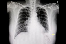
Instrumental diagnostics to visualize the pleural cavity include lung X-ray, endoscopic examination of the pleural cavity - thoracoscopy, contrast-enhanced ultrasonography - pleural ultrasound, and CT - chest computed tomography and CT pulmonary angiography.
An x-ray in hydrothorax will show darkening of the lung field or part of it.
Hydrothorax on ultrasound looks like a homogeneous anechogenic area, the boundaries of which are two anechogenic horizontal shadows of the posterior ribs and the pulmonary line - parietal and visceral pleura.
Computed tomography allows visualization of the lungs, pleura and pleural cavity; hydrothorax on CT is visualized as the presence of homogeneous water-like fluid in the pleural cavity. CT also provides information to quantify pleural effusion.
Differential diagnosis
Differential diagnosis is necessary to verify hydrothorax, primarily differential diagnosis of exudate and transudate.
It is necessary to differentiate hydrothorax and visceral pleural thickening, causing similar symptoms exudative pleurisy and hydrothorax, hydrothorax and edema in mediastinitis (resulting from infection, after endoscopy of the upper mediastinum or undergone chest surgery); air in the pleural cavity - pneumothorax and hydrothorax; thickening of lung tissue - atelectasis and hydrothorax; presence of blood in the pleural cavity - hemothorax and hydrothorax.Also require differentiation of hydrothorax and pulmonary emphysema.
Treatment of the hydrothorax
When treating hydrothorax, the underlying disease should also be treated, i.e. The underlying disease should be treated:
Treatment of hepatic hydrothorax may consist of salt and water restriction with the administration of diuretics. Drug treatment of hydrothorax in cirrhosis may include drugs to reduce portal hypertension: beta-blockers (Propranolol, Nadolol, etc.) and statins (e.g. Simvastatin).
Remove serous fluid from the pleural cavity with percutaneous thoracentesis (pleurocentesis), that is, drainage of the pleural cavity in hydrothorax under ultrasound control using a trocar - a fixed cannula through which the drainage tube is placed in the right place.
Transjugular intrahepatic portosystemic shunt (TIPS), a lower-pressure connection of the portal vein to neighboring vessels that reduces intrahepatic blood flow pressure and fluid outflow into the pleural cavity, has positive results in hepatic hydrothorax. [4]
Antibiotics in hydrothorax, given its non-infectious origin, are not prescribed.
Alternative - folk remedies for hydrothorax - use phytotherapy: Decoctions and/or infusions of roots and rhizomes of plants such as discurenia (Descurainia sophia), tuberous flipper (Asclepias tuberosa), Kansui milkvetch (Euphorbia kansui) or Peking milkvetch (Euphorbia pekinensis), laconos (Phytolacca americana), forest dudnik (Angelica sylvestris), medicinal rhubarb (Rheum officinale).
Prevention
Prevention of hydrothorax is facilitated by timely treatment of etiologically related diseases.
Forecast
Successful thoracentesis and correct etiologic treatment of the underlying pathology create prerequisites for a favorable prognosis of the outcome of hydrothorax, except for the terminal stage of liver cirrhosis and autoimmune diseases.