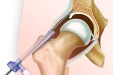
Arthroscopy of the hip joint
Last reviewed: 23.04.2024

All iLive content is medically reviewed or fact checked to ensure as much factual accuracy as possible.
We have strict sourcing guidelines and only link to reputable media sites, academic research institutions and, whenever possible, medically peer reviewed studies. Note that the numbers in parentheses ([1], [2], etc.) are clickable links to these studies.
If you feel that any of our content is inaccurate, out-of-date, or otherwise questionable, please select it and press Ctrl + Enter.

Arthroscopy of the hip joint is performed under endotracheal anesthesia. The position of the patient on the operating table is lying on a healthy side.
With the help of special additional supports, the traction system is adjusted. The operated joint is in the position of extension and neutral rotation, while the lower limb is withdrawn by 25 °. The joint gap is stretched to 10-15 mm. To monitor the expansion of the joint space in the operating room after the application of the traction system, a radiograph of the hip joint is performed in a direct projection. If the joint is not stretched enough on the control radiograph, the distraction is continued and the X-ray of the joint is repeatedly performed.
Before the beginning of arthroscopy, external reference points are applied and the projection of the intended accesses is noted. The marking of the joint is necessary for better orientation of the surgeon during the operation. After the preparation of the surgical field, external guidelines are applied to the skin: the contours of a large trochanter of the femur, the anterior superior iliac spine, the upper margin of the pubic articulation. Determine the pulsation of the femoral artery and mark the projection of the femoral neurovascular bundle. There are also places of standard access to the joint.
Through anterolateral access, perpendicular to the femur surface in the direction of the femoral head, 30-40 ml of physiological saline with epinephrine (1: 1000 dilution) is injected into the joint cavity using a syringe and a long spinal injection needle, which contributes to an additional enlargement of the intraarticular space. If the procedure is performed correctly, after removing the syringe through the needle located in the joint cavity, the injected liquid flows out under pressure with a trickle. After removing the needle at the site of its entrance with a scalpel, a crushed skin incision is made about 5 cm in length. A blunt trocar is placed in the joint, placed in the shaft of an arthroscope. It passes directly over the large trochanter on the outer surface of the femoral head under the lateral section of the acetabular lip. Due to the normal anteversion of the femoral neck, with a neutral rotation of the hip joint, the trocar block runs parallel to the anterolateral margin of the acetabulum. As the block moves to the joint after perforating the capsule, the end of the trocar is raised slightly to avoid damage to the joint surface of the femoral head. Troakar is removed, a 30-degree arthroscope with a diameter of 4.2 mm is inserted into the shaft. The arthroscopic chamber and the light guide are connected, as well as the irrigation system. It is preferable to use an inflow and outflow irrigation system with a roller pump, which allows to control and maintain the optimal intra-articular pressure (100-150 mm of water) at a constant level.
After the introduction of an arthroscope, a front access is made to the joint cavity. In his projection, a scalpel is made with a scalpel and an arthroscopic control is made (using a 70-degree arthroscope for this purpose). In the joint, a trocar in the shaft of the arthroscope is guided into the joint in the shaft of the arthroscope towards the midline of the body at an angle of 45 "to the front (cranial direction) and 30 ° to the sagittal plane (in the medial direction) Similarly, posterolateral access is performed, to the shaft of which tubing is connected to the inflow of liquid.After creating all three accesses, the hip joint cavity are examined through three interchangeable shafts using 30-degree and 70-degree optics.With a 70-degree arthroscope, it is convenient to examine the acetabular tube, the peripheral part of the acetabulum and the femoral head, and the deep pockets of the acetabulum and the round ligament. 30-degree optics, the central parts of the acetabulum and the femoral head are better visualized, as well as the upper part of the acetabulum.
Revision of the hip joint cavity begins with examination of the pit of the acetabulum and the fat pad located in it, surrounded by a semilunar cartilage.
With the advance of the arthroscope, a bundle of the femoral head is visualized in the cavity; it is possible to observe a transverse ligament, but not in all cases, since its fibers are more often intertwined into the capsule of the joint. Rotating the arthroscope clockwise, inspect the anterior margin of the acetabular lip and the ileum-femoral ligament from it (Y-shaped bundle Bigelow), it is tightly attached to the anterior part of the capsule of the joint above the upper part of the femoral neck. Continuing to turn the arthroscope, somewhat pulling it back, inspect the middle upper part of the semilunar surface and the lips of the acetabulum. As the arthroscope moves forward, the posterior section of the acetabular lip and the sciatic-femoral ligament split with it are accessible through a joint slit.
Sometimes, in the posterior compartment, using posterolateral access and 70-degree optics, it is possible to visualize the Weitbrecht sheath from the joint capsule to the head and the posteriorly superior femoral neck in the form of a flattened strand.
Advancing the arthroscope further down, sliding down the neck of the femur, examining the zona orbicularis - a circular ring forming a cushion around the neck of the femur.
Its fibers do not attach to the bone and stretch when the thigh is in the internal rotation position. Their tight tension around the neck of the femur can be mistaken for the acetabular lip. To avoid this, the hip needs to be given an external rotation position, which allows the fibers of the zona orbicularis to relax and move away from the neck of the femur. In this case, from the arbi cular fibers, as they relax, the synovial villi protrude, clearly differentiating them from the acetabular lip.
The surgeon's assistant, alternately using the external and internal rotation of the thigh, gives the necessary position to the head of the femur, in order to provide a better visualization of all parts of the joint and the articular surface of the femoral head.
Since the soft tissues of the joint, its muscles, articular-ligamentous apparatus were previously stretched and relaxed, special efforts are not required to stretch the joint on the part of the assistant.
When performing the operative stage of arthroscopy of the hip joint, use arthroscopic instruments with a diameter of 2 to 3.5 mm, as well as a shaver with a diameter of a nozzle of 2.4 mm for removal of intraarticular bodies, dissection of adhesions and treatment of zones of damaged cartilage.
After the end of arthroscopy, after revision and sanation of the hip joint cavity, the remaining liquid is aspirated from the joint cavity and 0.25% solution in an amount of 10-15 ml is injected with bupivacaine + epinephrine, the threaded rods are removed. On the field of arthroscopic accesses, sutures are removed, removed after 5-7 days, and aseptic dressings.
Indications and contraindications for arthroscopy of the hip joint
Indications for the therapeutic and diagnostic arthroscopy: the presence of intra-bodies, damaging the labrum of the acetabulum, osteoarthritis, injury of articular cartilage, avascular necrosis of the femoral head round ligament rupture, chronic synovitis, joint instability, septic arthritis, post previously conducted arthroplasty of the hip joint , the presence in the history of surgical interventions on the hip joint.
The most typical contraindication to performing arthroscopy is ankylosis of the hip joint. With this pathology, it is not possible to expand the intraarticular space, which creates an obstacle to the introduction of instruments into the joint cavity. Significant abnormalities in normal bone anatomy or surrounding soft tissue as a result of a previous injury or surgery also exclude the possibility of performing arthroscopy.
Severe obesity is a relative contraindication to arthroscopy of the hip joint. At an extreme density of soft tissues, even with long instruments, it is impossible to reach the joint cavity.
Diseases manifested by destruction of the hip joint are also considered a contraindication to arthroscopy.
Possible complications in arthroscopy of the hip joint and precautionary measures
- Intra-articular infection (suppuration of arthroscopic wound, coxitis, sepsis ).
- During the surgery to prevent the development of suppuration in the postoperative period, you must strictly follow the rules of asepsis and antiseptics.
- In the preoperative and early postoperative periods, it is possible to prescribe broad-spectrum antibiotics.
- Damage to articular cartilage during the introduction of arthroscopic instruments.
- To avoid this complication, it is necessary to insert instruments into the hip joint cavity without sudden movements and efforts.
- Temporary pain syndrome.
- To stop pain in the early postoperative period (the first day) prescribe narcotic analgesics.
- In the future, patients are shown anti-inflammatory non-steroid drugs for 5-7 days.
- During the arthroscopy there is a risk of breakage of arthroscopic instruments, which leads to the need to remove the foreign body from the joint cavity.
- To prevent this complication, it is necessary to ensure sufficient stretching of the joint gap - up to 10-15 mm.
- If a free foreign body forms in the joint during the breakdown, it is very important to keep the position of the joint unchanged so as not to lose sight of the broken fragment and be able to grab and remove it with a clamp as soon as possible.
- Tractional damage of the neurovascular bundle and capsular-ligament apparatus.
- To prevent this complication, avoidance of distraction should be avoided. Before the operation, the patient lies for 15-20 minutes on the operating table with minimal distraction effort.
- Extravasation of liquid.
- To ensure that the wash fluid does not enter the subcutaneous tissue, the following rules must be observed:
- do not allow the pressure in the washing system to increase above the normal level;
- shut off the supply of fluid on the washing system with an accidental exit of the end of the arthroscope from the joint cavity.
Postoperative rehabilitation of patients after arthroscopy of the hip joint
In the early postoperative period it is important to provide the patient with adequate anesthesia. The intensity of pain sensation depends on the specific pathology and the amount of surgical intervention performed during arthroscopy of the hip joint. For example, after the removal of free intraarticular bodies, the pain after surgery is practically unaffected by the patient, the discomfort after surgery is much less than before. Conversely, after abrasive arthroplasty in the case of cartilage damage immediately after surgery, the patient experiences pain of a more intense nature. In the first day after the operation, analgesia is provided with the help of narcotic analgesics, and in future patients are prescribed non-steroidal anti-inflammatory drugs for 5-7 days (ketoprofen 100 mg 2-3 times a day).
Immediately after arthroscopic surgery, a bag of ice is placed on the hip joint area. At the same time, the body's attempts to retain heat by narrowing the superficial cutaneous vessels lead to a decrease in capillary permeability and a decrease in bleeding. This changes the biological response of tissues to trauma, reducing inflammation, edema and pain. Ice is used for 15-20 minutes every 3 hours during the first day, and sometimes within 2-3 days.
Change the bandages performed on the day after the operation. Dressings are produced every other day. Seven days after surgery, seams are removed. In the early postoperative period, patients are allowed to sit down. This is due to the fact that when flexing the hip joint, the capsule relaxes, so patients feel more comfortable sitting. Get up using crutches, recommend in the first 2 days after the operation, but without the load on the operated limb. Functional restorative treatment begins from the 2nd day after the operation. The rehabilitation program is individual for each patient, it depends on the pathology and the amount of surgical intervention.


 [
[