Medical expert of the article

New publications
Radiologic diagnosis of osteoarthritis
Last reviewed: 06.07.2025

All iLive content is medically reviewed or fact checked to ensure as much factual accuracy as possible.
We have strict sourcing guidelines and only link to reputable media sites, academic research institutions and, whenever possible, medically peer reviewed studies. Note that the numbers in parentheses ([1], [2], etc.) are clickable links to these studies.
If you feel that any of our content is inaccurate, out-of-date, or otherwise questionable, please select it and press Ctrl + Enter.
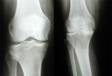
Despite the rapid development in recent years of such modern methods of medical imaging as MRI, X-ray computed tomography, expansion of ultrasound diagnostic capabilities, X-ray diagnostics of osteoarthrosis remains the most common objective method of diagnosing and monitoring the effectiveness of osteoarthrosis treatment. This is due to the availability of this method, simplicity of research, cost-effectiveness and sufficient information content.
In general, radiographic diagnostics of osteoarthrosis is based on the detection of joint space narrowing, subchondral osteosclerosis and osteophytes (OF), with the degree of narrowing of the radiographic joint space being of primary diagnostic importance. Joint radiographs may show areas of ossification of the joint capsule (late osteoarthrosis). In the nodular form of osteoarthrosis, the greatest diagnostic importance is the detection of osteophytes, sometimes accompanied by severe destruction of the joint surfaces (the so-called erosive arthrosis).
The X-ray joint space, being filled with cartilage and a layer of synovial fluid, which do not produce an image on X-rays, has the appearance of a more transparent strip between the articular surfaces.
The total thickness of articular cartilage on radiographs is determined by measuring the width of the radiographic joint space between the articular surfaces of the bone epiphyses. It should be noted that the width of the radiographic joint space is still used as the main indicator in the diagnosis of osteoarthritis, and standard radiography of the knee joints in direct and lateral projections is recommended by WHO and ILAR as the method of choice for assessing the dynamics of changes in the articular cartilage during clinical trials of drugs. Narrowing of the radiographic joint space corresponds to a decrease in the volume of articular cartilage, and subchondral osteosclerosis and osteophytes on the edges of the articular surfaces are considered by most researchers as a response of bone tissue to an increase in the mechanical load on the joint, which in turn is the result of degenerative changes and a decrease in the volume of articular cartilage. The above is important not only for the diagnosis of osteoarthritis, but also for assessing the progression of the disease and the treatment.
The indicated radiological symptoms are considered specific for osteoarthritis and are included in the list of radiological criteria for diagnosing this disease along with clinical ones.
Methods for optimizing radiological diagnostics of osteoarthritis
As already mentioned, the methods for assessing the progression of osteoarthritis are based on identifying the radiographic dynamics in the joints. It should be taken into account that the dynamics of radiographic changes in osteoarthritis is characterized by a slow rate: the rate of narrowing of the radiographic joint space in patients with gonarthrosis is approximately 0.3 mm per year. The results of long-term studies of radiographic changes in patients with osteoarthritis in the knee joints who received non-hormonal anti-inflammatory treatment showed the absence of radiographic progression of the disease after 2 years of observation and minimal differences between the groups of patients receiving treatment and the control. The absence of reliable changes in long-term studies give reason to assume that the radiographic symptoms of osteoarthritis in standard radiography of joints remain relatively stable for a long time. Therefore, to assess the dynamics of changes, it is preferable to use more sensitive X-ray technologies, one of which is microfocus radiography of joints.
Microfocus X-ray machines use special X-ray tubes with a point source of radiation. Quantitative microfocus radiography with direct magnification of the image shows sufficient sensitivity in detecting small changes in bone structure. With this method, the progression of osteoarthritis and the effect of the treatment can be recorded and accurately measured in a relatively short time between examinations. This is achieved by standardizing the examination and using a radiographic measurement procedure, improving the quality of the obtained radiographs of joints with direct magnification of the image, which allows recording structural bone details invisible on standard radiographs. WHO/ILAR recommend measuring the width of the radiographic joint space manually using the Lequesne method using a magnifying lens and calculating the width of the radiographic joint space at different points. Such measurements show that the coefficient of variation with repeated measurements is 3.8%. The development of microcomputer and image analysis technology provides a more accurate assessment of changes in joint anatomy than manual methods. Digital processing of the X-ray image of the joint allows automatic measurement of the joint space width using a computer. The researcher's error is practically excluded, because the accuracy of repeated measurements is set by the system itself.
From the point of view of diagnostic efficiency, simplicity and ease of use, mobile X-ray diagnostic devices with a multi-position C-arm stand, widely used in world practice, are of particular interest. Devices of this class allow for examination of the patient in any projections without changing his position.
Worthy of attention is the method of functional radiography of knee joints, consisting of performing 2 consecutive X-ray images of the knee joint with the patient standing in a direct anterior projection with predominant support on the examined limb (the 1st image - with a fully straightened knee joint, the 2nd - with flexion at an angle of 30 °). The contours of the bone elements forming the X-ray-joint gap from the 1st and 2nd radiographs were transferred to paper and sequentially entered into a computer using a scanner, after which the degree of damage to the hyaline cartilage of the knee joint was determined by the difference in the ratio of lateral and medial areas between the 1st and 2nd radiographs (the stage of osteoarthrosis was assessed according to Hellgen). Normally, it was 0.05 ± 0.007; for stage I - 0.13 ± 0.006; for stage II - 0.18 ± 0.011; for stage III - 0.3±0.03. There is a significant difference between the normal values and those at stage I (p<0.001): between stages I and II the difference is reliable (p<0.05), between stages II and III of osteoarthritis - a significant difference (p<0.001).
The obtained data indicate that X-ray planimetry of the knee joint during functional radiography objectively displays the stage of osteoarthrosis of the knee joint.
The method of functional radiography with load allowed to establish that in 8 patients, in whom pathological changes were not detected by traditional radiography, there is an initial decrease in the height of the radiographic joint space. In 7 patients, a more severe degree of damage was established. Thus, the diagnosis was changed in 15 (12.9+3.1%) patients.
Along with the traditional method of X-ray examination of the knee joint - examination of the knee joint in standard projections with the patient in a horizontal position - there is a method of examining this joint in a vertical position. According to V. A. Popov (1986), a picture of the knee joint taken in a horizontal position does not reflect the real mechanical conditions of the joint under body weight load. He proposed to conduct an examination of the knee joint in an orthostatic position with predominant support on the limb being examined. SS Messich et al. (1990) suggested that the best position for diagnosing osteoarthritis is knee flexion by 28° with the patient in an upright position, also with predominant support on the limb being examined, since biomechanical studies have shown that the initial lesion of the hyaline cartilage of the knee joint is noted in the posterior parts of the femoral condyles, located at an angle of 28° in the sagittal plane, since it is in this position that the main mechanical load on the cartilage acts (the physiological position of the knee joint). H. Petterson et al. (1995) proposed a technique for radiography of the knee joint with a load, in which the lower part of the leg is at an angle of 5-10° to the plane of the film and the joint is additionally flexed at an angle of 10-15°. According to the authors, in this position the central ray is directed tangent to the plane of the tibial condyle and the joint space will be correctly represented in the image.
Thus, the targeted use of classical radiography capabilities, taking into account clinical manifestations, allows in many cases to confirm or at least suspect the presence of damage to a particular structure of the ligament-meniscus complex of the knee joint and to decide on the need for additional examination of the patient using other means of medical imaging.
Radiographic symptoms required to establish a diagnosis of primary osteoarthritis
Narrowing of the radiographic joint space is one of the most important radiographic symptoms, which has a direct correlation with pathological changes occurring in the articular cartilage. The radiographic joint space in different parts of the joint has different widths, which is due to the uneven decrease in the volume of articular cartilage in different areas of the articular surface. According to WHO/ILAR recommendations, the width of the radiographic joint space should be measured in the narrowest area. It is believed that in a pathologically changed joint, this area experiences the maximum mechanical load (for the knee joint, these are most often the medial sections, for the hip joint - the superomedial, less often - the superolateral sections). The anatomical landmarks used to measure the joint space on radiographs of large joints include:
- for convex surfaces (head and condyles of the femur) - the cortical layer of the endplate of the articular surface of the bone;
- for concave surfaces (edge of the acetabulum, proximal condyles of the tibia) - the edge of the articular surface at the base of the glenoid cavity.
Subchondral osteosclerosis is a compaction of bone tissue located directly under the articular cartilage. Usually, this radiographic symptom is a consequence of friction of exposed articulating uneven articular bone surfaces against each other. It is detected in the late stages of osteoarthrosis, when the joint space is sharply narrowed. This symptom indicates a deep degenerative-destructive process in the articular cartilage or even the disappearance of the latter. Violation of the integrity of the articular cartilage, preceding its quantitative reduction, may be the result of compaction of the cortical and trabecular bone tissue located directly under the cartilage. Compaction of the subchondral bone tissue in the area of the articular surfaces of the bones is measured at three equally spaced points along the articular edge; the measurement results can be averaged.
Osteophytes are limited pathological bone growths of various shapes and sizes that occur with productive inflammation of the periosteum at the edges of the articular surfaces of bones - a characteristic radiographic symptom of osteoarthritis. In the initial stages of osteoarthritis, they look like sharpenings or small (up to 1-2 mm) bone formations at the edges of the articular surfaces and at the attachment points of the joints' own ligaments (in the knee joints - along the edges of the intercondylar tubercles of the tibia, at the attachment points of the cruciate ligaments; in the hip joints - along the edges of the fossa of the femoral head, on its medial surface, at the attachment point of the femoral head's own ligament).
As the severity of osteoarthritis increases and the narrowing of the joint space progresses, osteophytes increase in size, acquire various shapes in the form of "lips" or "ridges", rectilinear or "lush" bone growths on a wide or narrow base. In this case, the articular head and socket can significantly increase in diameter, become more massive and "flattened". The number of osteophytes can be counted separately or in total in both joints, and their sizes can be determined by the width at the base and length. Changes in the number of osteophytes and their sizes are a sensitive indicator of the progression of osteoarthritis and monitoring the effectiveness of its treatment.
Radiographic findings not required for diagnosis of primary osteoarthritis
Periarticular marginal bone defect. Although this radiographic finding, which may be seen in osteoarthritis, was defined by RD Altman et al. (1990) as "erosion of the articular surface," the term "periarticular marginal bone defect" is preferable because there is no precise histologic characterization of these radiographically detectable changes. Marginal bone defects may also be seen in the early stages of osteoarthritis, and their appearance may be caused by inflammatory changes in the synovial membrane. Similar changes have been described in large joints and in the joints of the hands. Typically, in osteoarthritis, these defects are small in size, with an area of osteosclerosis at the base. Unlike true erosions detected in rheumatoid arthritis, which do not have sclerotic changes at the base and are often determined against the background of periarticular osteoporosis, the bone tissue surrounding the periarticular marginal defect is not rarefied in osteoarthrosis.
Subchondral cysts are formed as a result of bone tissue resorption in areas with high intra-articular pressure (at the site of the greatest load on the articular surface). On radiographs, they look like ring-shaped defects of trabecular bone tissue in the subchondral bone with a clearly defined sclerotic rim. Most often, subchondral cysts are located in the narrowest part of the joint space and occur during an exacerbation of the disease. They are characteristic of osteoarthritis of the hip joints, and can be found both in the head of the femur and in the roof of the acetabulum. The dynamics of changes in subchondral cysts are judged by their number and size.
Intra-articular calcified chondromas are formed from areas of necrotic articular cartilage and may also be a fragment of bone tissue (osteophytes) or produced by the synovial membrane. They are usually small in size, located between the articular surfaces of bones or on the side of the bone epiphyses, have different shapes (round, oval, elongated) and an uneven speckled structure, which is due to the deposition of calcium-containing substances in the cartilaginous tissue. Usually no more than 1-2 chondromas are found in a joint.
In the knee joint, the sesamoid bone (fabella) in the popliteal fossa can be mistaken for a calcified chondroma, which also changes its shape, position and size in osteoarthritis of the knee joint. Fabella deformity is one of the symptoms of osteoarthritis of the knee joint.