
Medical expert of the article

New publications
Swollen Legs: What to Do and When It's Dangerous
Last updated: 12.03.2026

All iLive content is medically reviewed or fact checked to ensure as much factual accuracy as possible.
We have strict sourcing guidelines and only link to reputable media sites, academic research institutions and, whenever possible, medically peer reviewed studies. Note that the numbers in parentheses ([1], [2], etc.) are clickable links to these studies.
If you feel that any of our content is inaccurate, out-of-date, or otherwise questionable, please select it and press Ctrl + Enter.
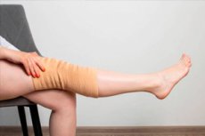
Leg edema is a symptom, not a diagnosis in itself. Pathophysiologically, it occurs when the balance between hydrostatic pressure, oncotic pressure, vascular permeability, and lymphatic drainage is disrupted, causing fluid to accumulate in the interstitial space. This is why the same external symptom can be associated with venous hypertension, heart failure, nephrotic syndrome, and lymphatic insufficiency. [1]
The first clinical question is always the same: is the swelling in one leg or both? The second question is how quickly it developed. Acute unilateral edema requires first ruling out deep vein thrombosis, cellulitis, trauma, hematoma, and other conditions where delay could be dangerous. Chronic bilateral edema is more often associated with chronic venous disease, systemic causes, drug effects, lymphedema, or obstructive sleep apnea. [2]
The next important clue is the nature of the edema itself. Pastous edema is more common in chronic venous disease, systemic causes, deep vein thrombosis, and early lymphedema. Non-pastous edema is more likely to suggest late lymphedema or pretibial myxedema. However, there are no absolute rules here: early lymphedema may still leave a pit, while late lymphedema becomes dense and fibrous. [3]
It's crucial not to confuse true edema with lipedema. Lipedema isn't simply "fat and water" or simple fluid retention, but a chronic disease of the subcutaneous fat tissue characterized by disproportion, pain, and, typically, preservation of the feet. This is why lipedema is included in the differential diagnosis of leg edema, but is not limited to it. [4]
For clinical practice, it's helpful to remember another rule: leg swelling should not be assessed in isolation from the patient's overall condition. Shortness of breath, orthopnea, chest pain, fever, sudden calf pain, rapid increase in limb circumference, nocturnal worsening of breathing, new asymmetry, and medication history are often more important than the swelling itself. Therefore, a competent article on leg swelling should always focus on the diagnostic process, not just the symptom.
Table 1. What most often causes leg swelling
| Group of reasons | Typical mechanism | What the diagnosis often suggests |
|---|---|---|
| Chronic venous disease | Venous hypertension and congestion | Heaviness in the evening, varicose veins, hyperpigmentation |
| Deep vein thrombosis | Acute venous outflow obstruction | Sudden one-sided swelling, pain, warmth |
| Lymphedema | Insufficiency of lymphatic drainage | Dense edema, thickening of the skin, Stemmer's sign |
| Heart failure | Increased venous pressure and fluid retention | Dyspnea, orthopnea, bilateral edema |
| Kidney and liver diseases | Decreased oncotic pressure and sodium retention | Generalized edema, changes in test results |
| Drug-induced edema | Pharmacological vasodilation or sodium retention | Connection with the new drug |
| Lipedema | Pathology of subcutaneous adipose tissue | Symmetry, pain, relative preservation of the feet |
The table is based on the peripheral edema review, chronic venous disease guidelines, lymphedema consensus, and lipedema guidelines.[5]
Why do legs swell?
The most common cause of chronic swelling of the legs and feet in adults is chronic venous disease. Due to venous reflux, obstruction, or a combination of these factors, pressure in the venous system of the lower extremities increases, causing fluid to leak into the tissues. Clinically, this typically results in swelling that increases toward evening, a feeling of heaviness, burning, distension, and itching, and, over time, skin changes, including hyperpigmentation and lipodermatosclerosis. [6]
Lymphedema develops differently. Here, the main problem is not venous pressure, but rather impaired lymphatic transport. The International Society of Lymphology even identifies a stage 0, when clinically evident edema is not yet present, but lymphatic transport is already impaired. In stage 1, swelling may decrease with elevation of the limb; in stage 2, this is less pronounced, and then fibrosis, skin thickening, and persistent tissue deformation occur. [7]
Systemic causes are also very important. Bilateral leg edema is often seen in heart failure, nephrotic syndrome, cirrhosis, pregnancy, hypothyroidism, and some forms of pulmonary hypertension. In such cases, the legs are only the visible part of the overall problem, and the doctor's task is not to select the "best diuretic," but to find the underlying disease. This is why a basic examination includes biochemistry, liver function tests, thyroid-stimulating hormone, natriuretic peptide, and urine protein assessment. [8]
Drug-induced edema is one of the most underestimated causes, especially in elderly patients. The most common example is dihydropyridine calcium channel blockers, such as amlodipine and nifedipine. However, edema can also be caused by estrogens, progesterone, testosterone, nonsteroidal anti-inflammatory drugs, gabapentin, pregabalin, thiazolidinediones, and some other drugs. The key to diagnosis is the time relationship between the start of treatment and the onset of edema. [9]
In acute unilateral edema, urgent rather than chronic causes come to the fore. These include deep vein thrombosis, cellulitis, trauma, hematoma, and, less commonly, ruptured Baker's cysts or anatomical variants of venous compression, including May-Turner syndrome. It is in this group of conditions that the mistake of "waiting for a couple more days" can be most dangerous. [10]
Lipedema occupies a special place. Current guidelines emphasize that it is not a form of classic edema and is not a consequence of venous or lymphatic insufficiency per se. It is characterized by tenderness to pressure, a feeling of heaviness, symmetrical involvement of the extremities, and lack of involvement of the feet and hands. Therefore, diuretics are not considered as a treatment for lipedema unless there is another internal reason for their use. [11]
Table 2. Main causes of leg swelling and their symptoms
| Cause | Usually 1 leg or 2 | Pain | Leather and fabrics | Typical context |
|---|---|---|---|---|
| Deep vein thrombosis | More often than 1 | Often there is | Warmth, redness, swelling | Sudden onset, flight, surgery, immobilization |
| Chronic venous disease | Often 2 | Heaviness, burning sensation | Varicose veins, hyperpigmentation, lipodermatosclerosis | Worse in the evening, standing |
| Lymphedema | 1 or 2 | Often not the main complaint | Dense, thickened skin, hyperkeratosis | After surgery, radiation therapy, congenital forms |
| Heart failure | More often than 2 | There is usually no local pain. | Pastosity without pronounced local inflammation | Shortness of breath, orthopnea, fatigue |
| Drug-induced edema | More often than 2 | Usually no | Mild swelling without local inflammation | New drug in recent weeks |
| Lipedema | More often than not 2 and symmetrically | There is often pain | The feet are intact, but the tissues are painful. | Long-term course, figure disproportion |
| Cellulite | Usually 1 | Often expressed | Heat, redness, soreness | Fever, the entry point for infection |
The table is based on the peripheral edema review, deep vein thrombosis guidelines, lymphedema consensus, and lipedema guidelines.[12]
When leg swelling is dangerous
The most dangerous scenario is a combination of leg swelling with chest pain, shortness of breath, hemoptysis, severe weakness, dizziness, or fainting. In this situation, pulmonary embolism should be considered immediately and action taken immediately. Guidelines for venous thromboembolic diseases consider these symptoms a reason for urgent evaluation rather than home monitoring.
Sudden unilateral swelling, especially if accompanied by pain, local warmth, and redness, also requires prompt diagnosis. If deep vein thrombosis is suspected, the clinical probability is first assessed using the Wells scale, followed by rapid progression to D-dimer testing or compression ultrasound, depending on the risk level. If the likelihood of deep vein thrombosis is high, an ultrasound scan should be performed within 4 hours, and if this is not possible, therapeutic anticoagulation should be considered temporarily until imaging is performed. [13]
Edema is also dangerous, accompanied by high fever, increasing redness, severe pain, rapidly spreading inflammation, and general deterioration. In this case, one must consider not so much a venous problem as an infectious process, primarily cellulitis, and sometimes even soft tissue abscess formation. Home warming and massage in this situation only delay proper treatment. [14]
Rapidly increasing bilateral edema, combined with orthopnea, dyspnea on exertion, rapid weight gain, nocturnal worsening of breathing, or signs of pulmonary edema, requires special attention. This combination of symptoms necessitates the exclusion of decompensated heart failure. If decompensation is suspected, natriuretic peptide is measured and, if clinical data are available, echocardiography is performed, as further treatment depends on cardiac function. [15]
Finally, rapidly growing, dense swelling with thickening of the skin, recurrent inflammation, and persistent increase in limb size rarely requires an ambulance call but does require prompt referral to a lymphatic specialist. Early diagnosis of lymphedema is important because in later stages, fat and fibrosis are added to the fluid, making treatment significantly more difficult. [16]
Table 3. Red flags for leg swelling
| Situation | What should be excluded first? | Urgency |
|---|---|---|
| Leg swelling plus chest pain or shortness of breath | Pulmonary embolism | Immediately |
| Sudden one-sided swelling and pain | Deep vein thrombosis | Urgently |
| Swelling, heat, bright redness, fever | Cellulitis and other soft tissue infections | Urgently |
| Rapid bilateral edema plus orthopnea | Decompensation of heart failure | Urgently |
| Dense progressive edema with thickening of the skin | Lymphedema with chronicity and complications | Fast Track |
| Rapidly growing bruise and swelling due to anticoagulants or trauma | Hematoma and bleeding in tissue | Urgently |
The table is compiled based on the guidelines on venous thromboembolic diseases, the review on peripheral edema, and the consensus on lymphedema. [17]
Diagnostics
A proper diagnosis begins not with an analysis or ultrasound, but with a competent triage of the complaint. The doctor must immediately understand four things: whether the swelling is unilateral or bilateral, acute or chronic, pasty or dense, and whether it is accompanied by pain, shortness of breath, skin changes, or drug triggers. This approach allows for a dramatic reduction in unnecessary examinations while simultaneously preventing the possibility of dangerous thrombosis or cardiac decompensation. [18]
A physical examination provides a wealth of information. Swelling often suggests a venous or systemic cause. Symmetrical edema that is characteristic of the foot makes lipedema more likely. Stemmer's sign, the inability to take a fold of skin on the dorsum of the second toe, is considered pathognomonic of late lymphedema. Hyperpigmentation, a medial malleolar ulcer, lipodermatosclerosis, and prominent venous vessels suggest chronic venous disease. Hyperkeratosis and wart-like changes more strongly support a diagnosis of lymphedema. [19]
Basic laboratory testing for lower extremity edema should include B-type natriuretic peptide, thyroid-stimulating hormone, liver function tests, basic biochemistry, and urine protein-to-creatinine ratio. This panel helps quickly differentiate a localized problem from cardiac, renal, hepatic, or endocrine causes. For this reason, attempting to "treat only the legs" without minimal laboratory testing is often a mistake. [20]
If the swelling is acute and unilateral, the primary concern is deep vein thrombosis. For such patients, the Wells scale is used. If the probability is low, a D-dimer test is performed. If the probability is intermediate or high, a compression ultrasound is performed immediately. If the initial ultrasound is negative, but the D-dimer is positive or clinical suspicion remains high, a repeat ultrasound is indicated in 6-8 days. [21]
If the swelling is chronic and a venous origin is suspected, duplex ultrasound of the lower extremity veins becomes the primary imaging method. European vascular guidelines recommend it as the primary diagnostic and treatment planning method for chronic venous disease. It allows for the visualization of not only thrombi or obstruction, but also venous reflux, deep vein patentness, and the anatomy of the superficial system. [22]
When the clinical picture more closely resembles lymphedema, diagnosis often remains clinical. However, if the cause is unclear, if clarification is needed before treatment, or if primary lymphatic pathology must be differentiated from other conditions, the International Society of Lymphology recommends lymphoscintigraphy. Many centers initially use ultrasound to rule out venous disease, and then move on to lymphatic imaging if necessary. [23]
In some patients, chronic bilateral edema is associated with obstructive sleep apnea syndrome. Therefore, in individuals with obesity, snoring, somnolence, and persistent bilateral edema, after excluding obvious cardiac, renal, hepatic, and venous causes, it is reasonable to use a validated questionnaire for the initial screening of this condition. [24]
Table 4. Step-by-step diagnostic route
| Step | What is being assessed? | What does this give? |
|---|---|---|
| 1 | 1 leg or 2, acute or chronic | Narrows the range of causes |
| 2 | Pain, shortness of breath, fever, medications, injury | Identifies red flags |
| 3 | Pastosity, feet, skin, veins, Stemmer's sign | Helps in differential diagnosis |
| 4 | Biochemistry, liver function tests, thyroid stimulating hormone, urine protein, natriuretic peptide | Looks for systemic causes |
| 5 | Wells scale, D-dimer, compression ultrasound | Rules out or confirms deep vein thrombosis |
| 6 | Duplex ultrasound examination with reflux assessment | Confirms chronic venous disease |
| 7 | Lymphoscintigraphy when the picture is unclear | Clarifies lymphedema |
The table is based on the review of peripheral edema, the venous thromboembolic disease guidelines, the vascular guidelines for chronic venous disease, and the lymphedema consensus.[25]
Differential diagnosis
Chronic venous disease and lymphedema are most often confused. With venous edema, complaints typically worsen in the evening and after standing, the swelling is often pasty, and varicose veins, hyperpigmentation, itching, and sometimes medial ulcers and lipodermatosclerosis are present. With lymphedema, in the later stages, the swelling becomes denser, less easily relieved by elevated limbs, the skin thickens, and hyperkeratosis and characteristic fibrous changes appear. [26]
It's also important not to confuse lymphedema and lipedema. Lipedema is typically characterized by bilateral symmetry, marked tenderness to pressure, subjective heaviness, and no involvement of the feet. In contrast, lymphedema often involves the foot, and marked tenderness is not the primary symptom. Furthermore, modern guidelines on lipedema explicitly emphasize that it is not a classic edema or a manifestation of pure venous or lymphatic dysfunction. [27]
Acute unilateral edema requires distinguishing deep vein thrombosis from cellulitis, hematoma, trauma, and Baker's cyst rupture. Deep vein thrombosis is more typically characterized by the entire leg or lower leg, tenderness along the deep veins, swelling, and sometimes dilated superficial collaterals. Cellulitis is characterized by intense redness, heat, tenderness, and fever. A ruptured Baker's cyst may be characterized by a crescent-shaped ecchymosis in the ankle area. [28]
Systemic edema is characterized by the legs rarely being the only complaint. Heart failure can cause orthopnea, jugular venous distension, shortness of breath on exertion, and often rapid weight gain. Cirrhosis can also cause ascites and spider veins. In nephrotic syndrome or other renal pathologies, proteinuria and generalized fluid retention are significant. Therefore, in the case of bilateral leg edema without localized vascular signs, examination of the extremities alone is not sufficient. [29]
Drug-induced edema is especially important to consider in patients without compelling evidence of thrombosis, venous insufficiency, or lymphedema. If edema develops 1-4 weeks after starting a new medication, especially amlodipine or nifedipine, the likelihood of a drug-induced cause is high. This is of practical importance because diuretics do not address the underlying mechanism of this edema.
Table 5. How to distinguish the main forms of leg swelling
| Sign | Chronic venous disease | Lymphedema | Lipedema | Deep vein thrombosis | Systemic edema |
|---|---|---|---|---|---|
| 1 leg or 2 | Often 2 | 1 or 2 | Usually 2 | More often than 1 | Usually 2 |
| Pastosity | Often there is | Maybe sooner, maybe less later | Not a leading feature | Often there is | Often there is |
| Pain | Heaviness, burning sensation | Usually not the main one | Often severe pain | Often there is | Usually there is no local pain. |
| Feet | Often involved | Often involved | Often preserved | Involved | Involved |
| Leather | Hyperpigmentation, lipodermatosclerosis | Thickening, hyperkeratosis | Without typical venous trophic signs | Warmth, redness | Usually without local inflammation |
| Key test | Duplex ultrasound examination of veins | Lymphoscintigraphy in case of doubt | Clinical diagnosis | Wells scale plus ultrasound examination | Laboratory and cardiological search |
The table is compiled from the peripheral edema review, lymphedema consensus, lipedema guidelines, and thrombosis guidelines.[30]
Treatment
The main principle of treating leg swelling is to treat the cause, not just the fluid. This is especially important because diuretics are only helpful for systemic forms of fluid retention and should not be used as a general treatment for venous disease, lymphedema, lipedema, or drug-induced edema. A recent review of peripheral edema clearly emphasizes that there is no evidence of benefit from diuretics for non-systemic causes. [31]
For chronic venous disease, compression remains the mainstay of treatment. European vascular guidelines indicate that in patients with chronic venous disease and edema, below-knee compression therapy with ankle pressure of 20-40 mmHg reduces swelling. In addition, conservative management includes exercise, weight loss, elevated leg positioning at rest, and, in some patients, venotonic medications. However, if venous insufficiency is obvious and symptoms persist, interventional treatment may be required. [32]
For lymphedema, the primary non-surgical treatment is complex decongestant therapy. The International Society of Lymphology describes a two-phase approach: first, skin care, gentle manual lymphatic drainage techniques, exercises, and multilayer bandaging, followed by a maintenance phase with compression stockings, continued physical activity, and skin care. This requires patient education and specialist involvement, and goes beyond simply advising "to wear stockings." [33]
If deep vein thrombosis is confirmed, treatment should be initiated promptly. NICE recommends prompt ultrasound imaging if deep vein thrombosis is suspected, and if imaging is delayed, temporary therapeutic anticoagulation should be considered until imaging is performed. Once the diagnosis is confirmed, anticoagulation becomes the mainstay of treatment, and further management depends on the location of the thrombus, the risk of bleeding, and the individual patient profile. [34]
In drug-induced edema, the primary decision is to review the medication. Sometimes, reducing the dose or discontinuing the offending medication is sufficient; sometimes, switching to a different class of medication is necessary. For patients receiving calcium channel blockers, a review of peripheral edema notes that adding an angiotensin-converting enzyme inhibitor can reduce peripheral edema. However, this decision is made based on the overall cardiovascular treatment plan, not just the leg complaint. [35]
Diuretics are not used as a treatment for lipedema. Current guidelines emphasize that compression for lipedema is primarily aimed at reducing pain and other subjective symptoms, not at "removing fluid." Treatment includes movement, exercise, pneumatic compression if necessary, psychosocial support, and, in some cases, surgical correction. If a patient has lipedema and another type of edema, such as venous edema, then therapy is tailored to address both processes. [36]
In systemic edema, the final outcome depends on control of the underlying disease. In heart failure, confirmation of the diagnosis and selection of cardiac therapy are important; in renal disease, monitoring of urine protein and renal function; and in cirrhosis, management of portal hypertension and hypoalbuminemia. Therefore, symptomatic reduction of leg volume without a systemic diagnosis is not considered a complete treatment. [37]
Table 6. Treatment depending on the cause
| Cause | What is the basis of treatment? | What not to do |
|---|---|---|
| Chronic venous disease | Compression, movement, body weight correction, venous intervention if indicated | Relying solely on diuretics |
| Lymphedema | Complex decongestant therapy, skin care, compression, exercises | Limit treatment to massage only without compression |
| Deep vein thrombosis | Urgent diagnostics and anticoagulation | Wait for spontaneous improvement |
| Drug-induced edema | Revision of the causative drug | Uncontrolled use of diuretics |
| Lipedema | Compression for symptoms, movement, comprehensive support | Treat with diuretics as usual for edema. |
| Cardiac, renal, hepatic edema | Treatment of the underlying systemic disease | Ignore the overall context and analysis |
The table is based on the peripheral edema review, chronic venous disease guidelines, lymphedema consensus, lipedema guidelines, and venous thromboembolic disease guidelines.[38]
Compression hosiery and home measures
Compression therapy is one of the most effective non-pharmacological treatments for chronic venous disease and an important part of the treatment of many other forms of edema. A review of peripheral edema and European vascular guidelines indicate that graduated compression is most often the first line of treatment for venous edema, and the 20-40 mmHg range has been particularly well studied for reducing swelling, pain, and trophic manifestations of chronic venous disease. [39]
However, compression is not suitable for everyone. Before prescribing it to patients at risk for peripheral arterial disease, the ankle-brachial index should be assessed. European guidelines list contraindications for this condition as severe lower extremity ischemia (an index below 0.6) or ankle pressure below 60 mmHg, severe functional class 4 heart failure, and functional class 3 heart failure without clinical and hemodynamic monitoring. [40]
Compression is also necessary for lymphedema, but it serves a different purpose. During the active phase of treatment, multilayer bandaging or other forms of intensive compression are used, followed by a transition to supportive hosiery. The International Society of Lymphology emphasizes that compression garments should be selected by a specialist, and the success of treatment depends largely on the quality of the fit and daily wear. [41]
Among home measures, walking, regular activation of the calf muscle pump, reduction of prolonged sitting and standing, and elevated leg positioning at rest offer the best balance of benefit and safety. European vascular guidelines note that the benefits of exercise for venous function are supported by indirect and clinical evidence, and that elevated leg positioning and increased activity of the foot and calf muscle pumps may be beneficial in venous hypertension. In lymphedema, physical activity is also supported by consensus, as it improves function and quality of life. [42]
Self-administration of diuretics, aggressive massage if thrombosis is suspected, and attempts to "warm up" acute, painful swelling cannot be considered safe. If the condition is a chronic venous or lymphatic process without any "red flags," home measures are acceptable in addition to an examination. However, if the swelling is new, rapidly increasing, or accompanied by pain and shortness of breath, diagnosis remains the priority, not home treatments. [43]
Table 7. When compression helps and when caution is needed
| Situation | The role of compression | Comment |
|---|---|---|
| Chronic venous disease with edema | Basic treatment | Often used below the knee |
| Lymphedema | An essential part of long-term therapy | Professional selection is needed |
| Post-thrombotic syndrome | May reduce the severity of symptoms | Selected individually |
| Severe leg ischemia | Contraindicated or severely limited | Blood flow assessment needed |
| Severe heart failure | May be contraindicated | Especially in cases of severe decompensation |
| Allergy to the material | Other material or refusal required | It is important to consider portability |
The table is compiled according to the European vascular guidelines, the international consensus on lymphedema and the international consensus on the risks of compression therapy. [44]
Prevention and prognosis
The prognosis for leg swelling depends entirely on the cause. With drug-induced edema, improvement often occurs after adjusting the treatment regimen. With chronic venous disease, symptoms can persist for years, but their severity is significantly reduced with good compression, exercise, and timely treatment of venous reflux. With lymphedema, the goal is often not a complete "cure," but rather long-term control of limb volume, skin, and quality of life. [45]
Prevention of acute thrombosis is based on the recognition of risk factors. Recent surgery, immobilization, active cancer, previous venous thrombosis, paralysis, and severe leg asymmetry are not just elements of a scale, but signs that should raise the threshold of suspicion and prompt prompt referral of the patient for ultrasound examination. [46]
Prevention of chronic venous edema is based on reducing venous hypertension. In practice, this means weight control, regular walking, minimizing prolonged standing and sitting, using compression as indicated, and treating chronic venous insufficiency before it progresses to skin changes and ulcers. European guidelines also emphasize the need to first rule out non-venous causes of bilateral edema before considering venous interventions. [47]
Early recognition and prevention of skin complications are particularly important for lymphedema. International consensus indicates that early detection and initiation of a conservative program, including compression garments, self-care training, and physical activity, offer a chance to prevent the disease from progressing to its earlier stages and delay fibrotic changes. [48]
The most practical conclusion is this: leg swelling is rarely harmless "in itself," but it doesn't always indicate a serious emergency. The most dangerous are not chronic, stable forms, but new asymmetry, rapid swelling growth, and a combination with pain, fever, shortness of breath, and a general deterioration in condition. These situations should prompt a change in the speed with which one seeks help. [49]
Frequently asked questions
Can leg swelling be caused solely by heat or sedentary work?
Yes, it can be, especially with chronic venous disease or functional venous congestion. But if swelling appears for the first time, becomes asymmetrical, is accompanied by pain, or does not subside after rest, it cannot be automatically attributed to heat or sitting. [50]
If both legs swell, is it definitely not thrombosis?
No. Bilateral edema is more often associated with systemic, medicinal, or venous causes, but thrombosis cannot be completely ruled out, especially with proximal venous obstruction or a combination of risk factors. It's just that with unilateral acute edema, the likelihood of deep vein thrombosis is significantly higher. [51]
Should everyone have a vein ultrasound?
No. In cases of acute unilateral edema and suspected deep vein thrombosis, yes; it's often one of the first tests. In cases of chronic edema, an ultrasound is especially useful if chronic venous disease is suspected. However, if the clinical picture suggests a systemic disease, laboratory and general assessment are more important first. [52]
Is it possible to start wearing compression stockings without a doctor's appointment?
Sometimes yes, but not everyone. If there is a risk of arterial disease, severe heart failure, severe foot ischemia, or unexplained new edema, a doctor's evaluation is necessary first. In patients with chronic venous disease, compression is often beneficial, but it must be safe and properly fitted. [53]
Do diuretics help with all types of leg swelling?
No. Diuretics are primarily indicated for systemic causes of fluid retention, such as heart failure or certain kidney and liver conditions. They do not address the underlying problem in cases of chronic venous disease, lymphedema, lipedema, and drug-induced edema. [54]
How can you differentiate lymphedema from lipedema at home?
It's impossible to make a definitive diagnosis at home, but there are some clues. With lipedema, the lesions are usually symmetrical, the feet are relatively spared, and the tissues are painful. With lymphedema, the feet are often also involved, and in later stages, the skin becomes dense and Stemmer's sign may become positive. A definitive diagnosis requires clinical evaluation. [55]
Is it true that amlodipine often causes leg swelling?
Yes. This is one of the most common drug-induced causes of peripheral edema. Importantly, the mechanism is not related to simple fluid retention, but to arteriolar vasodilation, so uncontrolled use of diuretics is not considered a good idea. [56]
When does leg swelling require emergency care?
When it is accompanied by shortness of breath, chest pain, hemoptysis, sudden weakness, high fever, a very rapid increase in leg size, or severe one-sided pain and redness. These signs suggest pulmonary embolism, deep vein thrombosis, and severe infection. [57]
Key points from experts
Peter Glowicki, MD, FACS, Distinguished Professor of Surgery at the Mayo Clinic, former Chief of the Division of Vascular and Endovascular Surgery, former President of the Society for Vascular Surgery and the American Venous Forum, is a leading expert in the field of vascular edema. The practice thesis that runs through current vascular guidelines and his long-standing work is that chronic venous edema requires not a cursory examination but a standardized venous assessment with duplex ultrasound and subsequent individualized decision-making between compression, observation, and intervention. [58]
Mark H. Meissner, MD, professor of surgery at the University of Washington, is a vascular surgeon whose clinical interests include acute deep vein thrombosis, chronic venous disease, and postthrombotic syndrome. His clinical emphasis reflects the modern approach to leg edema: new asymmetry and suspected thrombosis cannot be assessed "by feel," because early, accurate diagnosis changes both management and prognosis. [59]
Stanley G. Roxon, MD, is a professor of lymphatic medicine and lymphatic research at Stanford University, director of the Stanford Center for Lymphatic and Venous Disorders, and editor-in-chief of the Journal of Lymphatic Research. The key message of the lymphological community, which he embodies, is that lymphedema is a chronic, staged disease of lymphatic transport, not simply "solid swelling," and that success depends on early recognition, decongestant therapy, compression, and ongoing self-monitoring. [60]
Gregory Piazza, MD, MS, chief of the Section of Vascular Medicine in the Division of Cardiovascular Medicine at Brigham and Women's Hospital, is an associate professor at Harvard Medical School and an expert in thrombosis, thromboembolic disease, and venous insufficiency. His practical thesis for daily practice is this: acute leg swelling should be assessed against the risk of venous thromboembolism, because a missed deep vein thrombosis poses a risk not only to the leg itself but also to the potential for pulmonary embolism. [61]
Stavros Kakkos, MD, MSc, PhD, Professor and Head of the Department of Vascular Surgery, University of Patras School of Medicine, contributor to 26 guidelines and consensus documents, member of the European Society for Vascular Surgery Guidelines Committee 2013-2022. One of the main practical conclusions of the European School of Vascular Medicine is that in chronic venous edema, non-venous causes must first be excluded, especially in bilateral cases, and only then should venous intervention be considered. [62]