Medical expert of the article

New publications
Venous angioma
Last reviewed: 05.07.2025

All iLive content is medically reviewed or fact checked to ensure as much factual accuracy as possible.
We have strict sourcing guidelines and only link to reputable media sites, academic research institutions and, whenever possible, medically peer reviewed studies. Note that the numbers in parentheses ([1], [2], etc.) are clickable links to these studies.
If you feel that any of our content is inaccurate, out-of-date, or otherwise questionable, please select it and press Ctrl + Enter.
Doctors classify angioma as a benign pathological neoplasm. Venous angioma is a so-called birthmark, consisting of a plexus of blood vessels. Depending on the location, its danger may increase: from harmless to fatal.
ICD-10 code
According to the International Classification of Diseases, Tenth Revision (ICD code 10), benign neoplasms such as venous angioma have their own individual code – D18.0 – hemangioma (angioma) of any localization.
Causes of venous angioma
The most probable and common causes of venous angioma are the following:
- Genetic genesis of pathology.
- Consequences of infectious damage to the patient's body.
- Trauma received in the head area.
As observations show, if there is no trauma, heredity comes first. Infectious lesions leading to the development of the pathology of interest are rare, but they should not be excluded from the account.
 [ 1 ], [ 2 ], [ 3 ], [ 4 ], [ 5 ]
Pathogenesis
To select the most effective treatment, it is necessary to understand the pathogenesis of the disease. Angioma progresses on the basis of tissue proliferation by proliferation of vascular cells and vascular endothelium.
The essence of the difference between angioma is in the existing arterial and venous connections that capture the level of arterioles and venules. This fact contributes to the flow of blood fluid from the arterial system to the venous system, without passing through the capillaries. This is the picture that is characteristic of this pathological picture.
Small capillary or large venous vessels form a mesh, the elements of which are in fairly close contact with each other. Under certain circumstances, these capillaries grow together, forming sectors separated by stromal walls. This is how an angioma is formed. Or as it is called in this case, a hemangioma.
This pathology has a significant difference from a more classic neoplasm. Hemangioma can spontaneously regress. That is, reduce the speed of its growth, or even reverse the process. This is facilitated by the action of many factors.
Symptoms of venous angioma
Any neoplasm that forms in the internal space of the human body takes up a certain volume, which it “steals” from nearby organs. Therefore, the symptoms of venous angioma are as follows:
- Pain in the head area of varying frequency, intensity and nature.
- Increased frequency of dizziness, which provokes nausea and causes vomiting reflexes.
- The appearance of epileptic seizures.
- Depending on the location of the tumor, individual elements of the human body may be paralyzed.
- Fainting.
- Failures in the coordination center responsible for human movement.
- The emergence of speech problems.
- The occurrence of convulsions.
- Changes in taste preferences.
- A sharp decline in vision.
- Failure of mental activity.
- The appearance of a noise curtain in the head.
- Development of problems with the circulatory and cardiovascular systems.
First signs
If the disease is just emerging and beginning to develop, no pathological symptoms are observed. And only with time do the first signs of the disease appear, initially expressed in weak manifestations of dizziness and headache. Gradually, the intensity of their manifestations increases, and other signs of the disease are added.
It is very important in such a situation not to waste time and seek advice and help from a qualified specialist.
[ 6 ]
Venous angioma of the brain
The venous nature of the disease is a less aggressive nature of the pathology, but given the localization of the neoplasm, venous angioma of the brain is a disease that should not be ignored. It must be treated at an early stage of development. After all, the larger the lesion, the more intense the symptoms and the higher the risk of sudden hemorrhage into the brain tissue, which can end in the death of the patient.
Any tumor growth cannot go unnoticed by the patient's body. The tumor presses on areas of the brain, leading to disturbances and discord in the functioning of the human body.
There are a great many diseases that affect the human body. But there are pathologies that are especially dangerous for the human body. Venous angioma of the brain is one of them. If you ignore its symptoms, the outcome of the disease can be one - the death of the patient.
The tangled interweaving of venous blood vessels in the brain forms a fused monocolumn. Venous angioma is the least dangerous variant of the disease in question, but depending on the location of the problem (the brain), the situation worsens and worsens.
Intertwined vascular formations undergo expansion, provoking the emergence and progression of the inflammatory process in the tissue structures of the brain.
A growing angioma can be localized in various parts of the brain, leading to serious pathological changes and its destruction.
The degree of danger of this formation depends on the location and rate of tumor growth. The final outcome of particularly severe cases can be fatal.
Since the blood vessels grow together, the likelihood of cerebral hemorrhages increases. Their scale also increases the risk of death. In this situation, neither medication nor surgery can help.
Venous angioma of the frontal lobe
Depending on the location of the tumor, the patient experiences a slightly different set of symptoms. At the same time, a number of signs are common to all pathological manifestations, and a number give off individuality. Venous angioma of the frontal lobe can manifest itself:
- Dizziness and pain in the forehead.
- The appearance of convulsive syndrome.
- Epilepsy attacks are possible.
- Decreased sensitivity of the skin.
- Impaired attention and difficulties in logical thinking.
- Speech problems.
- Incorrect self-esteem.
- The emergence of apathy towards the surrounding life.
- Emotional instability of the patient.
- Inadequacy of behavior.
- Unawareness in actions.
- Problems with walking, vertical body stability.
The frontal lobes of the brain are responsible for the manifestation of interest, responsibility, the ability to make balanced decisions, and initiative. When this area changes, these capabilities are transformed, which is expressed by the pathological symptoms described above.
Venous angioma of the parietal lobe
If we talk about the parietal part of the brain, then this area is responsible for the following characteristics and capabilities of the body:
- The parietal lobes are designed to assess the level of sensitivity (thermal, pain threshold, etc.).
- They are responsible for human tactile sensations.
- Coordinate consistency in movements.
- They allow you to recognize symbols and signs, which makes it possible to learn about the world around you and learn to read.
- Not directly, but as a neighboring area, they can influence a person’s speech abilities.
Knowing this, it is possible to predict what failures will occur if the patient is diagnosed with venous angioma of the parietal lobe. The first signs of pathology are similar to the general symptoms of the disease in question.
Cerebellar venous angioma
When a brain area in the cerebellum is damaged or an angioma appears in its tissues, several other disturbances in the coordination and normal functioning of the body appear. Venous angioma of the cerebellum provokes the following pathological disturbances:
- A failure in the coordination of skeletal muscle function.
- Imbalance of motor coordination.
- An impairment in the body's ability to maintain its balance.
- Failure in the purposefulness of movements.
- Since the vegetative function directly affects the respiratory system and cardiovascular elements, angioma entails a disruption in their work.
- Control over maintaining the adopted posture is impaired.
- The connection with the human sensory system may be disrupted.
- There may be problems with the musculoskeletal system.
- Disruption of blood flow, with the ensuing consequences.
Venous angioma of the right hemisphere
If we are talking about the hemisphere - a layer of gray matter 1.3-4.5 mm thick, located on the periphery of the cerebral hemispheres, then venous angioma of the right hemisphere is fraught with the appearance of such negative symptoms:
- The patient loses the ability to move smoothly.
- The appearance of tremors in the lower and/or upper limbs.
- Changes in the speech apparatus are manifested by scanned speech. It becomes somewhat drawn-out and rhythmic.
- The writing style is violated.
- Movements become jerky and slow.
This pathology cannot be solved by medication; in this case, the only way to stop the problem is to perform surgery.
Venous angioma of the left hemisphere
The symptoms of this type of pathology are expressed by symptoms similar to all angiomas affecting the patient's brain. Venous angioma of the left hemisphere manifests itself, in addition to the main symptoms in the form of dizziness, noises and headaches, with such symptoms as:
- Gait disturbance.
- Incoordination of muscle function in the upper and lower extremities.
- Changes in taste preferences.
- Deterioration of vision.
- The appearance of convulsive syndrome in individual parts of the body.
- Partial paralysis.
- Impaired spoken language.
- The appearance of epileptic seizures.
- Nystagmus of the eye moving muscles.
- Deterioration of blood flow, which entails poor supply of nutrients and oxygen to tissues.
This pathology is characterized by a high rate of progression, therefore, at the slightest discomfort and suspicion of a developing disease, it is necessary to contact a qualified specialist.
Venous angioma in the basal ganglia
This type of pathological lesion - venous angioma in the basal ganglia - manifests itself with symptoms that have already been described above more than once. Therefore, only an experienced specialist should differentiate the problem and its localization. If such pathological symptoms appear, an urgent consultation with a doctor is necessary.
Consequences and complications
Venous angioma causes a lot of discomfort to the patient, but the consequences of its further progression should be alarming. The final result largely depends on the localization of the pathology and the level of neglect of the disease.
If you do not monitor your health, ignoring treatment, you can wait for a hemorrhage, the consequences of which are difficult to predict. If it is large, even a fatal outcome is possible.
Any pathology entails disturbances in the functioning of the patient's body. Complications of venous angioma are reduced to progressive symptoms that affect:
- Cardiovascular system.
- The work of the respiratory system.
- Problems with the vestibular system.
- Problems with speech and vision.
- Epileptic seizures.
- Changes in tactile and taste perception.
- The patient's condition is expressed by emotional instability.
- Partial paralysis.
- Deformation of surrounding tissues.
- Defect of attention and thinking.
- And many other pathological complications.
The main and most severe complication of the disease in question is cerebral hemorrhage.
Diagnosis of venous angioma
If the symptomatic picture of the disease allows the specialist to suspect the pathology in question in his patient, he prescribes studies that make it possible to clarify the picture and make the correct diagnosis. Diagnosis of venous angioma is a complex of such medical studies:
Laboratory tests:
- General and biochemical blood analysis.
- General and biochemical analysis of urine.
Instrumental studies:
- Angiography is a study of blood vessels to determine their patency using a special coloring agent injected into the bloodstream.
- Radiography – obtaining a picture of an area scanned with x-rays.
- Ultrasound examination. It is especially relevant for newborns, when the fontanelle on the head allows the ultrasound specialist to "look inside."
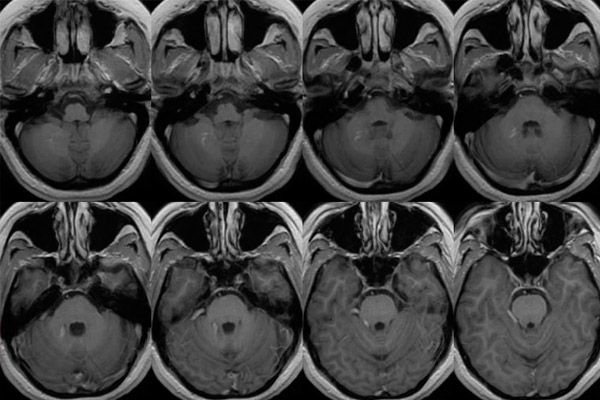
- Computer tomography of the brain. This is a technique for older patients.
Conducting an analysis of the obtained results, excluding other diseases and making the correct diagnosis.
[ 10 ], [ 11 ], [ 12 ], [ 13 ], [ 14 ]
Tests
Today, no diagnosis can be made without laboratory tests of the patient’s blood and urine.
A blood test allows us to diagnose the presence of an inflammatory process in the patient’s body and to assess the changes that inevitably affect the balance of blood components.
You can't do without a urine test. With such a pathology, protein inclusions may appear in the excreted fluid. Other changes may also appear that will allow the attending physician to make the right conclusions. After all, laboratory tests are quite informative for a specialist.
Instrumental diagnostics
Today, it is almost impossible to imagine a doctor without research conducted using specialized medical equipment. Instrumental diagnostics, in most cases, is the main one in the formulation of almost any analysis.
To establish the diagnosis discussed in this article, the attending physician usually refers to the results of the following studies:
- Angiography is an examination of various blood vessels using X-rays and a contrast agent injected into the bloodstream. This test allows us to evaluate the patency of the vessels, the locations of narrowing, dilation and pathological changes.
- X-ray is rarely used to determine venous angioma of the brain due to the impossibility of penetrating into the cranium. But this method is still acceptable in some cases for making a diagnosis. This analysis is necessary in case of head trauma or suspicion of it.
- Ultrasound examination. This method is acceptable when a disease is suspected in a newborn baby. This method of examination can be called more gentle for the baby and less expensive for its parents.
- But the main method of instrumental diagnostics in case of suspected brain pathology is still computed tomography or magnetic resonance imaging. These two methods allow obtaining layer-by-layer contrast images of the brain. The doctor has the opportunity to examine the brain in 3D as well. With the help of these studies, foci of altered density can be identified. The tomogram determines the exact localization and shape of the pathological focus in the brainstem. In this case, the information obtained with a package of frames is stored in the computer memory and can be used by the specialist repeatedly.
Differential diagnostics
Only after receiving all the results of the studies, an experienced specialist can begin their analysis. Differential diagnostics consists of excluding pathological diseases that can be expressed by such symptoms. This is the only way to make the only correct diagnosis and recognize accompanying diseases (if any).
Who to contact?
Treatment of venous angioma
In rare cases, a venous angioma can regress, resolving itself. This result is possible when certain factors coincide. For example, in the case of a sudden blockage of the vessels supplying blood to the angioma by a thrombus. Deprived of nutrition, it gradually dissolves.
But the most dangerous pathology is one that affects the human brain – this is a virtually unambiguous indication for immediate surgical intervention. The doctor may be forced to delay the operation and decide to monitor the neoplasm if there are no pathological symptoms and the benign tumor was discovered accidentally during an instrumental examination caused by other reasons. In such a picture, the doctor prescribes a repeat examination after a short period of time.
Only after the disease is confirmed are appropriate measures taken to alleviate the problem.
It is necessary to take urgent steps that justify the treatment of venous angioma in the following cases:
- Rapid increase in the size parameters of the neoplasm.
- Increasing the scale of the affected area.
- Detection of a brain hemorrhage.
- Any localization of the tumor under the patient's skull.
- Obvious disturbances in brain function.
- Destruction of tissues adjacent to the tumor.
A modern doctor is armed with a wide range of tools that allow him to fight the diagnosis. At the same time, treatment should be started immediately. After all, with such localization of the tumor, there is a high probability of hemorrhage in the brain. And these consequences are much more difficult to correct, and sometimes impossible.
With rapid growth of the neoplasm, drugs of the hormonal pharmacological group are introduced into the patient's treatment protocol. In the future and in other cases, the patient with the diagnosis of venous angioma is shown surgical intervention, but the method of its implementation is selected by the attending physician based on the obtained location of the tumor, its size, depth of localization and neglect of the progression process.
If, for example, the tumor is located deep enough and it cannot be excised with a classic surgical instrument without serious injury to the brain tissue, then a gamma knife can be used. Venous angioma of the brain is a very serious pathology that requires early diagnosis and the fastest possible treatment. After all, ignoring the problem can lead to irreparable consequences.
Drug treatment
To date, there is no panacea for the disease discussed in this article that would allow you to take a pill and the tumor will resolve on its own. In such a situation, full-fledged drug therapy is also impossible.
Only when the clinical picture and condition of the patient are such that surgical treatment is unacceptable, the attending physician prescribes medications that make up hormonal therapy.
These medications are also necessary in cases of high growth rate of the tumor's size parameters, dangerous localization, and also in cases where not one but several neoplasms are detected, located in different areas of the brain.
Prednisolone is the most common medicine of this kind. Analogues of this medicine are decortin, inflanefran, medopred, novo-prednisolone, prednigeksal, prednisol, prednisolone acetate, prednisolone hemisuccinate, solu-decortin, sherizolon.
The glucocorticosteroid prednisolone is prescribed both as oral tablets and as intramuscular injections.
In the light of replacement therapy, the patient is prescribed a daily dosage of 0.02 - 0.03 g. For maintenance therapy, these parameters are somewhat lower and amount to 0.005 - 0.01 g. If therapeutic effectiveness is not observed, the amount of the drug taken can be increased.
If the patient has a history of psychosis, the drug is taken under the monitoring of the attending physician.
For small patients, this daily dosage is calculated using the formula 1–2 mg per kilogram of the patient’s weight, divided into four to six doses; in the case of maintenance therapy, this value is 0.3–0.6 mg per kilogram of the baby’s weight.
In this case, the morning dosage should be taken higher, and the doses in the second half of the day should be lower.
In case of short-term use, a contraindication for prescribing this drug is individual intolerance of the patient's body to prednisolone or components of prednisolone.
Medicines of this pharmacological group inhibit the growth of neoplasms and dry out pathologically damaged blood vessels.
The treatment protocol also includes cytostatic (antitumor) drugs. These may include busulfan, streptozotocin, treosulfan, chlorambucil, vincristine, vinblastine, carmustine, mustoforan, ifosfamide, bendamustine, fludarabine, daunorubicin, epirubicin and many others.
Folk remedies
It is worth noting right away that any therapy should be carried out only with the consent of a specialist. This also applies to non-traditional methods of treatment. As practice shows, folk treatment of angioma can bring its positive results. But given the location of the problem discussed in this article, it is impossible to control the course of treatment independently. This can only be done with the help of special medical equipment.
Traditional medicine can be an auxiliary method of solving the problem, but not the main one. Most of these recipes are based on the use of minerals, herbs, and other plants.
It is worth remembering that you should not rely solely on folk methods. This disease cannot be cured in this way.
[ 24 ], [ 25 ], [ 26 ], [ 27 ]
Herbal treatment
In this article, we are ready to offer several recipes of traditional medicine that will support the body and allow you to solve the health problem faster. But using them as the only method of treatment is strictly prohibited.
Herbal treatment for angioma can be represented by the following recipe.
- Prepare a herbal mixture. It includes St. John's wort - 30 g, coltsfoot - 45 g, cat's paw - 30 g, tansy - 15 g, plantain - 60 g, comfrey leaves - 15 g, calendula flowers - 30 g, celandine - 30 g, cherry stalk - 30 g, yarrow - 15 g. Grind all the ingredients and mix well. Place a tablespoon of the herbal mixture in a vessel with 400 ml of boiled water. Put on the fire, bring to a boil and boil for five minutes. Leave for an hour. Strain the mixture. Take the medicine three to four times a day about twenty minutes before meals. The duration of treatment is about three weeks.
- Another recipe that shows good results in the fight against angioma is a herbal balm. First, you need to make a collection: pine buds - 100 g, chaga - 200 g, yarrow - 100 g, wormwood - 5 g, rose hips - 100 g. Grind all the ingredients and add to three liters of boiled water. Put on the fire and bring to a boil. Reduce the heat and keep for about two hours. Wrap the container with the balm and leave to infuse for a day. Strain and mix with a quarter liter of cognac, a glass of aloe juice and half a liter of honey. Leave to infuse for four hours. Drink the balm one tablespoon three times a day before meals.
Homeopathy
This section of traditional medicine is based on the principles of carefully developed compositions of drugs designed to stop a particular disease. Homeopathy in the treatment of angiomas involves not only the elimination of pathological symptoms, but also the impact on the cause of the pathology. In this case, homeopathic doctors recommend taking homeopathic medicines that are developed on the basis of sulfur, lime sulfur and sodium sulfate.
In this situation, homeopathy can offer Loma Lux Acne, Acidum Fluoricum, Condurango Cortex, Calcarea Fluorica, Lycopodium, Pulsatilla, Radium Bromine, Solanum Nigrum and a number of other medicines.
But it is worth immediately warning those wishing to be treated with non-traditional methods, self-prescribing medications is fraught with serious consequences, various complications (in some cases, irreversible pathological processes) and, most importantly, the loss of sometimes precious time for treatment. You should not take homeopathic remedies thoughtlessly, ignoring their side effects. Therefore, if you want to treat the problem in this way, you should first consult with your doctor. This may be a dermatologist, but a consultation with a specialist - a homeopath is also desirable.
But when taking homeopathic medicines, you need to carefully monitor your health. If your condition worsens or any negative side symptoms appear, you need to stop taking the medicine and consult a specialist. Perhaps he will adjust the dosage or replace the drug.
Surgical treatment
When diagnosing venous angioma of the brain, perhaps the main and sometimes the only way to relieve the problem is surgical treatment.
The doctor can refuse it or postpone the operation if the patient feels well, the pathology does not bother him - there are known cases when the neoplasm resolved on its own. Another option for refusing the operation is the patient's health condition, which does not allow doctors to decide on the operation. In this situation, the patient receives hormonal therapy.
In other cases, surgery is the basis for treating cerebral venous angioma.
Today, doctors have several methods of tumor removal at their disposal. Some methods involve step-by-step treatment, while others perform complete removal within one surgical intervention. The main goal of such treatment is complete removal of neoplasms, restoration of normal functioning of the vascular and lymphatic systems.
The most commonly used methods of treating angioma are:
- Cryotherapy is the removal of a neoplasm by cauterization using low temperatures (cold). In modern medicine, such a refrigerant as liquid nitrogen is used. Cryotherapy is effective, while preventing bleeding.
- Electrocoagulation, which is caused by cauterization of foreign bodies using electric current. This method has been used less frequently lately, although it is simpler and cheaper. The disadvantages of electrocoagulation are its painfulness and residual effects in the form of scars, which is especially unacceptable in brain surgeries.
- Sclerotherapy may be used. Its essence is in the use of special iodine salts, which allows normalizing the flow cross-section of the blood vessels of interest to the surgeon, which has a positive effect on blood flow. Such an injection is used in cases where the neoplasm is localized in a place difficult to access for classical surgical intervention.
- Laser treatment. The essence of the method is to stop the problem by laser cauterization. In some cases, the removal of the pathological neoplasm occurs in several stages, performing layer-by-layer excision of the tumor. Mutated tissues are removed until healthy cells appear. In this case, damage to healthy tissues is minimal.
- If the above methods do not give the desired result, or for some reason cannot be applied, doctors resort to excision of the tumor with a surgical scalpel.
- The most innovative method of angioma removal today is the radio- and electric knife removal methods. They allow the patient to get rid of the problem, causing minimal damage to his body. Low trauma also lies in the fact that only mutated cells are excised, healthy tissues remain untouched. With this operation, the formation of classic colloid scars is not observed, which is also important. But these methods require special medical equipment, experience and high qualification of the doctor. Today, not every specialized institution can boast of such equipment. At the same time, this procedure is expensive and not every patient can afford it.
- Diathermoelectrocoagulation is used in isolated cases, only when diagnosing small point tumors, and if they are localized in places that are difficult to access using other methods.
The method of angiomas removal is mainly chosen by a doctor - a dermatocosmetologist. His decision is based on the results of a visual examination of the patient and the capabilities of the clinic. In most cases, sclerotherapy or removal of the neoplasm using a laser is prescribed. These two methods are painless and highly cosmetically effective, the result is obtained over several sessions.
Prevention
Based on the known medical reasons that can provoke the development of this disease, prevention of any angiomas, including venous ones, can be expressed by the following recommendations:
- Head injuries should be avoided.
- Lead a healthy lifestyle, avoiding the abuse of alcohol, nicotine and drugs.
- If a woman is planning a pregnancy, it would be a good idea to consult a doctor and undergo a full examination. The doctor may prescribe a course of folic acid and multivitamins.
- Avoid stressful situations.
- Protect your body from hypothermia and infection. If the disease is detected, timely and complete treatment is necessary.
- Monitor your diet. It should be rational and balanced in nutrients, vitamins and minerals. Minimize carbohydrate intake. Their daily norm should not exceed 450 g. The norm of fats is no more than 90 g per day. Avoid overeating: small portions, but five to six meals a day.
Forecast
The disease considered in this article is a serious pathology, and its localization makes the neoplasm even more dangerous. Therefore, the prognosis of venous angioma largely depends on the stage of its detection and the effectiveness of the measures taken. If the treatment was adequate and carried out at an early stage of the pathology, a person is able to live to a ripe old age, leading an active, high-quality life.
If the pathology is detected late, a hemorrhage occurs, or the patient’s health condition makes it impossible to provide adequate therapy, the result may be disastrous – fatal.
73% of angiomas are congenital, and only the rest are acquired. But this does not mean that you should give up and do nothing. Preventive measures will reduce the risk of acquired pathology. It should be remembered that venous angioma is practically not treated with medication or alternative medicine. Today, the main and so far the only effective method of getting rid of this disease is surgery. But before you decide on it and choose a method for excising the neoplasm, we advise you to choose the right clinic and doctor who has experience in such operations and the appropriate equipment. To do this, you should talk to patients who have undergone this procedure. You should be more attentive to yourself! And be healthy!