Medical expert of the article

New publications
Urethrography
Last reviewed: 29.06.2025

All iLive content is medically reviewed or fact checked to ensure as much factual accuracy as possible.
We have strict sourcing guidelines and only link to reputable media sites, academic research institutions and, whenever possible, medically peer reviewed studies. Note that the numbers in parentheses ([1], [2], etc.) are clickable links to these studies.
If you feel that any of our content is inaccurate, out-of-date, or otherwise questionable, please select it and press Ctrl + Enter.
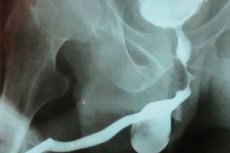
Urethrography is a medical procedure used to study the urethra (urethra) using X-rays. It is usually performed using a contrast agent that is injected into the urethra before x-rays are taken. Urethrography allows doctors to visualize the urethra and evaluate its structure, shape, and function. This procedure can be useful for diagnosing and evaluating various urethral problems such as urethral narrowing (strictures), trauma, infection, or other abnormalities. Urethrography can be performed in both men and women and can aid in the diagnosis and treatment planning for urethral diseases or conditions.
Indications for the procedure
Indications for urethrography may include:
- Urethralchanges: If a patient has symptoms or signs of urethral changes, such as pain during urination, burning, itching, urethral bleeding, or unusual discharge, urethrography may be ordered to detect possible abnormalities, strictures, tumors, or other abnormalities.
- Suspicion of urethral trauma: Urethrography may be used to evaluate the urethra when trauma such as fractures, sprains, or other injuries that may have occurred as a result of accidents or medical procedures are suspected.
- Urolithiasis: Urethrography may be performed to detect the presence of uroliths, which can block the urethra and cause pain and difficulty urinating.
- Preparation for surgical procedures: In some cases, urethrography may be ordered by your doctor before urethral surgery to better understand the structure and condition of the urethra.
- Examination of children with malformations: Urethrography can be used to diagnose and evaluate urethral malformations in children.
Preparation
Preparation for urethrography, also known as retrograde urethrography, may include the following steps:
- Consultation with your doctor: First, you will need to see a doctor or urologist who will order this test. The doctor will explain the purpose of the procedure, talk to you about possible risks and side effects, and answer your questions.
- Doctor's Warning: It is important to alert your doctor if you are allergic to medications, have specific medical conditions, or if you are pregnant, as this may require additional precautions.
- Medication Information: Per your doctor's instructions, you may need to temporarily stop taking certain medications before the procedure.
- Fasting: Your doctor may also ask you not to eat or drink for a certain period of time before the urethrography. This is usually required if the procedure will be done under general anesthesia or spinal anesthesia.
- Bladder: Your doctor may ask you to empty your bladder before the procedure to ensure better visibility and avoid constipation.
- Preparing for urethrography: On the day of the procedure, you must follow your doctor's instructions about what to wear, what medications to take, and other specific instructions.
The device for carrying out the procedure
The urethrography procedure is performed using an X-ray machine and a contrast agent that is injected into the patient's urethra. Here is an overview of the main components and equipment used in urethrography:
- X-raymachine: This is specialized equipment that is used to create X-ray images. An X-ray machine consists of an X-ray tube and a detector that records X-rays and creates images.
- Contrast agent: Urethrography uses a contrast agent that is injected into the patient's urethra. This substance makes the urethra visible on x-rays, allowing the doctor to evaluate its structure and function.
- Catheter: A catheter may be used to inject contrast agent into the urethra. The catheter is inserted into the urethra through the urethra and is used to deliver the contrast agent into the urethra.
- Computer: The computer is used to process and analyze the X-ray images created by the X-ray machine. It helps the doctor to get detailed pictures of the urethra.
- Screen and Monitor: The screen and monitor are used to visualize real-time x-ray images during the procedure.
- X-ray protection: The X-ray machine is equipped with shields and shields to protect personnel and patients from radiation.
- Sterilization equipment: Catheters and other instruments used in the procedure must be sterile, so sterilization equipment may be required.
Technique of the urethrography
Here are the basic steps in the technique of urethrography:
- Patientpreparation: The patient is dressed in a medical gown and lies down on the radiology table. The patient may be in the supine position with legs apart, and sometimes other positions may be required depending on the purpose of the study.
- Urethral catheterization: The doctor inserts a flexible urethral catheter into the urethra through the urethra. The catheter usually penetrates the bladder. This may cause some discomfort, but the procedure is performed in a controlled sterile environment.
- Contrastagent injection: After inserting the catheter into the bladder, the doctor injects contrast agent intravenously through the catheter. The contrast agent makes the structures of the urethra visible on x-rays.
- X-rays: The doctor takes X-rays of the urethra in different projections to get a complete picture of its structure and function. These images may be taken in real time (during the injection of the contrast agent) or after the procedure has been completed.
- CatheterRemoval: Once the study is complete, the catheter is removed and the patient can be monitored for any unpleasant symptoms or complications.
- Processing of the results: The radiologist interprets the images and prepares a report that is given to the prescribing physician.
Ascending urethrography
This is an X-ray imaging procedure of the urethra (urethra) in which a contrast agent is injected through the urethra and then recorded with a series of X-rays. This procedure is usually performed in men to evaluate the urethra in detail and may be used in the following cases:
- Suspicion of structural changes: Ascending urethrography may be indicated if strictures, deformities, or other structural changes in the urethra are suspected.
- Investigating the cause of pain or difficulty urinating: If a patient is experiencing pain, itching, bleeding, or other unusual symptoms related to the urethra, ascending urethrography can help find the cause of these symptoms.
- Assessment of surgical results: Ascending urethrography can be used to assess the results of urethral surgery and to verify its effectiveness.
- Preparation for surgical correction: Prior to urethral surgery, ascending urethrography can provide the physician with information about the structure and morphology of the urethra.
Retrograde urethrography
It is a medical procedure that is used to diagnose and visualize the urethra, i.e. The urethra, in a reverse method using X-rays and a contrast agent. This procedure allows doctors to evaluate the structure and function of the urethra and to detect abnormalities or problems in this area.
Here's how the retrograde urethrography procedure works:
- The patient usually lies on his or her back on the x-ray table.
- The urethra (urethra) inside the pelvis is thoroughly cleaned and disinfected.
- A thin, flexible catheter is then inserted into the urethra.
- Through this catheter, a contrast agent is injected into the urethra, which makes the urethra visible on x-rays.
- The radiologist takes a series of pictures showing the contour and structure of the urethra as the contrast agent passes through it.
After retrograde urethrography, the doctor can evaluate the condition of the urethra, detecting the presence of narrowings (strictures), polyps, tumors, or other abnormalities that may be the cause of symptoms or problems in this area. This procedure helps doctors make an accurate diagnosis and plan the necessary treatment.
The retrograde urethrography procedure is performed by specialists in radiology or urology and may require specific preparation and aftercare.
Contraindications to the procedure
Urethrography, like many medical procedures, can have contraindications and risks. Contraindications to urethrography may include the following conditions or circumstances:
- Allergy to contrast agent: If the patient has a known allergy to the contrast agent used in urethrography, this may be a contraindication.
- Active infection: If the patient has an active infection in the urethra or urethra, urethrography may not be desirable as it may spread the infection.
- Pregnancy: Urethrography can be an undesirable procedure in pregnancy, especially if it is not absolutely necessary. The doctor should carefully discuss the pros and cons of the procedure with the pregnant woman.
- Bleeding or clotting disorders: If a patient has urethral bleeding or clotting disorders, this may be a contraindication to urethrography.
- Other serious medical conditions: If a patient has other serious medical conditions that may make urethrography unsafe or unfeasible, this may also be a contraindication.
Normal performance
Normal urethrography values may vary depending on the specific situation and the purpose of the procedure. Urethrography is a method of visualizing the urethra, and normal values may be different for men and women, and may also depend on the purpose of the procedure. Here are some common aspects of normal values:
- Urethral patency: Urethrography can help visualize the urethra and confirm its patency without obstruction, narrowing, or other abnormality.
- Structure and shape of the urethra: The normal urethra has a certain structure and shape that should be shown on x-rays. The doctor can assess whether there are deformities, strictures (narrowings) or other abnormalities.
- Urethral function: Urethrography can be used to evaluate urethral function during urination. Normal urination and distribution of contrast agent may be important indicators.
- Absence of stones and tumors: Urethrography can also help detect the presence of urolithiasis or tumors that may be blocking the urethra.
It is important to note that interpretation of urethrography should be performed by a qualified medical professional, usually a radiologist or urologist. They will analyze the results and take into account the clinical context to conclude on the presence or absence of pathological changes.
Normal values may also vary depending on the age and gender of the patient, so specific norms must be established for each case.
Complications after the procedure
Some complications or unpleasant symptoms may occur after the urethrography procedure. However, these are usually rare and usually temporary. Here are some of the possible complications after urethrography:
- Pain or discomfort: After removal of the urethral catheter, the patient may experience slight pain or discomfort when urinating. This is usually temporary and disappears after a few hours.
- Infection: Although the procedure is performed under sterile conditions, there is a small risk of urinary tract or urethral infection. If you develop symptoms of infection, such as lower abdominal pain, frequent urination, burning when urinating, or fever, you should tell your doctor immediately.
- Allergic reaction: In rare cases, some patients may have an allergic reaction to the contrast agent used during urethrography. This may manifest as a skin rash, itching, redness, or even more serious allergic reactions. If you notice any allergic symptoms, notify the medical staff immediately.
- Bleeding: In rare cases, a small amount of bleeding from the urethra may occur after urethrography. This is usually minimal and stops on its own, but if bleeding continues or increases, you should see your doctor.
- Reaction to Anesthesia: If the procedure is performed under general anesthesia or spinal anesthesia, reactions to anesthesia such as nausea, vomiting, dizziness, or allergic reactions may occur.
Care after the procedure
After urethrography, it is important to follow some care guidelines to prevent possible complications and ensure comfort. Here are some general guidelines:
- Drink water: It is important to drink plenty of water after urethrography. This helps flush the contrast agent out of the urinary tract and reduces the risk of urethral irritation.
- Avoid infections: Try to avoid baths, pools, and whirlpools for a few days after the procedure to prevent possible infection.
- Avoid exertion: Try to avoid unnecessary physical exertion and heavy lifting for a few days.
- Avoid sexual activity: Your doctor may recommend abstaining from sexual activity for a few days after urethrography to avoid urethral irritation.
- Follow your doctor's recommendations: Your doctor may provide individualized recommendations that depend on your specific situation and circumstances. It is important to follow these recommendations and follow the prescribed treatment if necessary.
- Watch for symptoms: Pay attention to any unusual symptoms such as pain, bleeding, severe burning when urinating, or fever. If these symptoms occur, seek medical attention immediately.
- Keeping track of your medications: If your doctor prescribes medication, make sure you take it as prescribed.