Medical expert of the article
New publications
Transcranial magnetic stimulation of the brain
Last reviewed: 06.07.2025

All iLive content is medically reviewed or fact checked to ensure as much factual accuracy as possible.
We have strict sourcing guidelines and only link to reputable media sites, academic research institutions and, whenever possible, medically peer reviewed studies. Note that the numbers in parentheses ([1], [2], etc.) are clickable links to these studies.
If you feel that any of our content is inaccurate, out-of-date, or otherwise questionable, please select it and press Ctrl + Enter.

The transcranial magnetic stimulation (TMS) method is based on stimulation of nervous tissue using an alternating magnetic field. Transcranial magnetic stimulation allows to evaluate the state of the conductive motor systems of the brain, corticospinal motor pathways and proximal segments of nerves, excitability of the corresponding nerve structures by the value of the magnetic stimulus threshold required to obtain muscle contraction. The method includes analysis of the motor response and determination of the difference in conduction time between the stimulated areas: from the cortex to the lumbar or cervical roots (central conduction time).
Indications for the procedure
Magnetic stimulation of peripheral nerves and the brain allows, in clinical conditions, to monitor the state of the brain's motor system and to quantitatively assess the degree of involvement in the pathological process of the corticospinal motor pathways and various parts of the peripheral motor axons, including the motor roots of the spinal cord.
The nature of the disturbance of the processes of excitation conduction through the central structures of the brain and spinal cord is non-specific. Similar changes are observed in various forms of pathology. These disturbances include an increase in the latent time of the evoked potential, a decrease in the amplitude or absence of a response to stimulation of the motor zone of the cerebral cortex, its dispersion, as well as their various combinations.
Prolongation of central conduction time is observed in demyelination, degeneration of the corticospinal tract due to motor neuron pathology or hereditary disease, cerebrovascular disorders, glioma of the cerebral hemispheres, and discogenic compression of the spinal cord.
Thus, the indication for transcranial magnetic stimulation is considered to be pyramidal syndrome of any etiology. Most often in clinical practice, transcranial magnetic stimulation is used for various demyelinating lesions of the central nervous system (especially multiple sclerosis ), hereditary degenerative diseases, vascular diseases, tumors of the spinal cord and brain.
Technique transcranial magnetic stimulation
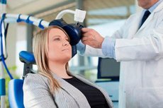
The patient is in a sitting position. The evoked motor potentials during magnetic stimulation are recorded using surface electrodes placed on the motor point area of the muscles of the upper and lower extremities in a standard manner, similar to the generally accepted procedure for recording the M-response during stimulation electromyography. Magnetic coils of two main configurations are used as a stimulating electrode: ring-shaped, having different diameters, and in the form of a figure 8, which are also called "butterfly coils". Magnetic stimulation is a relatively painless procedure, since the magnetic stimulus does not exceed the pain threshold.
Potentials recorded during stimulation of the cerebral cortex vary in latency, amplitude, and shape of the recorded curve. When studying healthy people, changes in evoked motor potentials during magnetic stimulation are observed in response to changing stimulation parameters (magnetic field strength, coil position) and depending on the state of the muscles being studied (relaxation, contraction, and minor voluntary motor activity).
Transcranial magnetic stimulation allows one to obtain a motor response of virtually any human muscle. By subtracting the latent time of formation of a motor response during stimulation of the cortical representation of the muscle and the exit point of the corresponding root in the region of the cervical or lumbar segments of the spinal cord, one can determine the time of impulse passage from the cortex to the lumbar or cervical roots (i.e., the central conduction time). The technique also allows one to determine the excitability of the corresponding nerve structures by the value of the magnetic stimulus threshold required to obtain muscle contraction. The registration of the evoked motor response is performed several times, and responses of maximum amplitude, correct shape, and minimum latency are selected.
Contraindications to the procedure
Transcranial magnetic stimulation is contraindicated in the presence of a pacemaker, if there is a suspicion of an aneurysm of the cerebral vessels, during pregnancy. The method should be used with caution in patients with epilepsy, as it can provoke an attack.
Normal performance
When performing transcranial magnetic stimulation, the following parameters are analyzed.
- Latency of evoked motor response.
- F-wave latency (when calculating radicular delay).
- Amplitude of the evoked motor response.
- Time of the central event.
- Radicular delay.
- Threshold for eliciting a motor response.
- Sensitivity of the studied structures to magnetic stimulus.
The most pronounced prolongation of central conduction time is observed in multiple sclerosis. In the presence of muscle weakness, changes in the parameters of the evoked motor potential and an increase in the threshold for inducing a motor response are found in all patients with multiple sclerosis.
In patients with ALS, significant changes in the functional state of the motor system are also detected; in most cases, sensitivity to magnetic stimuli decreases, the threshold for inducing a motor response increases, and central conduction time increases (but to a lesser extent than in multiple sclerosis).
In myelopathy, all patients show an increase in transcranial stimulation thresholds. The noted disorders are especially pronounced in the presence of a gross spastic component. In patients with spinocerebellar degeneration, clinically manifested by ataxia and spasticity, a decrease in the sensitivity of cortical structures to magnetic stimulation is observed. A response at rest is often not evoked even with a maximum stimulus.
When examining patients with cerebrovascular diseases, the entire spectrum of changes in central conduction time is observed - from the norm to a response delay of 20 ms and a complete absence of potential. The absence of a response or a decrease in its amplitude is a prognostically unfavorable factor, while a registered, albeit delayed, response in the early period after a stroke indicates the possibility of restoring the function.
Transcranial magnetic stimulation is successfully used in the diagnosis of spinal nerve root compression. In this case, asymmetry of central conduction time of more than 1 ms is detected. Even more informative in the diagnosis of radiculopathy is the "radicular delay" method.


 [
[