Medical expert of the article

New publications
Sensitivity study
Last reviewed: 07.07.2025

All iLive content is medically reviewed or fact checked to ensure as much factual accuracy as possible.
We have strict sourcing guidelines and only link to reputable media sites, academic research institutions and, whenever possible, medically peer reviewed studies. Note that the numbers in parentheses ([1], [2], etc.) are clickable links to these studies.
If you feel that any of our content is inaccurate, out-of-date, or otherwise questionable, please select it and press Ctrl + Enter.
The most common complaint associated with sensory disturbances is pain. If the patient has pain syndrome, the following aspects should be clarified:
- nature of pain (sharp, dull, burning, stabbing, shooting, etc.);
- localization and irradiation of pain;
- temporal characteristics (constant, paroxysmal, periods of increased/decreased pain) and their duration;
- severity of pain (the patient is asked to rate the pain on an 11-point scale, where 0 points corresponds to the absence of pain, 10 - the maximum possible);
- factors that contribute to the weakening/increasing of pain (movement, a certain posture, rest, stress, taking analgesics, etc.);
- accompanying symptoms (visual disturbances, muscle spasms, nausea or vomiting, etc.);
- onset of pain (date, circumstances surrounding the onset of pain, possible cause, etc.).
The sensitivity assessment is entirely based on the patient's self-report of their subjective sensations, so sensitivity is examined last during a neurological examination. Complaints and changes in the neurological status identified at previous stages of the examination largely determine the specifics of the sensitivity study for each individual patient. Thus, if the patient has no complaints and no neurological disorders have been detected before, screening sensitivity testing can be used, which includes a study of pain sensitivity on the face, limbs and torso, vibration and deep sensitivity on the limbs. On the contrary, if neurological disorders are detected and there are already assumptions about their cause, then sensitivity is examined taking into account the formed hypothesis. It can be quite difficult to interpret the results of a sensitivity study. In many cases (fatigue, anxiety, depression, decreased cognitive functions), self-assessment of sensory disorders does not reflect the real state of sensitive innervation of tissues and organs. Thus, an anxious patient with an analytical mindset is able to focus attention on the most insignificant sensations that have no clinical significance, while patients with a reduced level of wakefulness sometimes deny the most serious disorders.
There are simple and complex types of general somatosensory sensitivity. Simple types of general sensitivity are divided by their "receptor affiliation" into superficial (perception of signals from the exteroceptors of the skin analyzer) and deep (perception of signals from the proprioceptors of the motor analyzer). In turn, simple superficial (cutaneous or exteroceptive) sensitivity includes pain, temperature (cold and heat) and tactile (touch, feeling of light touch), and simple deep sensitivity - muscle-joint feeling (feeling of passive movement, feeling of position), kinesthesia of skin folds, feeling of pressure (strong touch), mass and vibration.
The results of the study of simple types of sensitivity reflect, first of all, the state of the receptor apparatus, the conductive part and the primary sensory (“projection”) fields of the cortex of the corresponding analyzers.
Complex types of sensitivity include the sense of localization, discrimination, two-dimensional and three-dimensional spatial sense. Sometimes the sense of mass is also considered complex types of sensitivity. Complex types of sensitivity are based on the analysis and synthesis of impulses of different modalities. Their study reflects the state of not only the conductive sections of analyzers and primary sensory fields of the cortex, but also secondary and tertiary cortical receptor fields (i.e., areas of the cortex that integrate information from various sense organs).
Surface sensitivity study
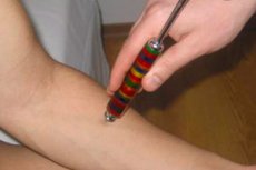
- Pain sensitivity is tested using a special safe needle soldered into a plastic case, and a new needle should be used for each new patient. The pressure of the needle should be strong enough to cause pain, but not traumatic. It is unacceptable to prick the patient "until blood" or leave scratches after testing. In response to the prick, the patient must report his sensation ("sharp" or "dull"), and not just state the fact of touching. A certain testing sequence should be followed: pain sensitivity is tested at symmetrical points on the right and left sides of the body, moving from the distal parts of the limbs to the proximal ones or from the area of one dermatome to another. If an increase in the pain threshold is detected, move in the direction from the area of reduced pain perception to the preserved area, starting from the center to the edges, in order to determine the boundaries of the disordered area. Damage to the trunk of a peripheral nerve causes a disturbance of sensitivity in the zone of its autonomic innervation, and damage to the spinal root causes a disturbance of sensitivity in the zone of the corresponding dermatome. In polyneuropathy, pain sensitivity disorders occupy the territory of "gloves" and "socks". We also note the presence of hyperalgesia.
- Tactile sensitivity is examined using light touches with a piece of cotton wool or a brush with soft hair. First, the patient is shown touches, applying them to the forehead area, and it is explained that he must report each touch he feels with the word "yes" or "I feel". Then the patient is asked to close his eyes and concentrate on analyzing the sensations he receives. The presence of hyperkeratosis in the area of the soles or palms increases the threshold of tactile sensitivity in these areas, which cannot be considered a neurological deficit.
- Thermal sensitivity (sensation of heat, cold) is usually examined only in patients with hypalgesia. Test tubes with hot (32-40 °C) and cold (no higher than 25 °C) water or other cold and warm objects (for example, a metal hammer and a doctor's finger) are used. First, the patient's ability to distinguish cold from hot is determined by alternately applying warm and cold objects to the area with presumably intact sensitivity. Normally, a difference of 2 °C is already noticeable to the patient. Then, a cold (or warm) object is applied alternately to symmetrical areas of the body, starting from the back of the foot, moving upwards and comparing the intensity of perception of the temperature stimulus on the right and left. Studies of cold and heat sensitivity are conducted separately, since they can be impaired to varying degrees. If necessary, temperature sensitivity is also examined in various dermatomes or in the zones of autonomic innervation of the affected nerves, finding the boundaries of altered sensitivity. A clear definition of the territory of impaired sensitivity, coinciding with a certain innervation, allows the patient's subjective sensation to be transformed into an objective neurological sign.

Deep Sensitivity Research
- The feeling of vibration occurs when deep receptors are stimulated by oscillations of a certain frequency and amplitude. A low-frequency (64-128 Hz) tuning fork is used for the study. It is advisable to independently test the tuning fork used on healthy people. Normally, the sensation of vibration on the ankles lasts from 9 (tuning fork 48 Hz) to 21 s (tuning fork 64 Hz). Vibration sensitivity is examined on the fingers and toes, ankles, patellas, pelvic bones, radius and ulna, collarbone, and skull. The leg of a vibrating tuning fork is applied to the area under study and the patient is asked to report when he or she stops perceiving vibrations. The threshold of vibration sensitivity is compared on the right and left limbs. If vibration sensitivity on the foot is impaired, it is checked in the area of the ankle, knee, and hip joint to determine the boundaries of the disorder. Vibration sensitivity in the fingers is examined in a similar manner. Vibration sensitivity is reduced in peripheral polyneuropathies and spinal cord diseases involving its posterior cords. In this case, vibration sensitivity may be reduced only in the distal parts of the legs and remain intact in the arms. A moderate increase in the threshold of vibration sensitivity in elderly people is observed even in the absence of any neurological pathology.
- Muscle-joint sense. The patient is first shown what passive movements will be made with his fingers and what to call them. Then the patient is asked to close his eyes, the nail phalanx of the finger is taken by the lateral surfaces and the finger is moved smoothly up, then down; the patient must report in which direction (up or down) his finger is moved. Normally, a person is very sensitive to even very subtle passive movements in the joints and is able to distinguish a movement at an angle of 1-2°. If the patient's muscle-joint sense is impaired in the distal parts of the limbs, the sensation of passive movements in the joints located more proximally is checked.
- The sense of position is examined by placing the limb in a certain position. The patient must determine this position with his eyes closed. If the sense of movement in a joint is perceived primarily by receptors localized in the tendons and joints, then the receptors located in the muscles, i.e. the afferents of the muscle spindle, are responsible for determining the static position of a body part in space.
Evaluation of research results
Based on complaints, anamnestic data and the results of the study of superficial types of sensitivity, it is possible to form an idea of the disorders present in the patient.
- Decreased/absent sensitivity is designated by the terms “hypesthesia” and “anesthesia” (for pain sensitivity - “hypalgesia” and “analgesia”; for temperature sensitivity - “thermohypesthesia” and “thermoanesthesia”; for deep sensitivity - “batianesthesia”).
- Increased sensitivity to normal non-painful stimuli is called hyperesthesia, increased sensitivity to pain is called hyperalgesia.
The above-mentioned disorders are designated as quantitative disorders; the following are classified as qualitative disorders of sensitivity.
- Polyesthesia (one injection is perceived as multiple).
- Allocheiria (the patient identifies irritation not in the place where it was applied, but on the opposite half of the body).
- Synesthesia (the sensation of perception both at the site of application of the stimulus and in another place where it was not applied).
- Paresthesia (spontaneous or evoked unusual sensations).
- Neuralgia (extremely severe, sharp pain that radiates along the course of one or more nerves).
- Causalgia (a feeling of intense burning pain).
- Dysesthesia (distorted perception of receptor affiliation). Variants of dysesthesia: temperature - the appearance of a sensation of heat in response to a prick; allodynia - the appearance of pain in response to irritation, which is not normally accompanied by them (sometimes allodynia is called only a pain reaction to touching with a brush, while pain sensations in response to temperature effects and pressure are designated by the terms "hyperalgesia to cold and heat" and "hyperalgesia to pressure", respectively).
- Hyperpathy (the appearance of excruciating pain in response to repeated painful and non-painful stimuli in combination with an increase in the threshold of perception of a single stimulus and difficulty in clearly localizing the irritation).
The study of simple types of general sensitivity also allows us to determine the type of distribution of sensitivity disorders.
- Damage to nerve trunks leads to a peripheral neural type of distribution of sensitivity disorders. It is characterized by a disorder of all types of sensitivity in the zone of innervation of peripheral nerves (in case of damage to the plexus - in the zone of innervation of the plexus; in case of damage to an individual nerve - in the zone of innervation of this nerve; in case of polyneuropathy - in the distal parts of the limbs). Sensory disorders are usually combined with paresis or paralysis of muscles innervated by the corresponding nerves.
- Damage to the posterior roots of the spinal nerves is accompanied by the development of a peripheral radicular type of sensory disturbance. All types of sensitivity in the dermatomes corresponding to the affected roots are impaired. However, since the cutaneous innervation zones of adjacent roots partially overlap, no loss of sensitivity is detected when one root is switched off (the area of the corresponding dermatome continues to be supplied by adjacent roots). Sensitivity is clearly impaired in the area of one dermatome only when three adjacent roots are affected. A decrease in sensitivity in this type of disorder is accompanied by severe pain and paresthesia in the corresponding dermatomes.
- Lesions of the posterior horns of the spinal cord may cause a spinal segmental type of sensory impairment: ipsilateral impairment of pain and temperature sensitivity in one or more dermatomes while tactile sensitivity in these segments is preserved. Such dissociated anesthesia may occur with intramedullary tumors, myeloischemia, hematomyelia, but is most typical for syringomyelia, which manifests itself in the formation of cavities in the gray matter of the spinal cord. Since the localization of syringomyelic cavities is typical in the cervical and upper thoracic regions of the spinal cord, the zone of sensory impairment has the appearance of a "half-jacket", and when the cavity spreads to the other half of the spinal cord or with the initial central location of the cavity - the appearance of a "jacket". When the nucleus of the spinal tract of the trigeminal nerve is involved in the process, pain and temperature sensitivity on the face in the outer zones of Zelder disappears; the middle and inner zones are involved later.
- The spinal conduction type of distribution of sensory disturbances occurs when the conduction pathways in the funiculi of the spinal cord are affected. When the lateral funiculus is affected with involvement of the lateral spinothalamic tract, there is a disturbance of temperature and pain sensitivity on the side opposite the lesion one to three dermatomes below the lesion level. When the posterior funiculus is affected, there is a disturbance of deep sensitivity (vibration sensitivity and muscle-joint sense) on the side of the lesion; however, pain and temperature sensitivity remain intact. This disorder is combined with ipsilateral sensory ataxia.
- Brown-Sequard syndrome occurs when one half of the transverse section of the spinal cord is damaged. On the side of the lesion below the level of the lesion, spastic paralysis (interruption of the pyramidal tract) and disturbance of deep sensitivity (disconnection of the posterior funiculus) occur, and on the opposite side from a level located several segments below the level of the lesion, a disorder of pain and temperature sensitivity of the conductive type occurs (disconnection of the spinothalamic tract in the lateral funiculus).
- The central type of distribution of sensory disorders occurs when the structures of the brain are damaged. Its manifestations vary depending on the level and which structures are affected, but in any case, with a unilateral localization of the lesion above the level of the medulla oblongata, sensitivity on the trunk is impaired on the side opposite the lesion.
- Damage to the lateral sections of the medulla oblongata (dorsolateral medullary Wallenberg-Zakharchenko syndrome) causes loss of pain and temperature sensitivity on the same side of the face (involvement of the spinal tract nucleus of the trigeminal nerve), decreased pain and temperature sensitivity on the half of the body and limbs opposite the lesion (damage to the spinothalamic tract) and decreased deep sensitivity on the side of the lesion in the limbs (involvement of the nuclei of the thin and cuneate fasciculi). Sensory disorders are combined with cerebellar ataxia on the side of the lesion (inferior cerebellar peduncle); dizziness, nystagmus when looking towards the lesion, nausea and vomiting (vestibular nuclei and their connections); Bernard-Horner symptom on the side of the lesion (damage to the descending tracts from the hypothalamus to the ciliospinal center in the lateral horns of C8 - T2 ); dysarthria, dysphagia, dysphonia, ipsilateral paralysis of the muscles of the soft palate, pharynx and vocal cord (lesion of the double nucleus of the IX-X pairs of cranial nerves).
- Damage to the thalamus (usually of vascular origin) results in loss of all types of sensitivity on the side of the body opposite the lesion. As a rule, sensitivity gradually improves, but on the same side of the body, burning ("thalamic") pains eventually occur, which are provoked by any stimuli, especially cold and emotional stress. These pains are of a painful, diffuse nature and can be observed against the background of an increase in the threshold of pain sensitivity. At the same time, sensory hemiataxia is detected in the extremities contralateral to the lesion and hemianopsia. A "thalamic hand" is often formed (the shoulder is pressed to the body, the forearm and hand are bent, the hand is pronated, the proximal phalanges of the fingers are bent, the rest are extended).
- When the posterior limb of the internal capsule is affected in its posterior third on the opposite side of the body, hemianesthesia occurs with impairment of all types of sensitivity (lesion of the thalamocortical fibers) and sensory hemiataxia, often combined with contralateral hemianopsia (involvement of the optic radiation). When the pathological process involves the entire posterior limb of the internal capsule, hemianesthesia and hemianopsia are combined with contralateral central hemiplegia.
- Damage to the primary sensory cortex (postcentral gyrus) causes some reduction in pain, temperature and tactile sensitivity on the opposite side of the body. Not the entire half of the body is affected, but only the area corresponding to the projection of the pathological focus. In addition, paresthesia (tingling, crawling and numbness) may occur in the affected limb.
Complex types of sensitivity reflect the analytical and synthetic work of the parietal lobe of the brain, integrating elementary sensory modalities. Therefore, it is advisable to study complex types of sensitivity only if simple types of general sensitivity are preserved. Thus, in a patient with peripheral neuropathy or spinal cord injury, it makes little sense to test cortical sensory functions.
- Discriminative sense is the ability to differentiate between two stimuli that are simultaneously applied to closely located areas of the body surface. A compass or two paper clips are used for the study. One or two stimuli are applied to the area under study, with the patient asked to report how many stimuli (one or two) he feels. The threshold of discriminative sensitivity (i.e. the minimum distance between the places where the stimulus is applied, at which it is perceived as double) varies significantly in different areas of the body: the fingertips are the most sensitive (4 mm), the back area is the least sensitive (7 mm).
- The sense of localization is tested by applying tactile stimulation to different parts of the body. The patient must determine the location of the touch.
- Stereognosis is the ability to recognize a familiar object by touching it with the eyes closed. The patient is asked to close his eyes, given a familiar object (a coin, a key, a matchbox) and asked to determine what it is. Normally, a person recognizes objects and is even able to determine the value of various coins. Destruction of the inferior parietal lobe of either hemisphere causes astereognosis. With left-sided damage, astereognosis occurs in the right hand, with right-sided damage, a bilateral decrease in tactile gnosis is noted. The patient retains the ability to feel an object in his hand, but is unable to recognize it by touch with his eyes closed. In addition, a defect in discriminatory sensitivity and a sense of localization may be observed.
- Two-dimensional spatial sense (graphesthesia). The patient is asked to close his eyes and identify a letter or number that the doctor draws on his palm with a blunt object. We compare perception on the right and left sides.
- Sense of weight (baresthesia). The patient compares the weight of two similar-sized objects placed in the palms of his outstretched hands. Typically, the object held in the affected hand feels lighter, regardless of its weight.
- The synchronous bilateral stimulation test is used in patients with parietal lobe lesions to detect unilateral spatial neglect (the phenomenon of ignoring one half of the space) on the side opposite the lesion. The subject is touched either on one side of the body (face or hand), or simultaneously on symmetrical areas on both sides. The subject is asked to report which side of the body (right, left, both) is being touched. If the subject correctly recognizes each side separately, but when both halves of the body are stimulated at once, he guesses the touch on only one side, hemi-spatial neglect is diagnosed.
 [
[