Medical expert of the article

New publications
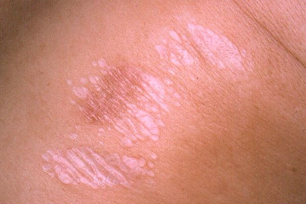
Scleroatrophic lichen
Last reviewed: 04.07.2025

All iLive content is medically reviewed or fact checked to ensure as much factual accuracy as possible.
We have strict sourcing guidelines and only link to reputable media sites, academic research institutions and, whenever possible, medically peer reviewed studies. Note that the numbers in parentheses ([1], [2], etc.) are clickable links to these studies.
If you feel that any of our content is inaccurate, out-of-date, or otherwise questionable, please select it and press Ctrl + Enter.

 [
[ Causes of lichen sclerosus
The causes and pathogenesis of lichen sclerosus are not fully understood. Pathologies of the nervous, endocrine and immune systems, infectious agents, etc. play an important role in the development of the disease.
Histopathology
In the epidermis, at the early stages of the disease, thickening, hyperkeratosis, horny plugs in the mouths of hair follicles are noted, in the late stages - atrophy. The dermis is edematous, lymphocytic infiltration is observed, capillaries are dilated, collagen fibers are homogeneous.
Symptoms of lichen sclerosus
Lichen sclerosus is more common in women. The rash is mostly localized on the neck, upper chest, axillary fossa, shoulders, genitals, sometimes on the back, abdomen, thighs. The primary element is a papule the size of a lentil or 3-5 mm in diameter, the color from chalky, similar to old ivory, to whitish-gray with a pearly tint. At the onset of the disease, the clinical picture resembles white spots. Sometimes a thin pink border is noted around the papule. Closely located papules merge into plaques, slightly elevated above the level of the surrounding skin. Later, they give the impression of being somewhat sunken. The clinical picture is like a miniature of plaque scleroderma. Sometimes telangiectasias, petechiae, and blisters are present on the surface of the plaques. In follicular location, hair follicles are enlarged, there is a large number of follicular horny plugs of a brownish-dirty color, somewhat reminiscent of comedones. Localization of scleroatrophic lichen in the vulva is called vulvar kraurosis, and on the foreskin and head of the penis - penile kraurosis. In this case, the lesion is characterized by dryness, sclerosis. In women, the entrance to the vagina narrows, unbearable itching is noted. In men, there are no subjective sensations. Narrowing of the foreskin leads to phimosis. Over time, skin atrophy occurs, pigmentation of the skin of the lesion is noted.
What do need to examine?
How to examine?
Who to contact?
Treatment of lichen sclerosus
Scleroatrophic lichen is treated with the use of general strengthening antimalarial drugs (delagyl, resorquine), corticosteroid ointments (corticosteroids are not prescribed in the atrophy stage), and agents that improve blood circulation and tissue turnover.