Medical expert of the article

New publications
Retrocerebellar cyst of the brain
Last reviewed: 29.06.2025

All iLive content is medically reviewed or fact checked to ensure as much factual accuracy as possible.
We have strict sourcing guidelines and only link to reputable media sites, academic research institutions and, whenever possible, medically peer reviewed studies. Note that the numbers in parentheses ([1], [2], etc.) are clickable links to these studies.
If you feel that any of our content is inaccurate, out-of-date, or otherwise questionable, please select it and press Ctrl + Enter.
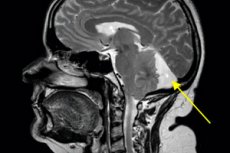
A retrocerebellar cyst in the brain is a specific type of cyst that is located in the back of the brain, in an area called the hindbrain or cerebellum. The cyst is a bubble, hollow vessel, or cavity filled with fluid, and it can vary in size.
The causes of retrocerebellar cysts can be varied, including:
- Congenital abnormalities: Some cysts can occur due to abnormalities in brain development while still inside the mother's body.
- Trauma: Head injuries can sometimes lead to cyst formation in brain tissue.
- Infections: The occurrence of a cyst can be associated with an infection or inflammatory process in the brain.
- Inflammation: Some diseases, such as meningitis (inflammation of the brain's membranes), can cause cysts to form.
- Other Causes: Cysts can occur for other reasons, which may be related to impaired drainage of fluid in the brain or other factors.
The symptoms and effects of retrocerebellar cysts can vary depending on their size and location. Cysts may be asymptomatic and discovered incidentally during a physical examination, or they may cause a variety of symptoms such as headaches, poor coordination, dizziness, vision problems, and more.
Treatment of a retrocerebellar cyst depends on its characteristics and symptoms. In some cases, surgical manipulation may be necessary to remove the cyst to relieve symptoms and prevent it from growing. Treatment always requires an individualized approach and consultation with a neurosurgeon or other specialist in the field of medicine. [1]
Causes of the a retrocerebellar cyst
Retrocerebellar cysts are usually caused by a variety of factors, and the exact causes can vary. Some of the possible causes of retrocerebellar cysts are listed below:
- Congenital abnormalities: Some cysts can occur due to brain abnormalities that developed while the fetus was still developing in the mother's body. This may be due to genetic factors or random mutations.
- Trauma: Trauma to the head, such as a blow, accident, or fall, can cause cysts to form in the brain. Traumatic injuries can damage brain tissue and lead to the formation of cysts.
- Infections: Some infections of the brain or its membranes (such as meningitis) can cause inflammation and cyst formation as a reaction to the infection.
- Inflammation: Inflammatory processes in the brain caused by various diseases or immune reactions can lead to the formation of cysts.
- Congenital cysts: In some cases, retrocerebellar cysts can be congenital, meaning they form in the brain before a person is born.
- Hydrocephalus: Hydrocephalus, a condition in which excessive amounts of fluid builds up inside the skull, can lead to the formation of cysts in various parts of the brain, including the retrocerebellar region.
- Other Factors: Cysts can also occur for other, less common reasons, which may include changes in the brain's blood flow or abnormalities in the drainage of brain fluid.
Pathogenesis
Pathogenesis describes the mechanisms that lead to the development of this condition. In the case of retrocerebellar cysts, the pathogenesis may be related to several possible factors:
- Congenital anomalies: Some retrocerebellar cysts may be genetically predisposed, meaning they can occur due to abnormalities in brain development while the fetus is still developing. These abnormalities may include abnormal formation of brain structures or abnormalities of cerebral fluid drainage.
- Trauma: Trauma to the head, such as a blow, accident, or fall, can cause damage to brain tissue and cyst formation in response to the injury. Traumatic injuries can disrupt normal blood and brain fluid flow, which can contribute to cyst formation.
- Infections and inflammation: Infections of the brain or its membranes, such as meningitis, can cause inflammation in the brain. Inflammation can lead to changes in brain tissue and possibly cysts.
- Hydrocephalus: Hydrocephalus, a condition in which excessive amounts of brain fluid accumulate in the skull, may be associated with the formation of cysts in various parts of the brain, including the retrocerebellar region.
- Other Factors: Cysts can also result from other mechanisms, such as changes in the blood supply to the brain, abnormalities in the drainage of brain fluid, or other medical conditions.
Understanding the exact pathogenesis of retrocerebellar cysts requires further research and patient evaluation. Often the formation of such cysts is multifaceted and can be associated with several factors simultaneously.
Symptoms of the a retrocerebellar cyst
Here are some of the possible symptoms of a retrocerebellar cyst:
- Headache: Pain in the head area can be one of the most common symptoms.
- Dizziness and unsteadiness: Retrocerebellar cysts can put pressure on brain structures responsible for coordination and balance, which can lead to dizziness and unsteadiness when walking.
- Visual impairment: The cyst can put pressure on the nerves or parts of the brain responsible for vision, which can lead to a variety of visual problems such as double vision, blurred images, or difficulty focusing.
- Head seizures: In some people, a retrocerebellar cyst can cause epileptic seizures.
- Hydrocephalus: If the cyst blocks the normal flow of fluid inside the skull, it can lead to hydrocephalus (a buildup of fluid inside the skull), which can cause headaches, vomiting, and worsening of the condition.
- Neurologic deficits: The cyst can put pressure on different parts of the brain, which can cause a variety of neurologic symptoms such as seizures, sensory disturbances, and changes in muscle strength and coordination.
Symptoms may vary depending on the individual patient and the characteristics of the cyst itself. [2]
Retrocerebellar cyst in a child
It is a condition in which a fluid-filled cavity forms in the back of the brain, in an area called the retrocerebellum. This medical condition can be congenital or acquired, and its diagnosis and treatment require specialist attention.
It is important to remember that retrocerebellar cysts can vary in size and symptoms, and they do not always cause problems. In some children they may be asymptomatic and discovered incidentally during a physical examination, while other children may have symptoms such as headaches, dizziness, coordination problems, vision problems, and other neurological symptoms.
The following steps should be followed to diagnose and manage a retrocerebellar cyst in a child:
- Physical examination: A pediatrician or neurologist will examine the child and identify symptoms and possible signs that may be associated with the cyst.
- Diagnostic tests: Magnetic resonance imaging (MRI) of the brain is usually performed to confirm the presence and evaluate the characteristics of the cyst. MRI provides detailed images of the brain and the cyst, determining its size, location, and other characteristics.
- Specialist consultation: Depending on the diagnostic findings and the child's symptoms, a neurosurgeon or other specialist may need to be consulted to determine the next steps in treatment and care.
- Treatment: Treatment of a retrocerebellar cyst in a child depends on the characteristics of the cyst and symptoms. In some cases, surgical removal of the cyst may be necessary, especially if it causes severe symptoms or threatens health. In other cases, there may be observation and monitoring without surgery.
The treatment and care of a child with a retrocerebellar cyst should be guided by doctors and specialists who can recommend the best plan of care for the situation.
Complications and consequences
Retrocerebellar cysts, like other brain cysts, can cause a variety of complications and problems, especially if left untreated or if their symptoms are not controlled. Complications can vary depending on the size, location, and characteristics of the cyst. Some of the possible complications are listed below:
- Nervous System Disorders: Retrocerebellar cysts can put pressure on surrounding brain and spinal cord tissue, which can cause a variety of neurological symptoms. These can include headaches, dizziness, coordination disorders, muscle weakness, sensory disturbances, and other problems.
- Hydrocephalus: In some cases, retrocerebellar cysts can interfere with the normal drainage of brain fluid, which can lead to hydrocephalus (fluid buildup inside the skull). Hydrocephalus can cause increased head volume, headaches, visual disturbances, and other symptoms.
- Compression of surrounding structures: Large or rapidly growing cysts can put pressure on nearby brain structures, which can cause serious neurological complications including paralysis, impaired consciousness, and other problems.
- Visual disturbances: Retrocerebellar cysts that put pressure on the visual pathways or periocular structures can cause visual disturbances including double vision, narrowing of the visual field, or even loss of vision.
- Increased intracranial pressure: Cysts can increase pressure inside the skull, which can lead to headaches, nausea, vomiting, and other symptoms of increased intracranial pressure.
Diagnostics of the a retrocerebellar cyst
Diagnosing a retrocerebellar cyst involves a number of medical procedures and examinations that will help establish the presence and characteristics of this cyst. Here are some of the main methods used to diagnose retrocerebellar cysts:
- Magnetic resonance imaging (MRI): Brain MRI is the primary method for detecting and characterizing retrocerebellar cysts. It is a non-invasive study that provides detailed images of the brain in different projections. MRI helps to determine the size, location, and structure of the cyst, as well as assess its impact on surrounding tissues.
- Computed tomography (CT): A CT scan of the brain may be performed when an MRI is unavailable or inappropriate. It may also be useful to further evaluate the cyst and its impact on surrounding structures.
- Liquorography: This is a procedure in which the doctor injects a contrast agent into the spinal canal and performs x-rays or MRIs to evaluate cerebral fluid drainage. Liquorography can be useful in evaluating the impact of a cyst on cerebral fluid drainage.
- Ultrasound: In rare cases, ultrasound may be used to diagnose cysts, especially in newborns or infants.
- Clinical examination and history: The physician may examine the patient, inquire about his or her medical and family history, and discuss symptoms that could indicate the presence of a cyst.
Once a retrocerebellar cyst has been diagnosed, it is important to perform further evaluation and assess the patient's symptoms. This will help determine the need for treatment and develop an individualized plan of care for the patient, which may include medical monitoring, treatment, or surgery, depending on the characteristics of the cyst and the clinical situation.
Differential diagnosis
The following conditions and diseases should be considered for differential diagnosis of retrocerebellar cysts:
- Epidural cyst: Epidural cysts are located in the spine and can cause compression of the spinal cord. Similar symptoms can also occur due to compression of the spinal cord by a retrocerebellar cyst.
- Traumatic cyst: After a head or spinal cord injury, a fluid cyst may occur as a result of a tear in the brain or spinal membranes.
- Arnold-Chiari Malformation: This is a congenital disorder of brain anatomy in which brain tissue may protrude down into the spinal canal, which may be mistaken for a cyst.
- Osteophytes or spinal tumors: These changes in the structure of the spine can compress the spinal cord and cause symptoms similar to those of a retrocerebellar cyst.
- Inflammatory or infectious processes: Infections such as meningitis or brain abscesses can cause similar symptoms.
It is important to perform a comprehensive examination, including a magnetic resonance imaging (MRI) or computed tomography (CT) scan of the head and/or spine, and consultation with a neurosurgeon or neurologist to make an accurate differential diagnosis and establish a definitive diagnosis.
Who to contact?
Treatment of the a retrocerebellar cyst
Treatment for a retrocerebellar cyst (or Darwin's cyst) depends on several factors, such as the size of the cyst, the symptoms it causes, and its potential complications. The following treatments are usually considered:
- Dynamic observation (waiting): If the retrocerebellar cyst is small and not causing symptoms or complications, doctors may recommend simply observing it with regular physical exams and monitoring. This may be a safe option for patients who have no pain or other uncomfortable symptoms.
- Symptom management: If the cyst is causing headaches, dizziness, poor coordination, and other symptoms, treatment may be aimed at relieving these symptoms. This may include taking painkillers, antiemetics, and rehabilitation.
- Surgery: In cases where the cyst becomes large, severely compresses surrounding tissue, or causes serious symptoms, surgical removal may be necessary. The procedure is called a "craniectomy" or "cystectomy." During this surgery, the surgeon removes the cyst and, if necessary, reconstructs the surrounding tissue.
- Drainage: Sometimes drainage techniques may be used, in which fluid is removed from the cyst to relieve symptoms. This may be a temporary solution.
Treatment of retrocerebellar cysts should be supervised by experienced specialists such as neurosurgeons or neurologists. They can assess the individual characteristics of each case and decide on the best treatment method for the specific situation.
Forecast
The prognosis for patients with retrocerebellar cysts can vary depending on several factors:
- Cyst size: Small cysts may be asymptomatic and not cause serious problems, while large cysts may press on surrounding structures and cause symptoms.
- Symptoms: The prognosis depends on what symptoms the cyst is causing. For example, cysts that cause headaches, dysarthria (impaired articulation of speech), coordination problems, and other neurologic symptoms may require more serious treatment.
- Treatment: Treatment can range from drug therapy to surgery. In some cases, especially large and symptomatic cysts, surgical removal may be necessary.
- Age and general health of the patient: The prognosis may also depend on the age and general health of the patient. Young and healthy patients may have a more favorable prognosis.
It is important to note that retrocerebellar cysts do not always cause serious problems, and many people can successfully manage these cysts with medical supervision and, in some cases, treatment. However, only a physician can provide an accurate assessment of prognosis based on the individual characteristics and clinical presentation of a particular patient. If you or a loved one suspects a retrocerebellar cyst, it is important to see a physician for evaluation and a treatment plan.
Retrocerebellar cyst and the army.
Acceptance into the military depends on many factors, including medical and physical fitness. The decision to enlist in the military with a retrocerebellar cyst will depend on a variety of circumstances:
- Size and nature of the cyst: If the retrocerebellar cyst is small, asymptomatic, and does not adversely affect the servicemember's health and abilities, it may not pose an obstacle to military service.
- Symptoms and Complications: If the cyst causes symptoms such as neurologic impairment, headaches, coordination problems, or other serious problems, it can affect military acceptance.
- Physician's Decision: The Armed Forces Medical Board will make an enlistment decision based on a medical evaluation of each individual case. If physicians believe that a retrocerebellar cyst represents a significant medical or neurological impairment, it may result in temporary or permanent exemption from military service.
It is important to emphasize that each case is evaluated individually and the decision is made by a medical committee based on specific medical data.