Medical expert of the article

New publications
Heel spur removal: basic methods
Last reviewed: 04.07.2025

All iLive content is medically reviewed or fact checked to ensure as much factual accuracy as possible.
We have strict sourcing guidelines and only link to reputable media sites, academic research institutions and, whenever possible, medically peer reviewed studies. Note that the numbers in parentheses ([1], [2], etc.) are clickable links to these studies.
If you feel that any of our content is inaccurate, out-of-date, or otherwise questionable, please select it and press Ctrl + Enter.
In 95% of cases, heel spurs are successfully treated with conservative methods, and surgical removal of heel spurs is a last resort.
The criterion for its implementation is a serious degree of the disease, when the intensity of pain cannot be reduced by all tried methods and means of treatment, and the quality of life of patients is significantly reduced.
Indications for the procedure
If your first steps in the morning are accompanied by a stabbing pain in the heel, and this continues day after day (with the acute pain increasing with increasing load on the feet), then most likely the cause is plantar fasciitis - inflammation of the plantar ligament that supports the arch of the foot at the point of its attachment to the heel bone.
It is in this place, as a result of deformation and inflammation of the fibers of the tendon of the calcaneal aponeurosis, that scar tissue first forms, which gradually ossifies with the formation of a growth on the bone - a marginal osteophyte, which is called a heel spur. Its pressure on the tissues surrounding the calcaneal tubercle and the endings of the lateral and medial plantar nerves leads to the appearance of acute pain.
The location of the marginal osteophyte is the area of the calcaneal tubercle on the plantar side or the zone of attachment of the Achilles tendon, and sometimes the lateral surface of the heel. By the way, bone spurs (large and very painful) can develop even on the front of the ankle joint due to arthritis of the ankle.
It should be borne in mind that the presence of a heel spur is not necessarily accompanied by pain: according to some estimates, up to 15-20% of people have an asymptomatic marginal osteophyte, which can be discovered by chance - during an X-ray of the foot for a completely different reason.
Therefore, only severe pain syndrome that cannot be relieved within 6-9 months from the start of treatment (taking non-steroidal anti-inflammatory drugs, glucocorticoid injections, physiotherapy procedures, exercise therapy for stretching the plantar ligament, etc.) can become an indication for surgery, in which the removal of the heel spur is performed surgically.
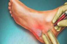
Surgical removal is controversial, and clinical studies show a high risk of complications after surgery. An alternative to surgery is non-invasive heel spur removal with shock wave therapy (extracorporeal shock wave therapy, ESWT). Laser removal of heel spurs is also possible.
 [ 4 ]
[ 4 ]
Preparation
Usually, preparation for surgery for heel spurs requires a complete blood count and coagulogram.
To identify a plantar osteophyte, an X-ray of the foot is necessary, but the doctor may order an ultrasound or MRI for better visualization of the defect, since it is very important to differentiate fasciitis from ankylosing spondylitis, Reiter's syndrome or rheumatoid arthritis.
One to two weeks before surgery, laser or shock wave treatment, you should not take anticoagulants, have GSK injections or use non-steroidal anti-inflammatory drugs to relieve pain.
[ 5 ]
Technique heel spur removal
Surgical procedures to remove heel spurs involve partial dissection of the plantar fascia, which results in decreased tension and improved foot function.
The technique of performing such an operation includes simultaneous removal of the marginal osteophyte. Plantar fasciotomy can be performed through direct access to the ligament with tissue dissection on the inner side of the leg (medial approach) or through an incision on the plantar surface. The plantar incision is used more often, as it provides easy access to the bone spur on the lower part of the heel with its direct visualization (absent with the medial approach).
Minimally invasive removal of heel spurs practiced in modern orthopedic surgery is percutaneous endoscopic fasciotomy. This is an endoscopic method of eliminating constant overstretching of the plantar fascia by dissecting it from the lower surface of the calcaneus (at the site of osteophyte localization) and removing the bone growth through two ports (small incisions). Like conventional fasciotomy, this operation is performed under spinal anesthesia.
The heel spur is also removed through one small incision - with X-ray monitoring of the manipulations. First, a fasciotomy is performed with a special mini-scalpel, and then the growth is cut off from the bone with a miniature cutter.
Foreign orthopedists use the TX MicroTip percutaneous fasciotomy technique, which combines traditional ultrasound technology and microsurgery.
ESWT treatment uses one to two thousand high-energy pulses generated by special equipment in one 20-30 minute session; four to five procedures are usually performed at weekly intervals. The microscopic traumatic effect of these waves on the tissues of the plantar fascia stimulates the process of natural regeneration of damaged cells (by activating growth factors), which begins with improved blood supply and tissue trophism, helping to relieve inflammation and pain. Read also - Ultrasound for heel spurs
Removal of heel spurs by shock wave therapy is performed under intravenous sedation and local anesthesia. The average shock wave value in this case is higher (up to 20.6 kV), and the number of pulses reaches 2.5 thousand.
Heel spur removal with a low-frequency laser is performed on an outpatient basis in several procedures: twice a week for 4-5 minutes each. According to foreign clinical statistics, complete recovery after laser removal of osteophyte on the heel bone is noted in 32% of cases, significant improvement - in 16%, moderate - in 24%, and no result is recorded in 28% of patients. Nevertheless, this is a good method of primary treatment of plantar fasciitis.
Contraindications to the procedure
Laser removal and treatment of heel spurs with the radiation of an optical quantum generator is contraindicated for patients with tumors of any etiology and localization, in the presence of metal bone implants in the corresponding limb, with hyperthyroidism, severe heart failure, vascular or dermatological diseases of the lower limbs. This procedure cannot be performed on pregnant women.
Shock wave therapy (using acoustic waves) is not used for cancer patients, patients with diabetic polyneuropathy or an implanted pacemaker, acute infections, blood clotting disorders, any problems with the heart or circulatory system, or during pregnancy.
Contraindications to surgical removal of the spur include all of the listed cases.
Consequences after the procedure
Possible consequences after the procedure of spur removal by surgical fasciotomy include increased pain in the ankle joint (metatarsalgia), swelling, hematomas and bleeding. And among the postoperative complications, there is a possibility of infection with the development of inflammation, a decrease in the height of the arch of the foot, the development of compartment syndrome (increased pressure in the tissues under the plantar ligament, leading to their necrosis), damage to nerve fibers and numbness of part of the foot (often with weakness in the limb).
[ 9 ]
Complications after the procedure
Complications after the laser spur removal procedure are manifested in swelling of the foot tissues, temporary local (in the heel area and sole) hyperthermia and hyperemia.
There are virtually no complications after the ESWT spur removal procedure: there may be slight and fairly quickly passing swelling of the foot.
Care after the procedure
It is clear that care after open fasciotomy requires antiseptic treatment of the suture, which is removed about a week after the operation. And the recovery of patients - until they can walk independently - usually lasts about 6-10 weeks.
If endoscopic minimally invasive removal of the heel spur was performed, patients recover faster: in 3-6 weeks. But, as surgeons say, everything depends on the characteristics of the patient's body and the specific clinical situation.
After removing the heel spur with a laser or shock wave method, no special care is required; it is necessary to reduce the load on the foot in the first two weeks and be sure to use orthopedic insoles, which are recommended to be worn constantly.
Folk remedies for removing heel spurs
From the point of view of evidence-based medicine, folk remedies for removing heel spurs can, in fact, reduce pain, but are not capable of destroying the marginal osteophyte.
Recipes with vinegar are especially popular for removing heel spurs that occur on the outer side of the heel (at the back), this is the so-called posterior heel - Achilles spur or Haglund's deformity.
One of these recipes involves preparing a mixture of 100 ml of vinegar (regular or apple) and the same amount of melted butter, into which one raw egg (in the shell) is placed. The mixture should be kept in a dark place for three days, then mixed and applied daily, at night, to the sore heel, tying and winding a sock. They say that the pain goes away after two to three weeks of such procedures.
It is recommended to use an ointment consisting of vegetable oil, vinegar essence and mustard powder, taken in equal proportions.
There are good reviews regarding the pain-relieving effect of paraffin compresses, compresses of cabbage leaves with honey, with warm linseed oil and turpentine, and foot baths with salt and iodine.
[ 12 ]