Medical expert of the article

New publications
Heel spur treatment with X-ray therapy
Last reviewed: 06.07.2025

All iLive content is medically reviewed or fact checked to ensure as much factual accuracy as possible.
We have strict sourcing guidelines and only link to reputable media sites, academic research institutions and, whenever possible, medically peer reviewed studies. Note that the numbers in parentheses ([1], [2], etc.) are clickable links to these studies.
If you feel that any of our content is inaccurate, out-of-date, or otherwise questionable, please select it and press Ctrl + Enter.
Treatment of plantar fasciitis with X-rays or X-ray therapy of heel spurs is an effective method of significantly reducing pain symptoms, and often completely eliminating it.
Numerous randomized clinical trials conducted by European specialists over the past three decades have proven that in 68-82% of cases after such treatment there is a very noticeable relief of pain, and in 27-36% of patients the pain completely stops within at least two years.
Indications for the procedure
Treatment of heel spurs with X-ray therapy, as well as some other enthesopathies and joint diseases caused by degenerative and dystrophic processes in the structures of the musculoskeletal system, has strict indications for implementation: intense, intractable pain and problems with movement.
The main criterion for prescribing radiation therapy for plantar fasciitis is the ineffectiveness of standard methods used for at least six months: local injections of glucocorticosteroids, pain-relieving ointments, massage, exercise therapy and hardware physiotherapy (subject to the use of insoles and orthopedic insoles).
Preparation
Since patients with heel spurs are treated by an orthopedist or podiatrist, all necessary examinations that are required to prepare for X-ray therapy are prescribed by the attending physician. The main thing is the availability of an X-ray image (in two standard projections) and/or the results of the latest MRI of the affected foot.
It is necessary to take a general blood test. And in unclear clinical cases, additional scintigraphy of the bone structures of the foot may be required.
10-12 days before the start of X-ray therapy sessions, any physical therapy procedures are cancelled and the use of local agents is discontinued.
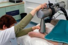
Technique heel spur X-ray therapy.
X-ray therapy for heel spurs can be short- and long-focus. The technique for short-focus X-ray therapy involves exposing the spur to rays generated by an X-ray therapy apparatus (penetrating the skin) no more than 60-70 mm deep into the tissues of the plantar fascia.
In this case, the choice of voltage and current of the X-ray unit (i.e. optimal technical parameters), focal length, size of the irradiated area, the value of a single focal and total (total) absorbed dose of ionizing radiation is carried out by specialists taking into account the depth of the location of the marginal plantar osteophyte and the condition of the surrounding tissues.
The fractionation regimen is also determined individually: the number of sessions, the duration of one irradiation session and their frequency.
There may be just one irradiation (if the pain-relieving effect is achieved quickly), two procedures (with a long interval) or 5-10 irradiations (every two to three days).
According to the recommendations of the German Society for Radiation Therapy and Oncology (DEGRO), updated in 2013, treatment of heel spurs with X-ray therapy should be carried out in two or three fractionations with a single focal dose of 0.5-1.0 Gy and a total absorbed dose within 3.0-6.0 Gy.
If pain persists or pain relief is insufficient, repeat radiation sessions may be recommended 6-12 weeks after the first treatment.
Contraindications to the procedure
X-ray therapy of heel spurs is absolutely contraindicated in patients with poor general health: severe cardiac, vascular and pulmonary pathologies (including thrombophlebitis of the lower extremities and pulmonary tuberculosis); hematological diseases; oncology; immunosuppression; pregnancy and lactation.
Also, temporary contraindications to this treatment are associated with the presence of acute inflammatory processes or infectious diseases.
It is not advisable to use orthovoltage radiation therapy in patients under forty years of age.
Consequences after the procedure
It is believed that such consequences after the procedure of irradiation of the foot area as the development of an oncological disease (skin or bone marrow cancer) in the distant period are unlikely. At least, as the European Journal of Orthopaedic Surgery & Traumatology writes, the risk of developing oncology as a result of this treatment is very low, and there were no documented cases of radiogenic acute or chronic side effects among patients of Western European medical institutions.
But complications after the procedure may manifest themselves as local hyperemia of the skin (immediately after irradiation), its swelling and some soreness. Radiation therapy can cause increased dryness and peeling of the skin on the sole of the foot (like exfoliative dermatitis), thinning of the epidermal layer of the skin and a decrease in its elasticity, cracking of the skin at the site of exposure to radiation - with the release of exudate.
 [ 8 ]
[ 8 ]
Reviews
Although in domestic orthopedics, unlike foreign orthopedics, X-ray treatment of heel spurs is not as widespread (due to the lack of a single protocol for its implementation and irrefutable evidence of safety), the feedback from most patients after such therapy testifies in favor of this method.
However, it should be borne in mind that acute pain in the foot may recur after some time, since low doses of ionizing radiation do not destroy the plantar osteophyte and are symptomatic treatment.