Medical expert of the article

New publications
Pleural cavity puncture
Last reviewed: 03.07.2025

All iLive content is medically reviewed or fact checked to ensure as much factual accuracy as possible.
We have strict sourcing guidelines and only link to reputable media sites, academic research institutions and, whenever possible, medically peer reviewed studies. Note that the numbers in parentheses ([1], [2], etc.) are clickable links to these studies.
If you feel that any of our content is inaccurate, out-of-date, or otherwise questionable, please select it and press Ctrl + Enter.
Pleural puncture is a procedure during which the pleural cavity is punctured in order to conduct diagnostics or perform a therapeutic manipulation that can save a person's life. Most often, it is used if a person has accumulated pleural fluid, or effusion. Pleural puncture is often needed for edema, wounds, severe inflammation, cardiac edema.
Purpose of pleural puncture
It all depends on the goal the doctor is pursuing when performing the procedure. During diagnostics, the insides are examined, liquid media are taken for further analysis. This gives a chance to fully study the pathology, conduct additional laboratory and instrumental studies. If the doctor suspects a malignant neoplasm, a biopsy is performed, during which biological material is collected for the purpose of further histological examination. Therapeutic puncture is used if there is a need to provide a therapeutic effect on a person, for example, the introduction of medical drugs, the removal of air. The effectiveness of therapy increases dramatically, since with this method, drugs are administered directly into the inflamed area, which increases the accuracy and bioavailability of any agent, the speed of action increases, the need and dosage are significantly reduced.
Indications for the procedure
The main indication is the accumulation of liquid exudate in the pleural cavity. The diagnosis is based on ultrasound, percussion, andX-ray data.
It is mandatory to perform it in cases of pleural inflammation, intrapleural bleeding, and severe edema. Sometimes the need for performing it arises when effusion appears against the background of heart failure.
Pleural puncture for pneumothorax
If a person has developed pneumothorax, a puncture must be performed immediately. It is performed according to the standard method, without any differences or limitations. The only exception is that fluid and air are pumped out of the cavity using a special syringe or pleuroaspirator.
Sometimes there is a need to perform only air drainage. For example, in the case of valve pneumothorax, when a person inhales, air continuously enters the pleural cavity. There is no provision for reverse drainage, so the tube is not clamped after the operation is performed. The patient needs to be hospitalized.
 [ 1 ]
[ 1 ]
Pleural puncture for hydrothorax
Hydrothorax is a serious condition in which pleural fluid accumulates in the pleural cavity. This can be either transudate or exudate. Other pathological fluids, including blood, can also accumulate. Fluid can accumulate if a person has been injured or during an intense inflammatory process. Sometimes a tumor can stimulate excessive fluid production. It is advisable to subject the pumped-out fluid to histological and bacteriological analysis in order to assess its composition and main parameters. This is an important diagnostic procedure that makes it possible to clearly think through the tactics and strategy of further treatment. After removing the fluid, the pleural cavity must be thoroughly washed. To provide a therapeutic effect, the necessary drugs and antiseptics are administered.
Pleural puncture for pleurisy
It is a necessary, often even urgent procedure, especially when the volume of inflammation is massive. It is mandatory to perform it when severe shortness of breath appears. If the median shadow on the radiograph shifts, it is also necessary to perform a puncture. In an emergency, no preliminary examination is required, and there is practically no preparation. The main goal of the procedure is to reduce the amount of fluid.
Pleural puncture in exudative pleurisy
Exudative pleurisy is a type of inflammation that is accompanied by intense accumulation of fluid. During the operation, special instruments are used to suck out the contents and wash the cavity with antiseptic solutions. Special medications are also administered. The procedure is performed with the patient in a sitting position. If the patient feels unwell or is in serious condition, it is advisable to lay him on his healthy side. The operating table is slightly lowered from the head side, which helps prevent the development of air embolism.
Indications for pleural puncture in chronic heart failure
Patients suffering from various pathologies of the heart and vascular system are at risk for developing pleural effusion. The operation can be performed urgently or on a planned basis. During a planned puncture, preliminary preparation is carried out, which includes an electrocardiogram and other studies of the heart and blood vessels, which makes it possible to prepare the body and avoid many complications, such as hemothorax, hydrothorax. Manipulations for a person with heart failure are necessarily carried out if pain occurs during deep inhalation and exhalation, severe cough, a feeling of distension and burning. The appearance of frequent reflex cough and asymmetric displacement of the chest, respiratory failure and displacement of the mediastinum, are a sign that a person is developing pleural fluid, and urgent measures are required. The development of severe edema and congestion, empyema, requires urgent removal of fluid. Local administration of drugs is carried out.
Preparation
Psychological preparation for the procedure is of primary importance. This is the responsibility of the nurse. She must explain the essence, set him up for a positive outcome, and maintain confidence in him.
It is important to be kind and nice to the patient, showing respect, addressing him by name and patronymic. If a person knows the essence of the procedure, has a rough idea of how and in what sequence it will take place, it will be much easier to endure it. Ignorance always causes more fear and panic. The patient must understand what results the doctor plans to obtain and what the procedure is actually needed for. The patient must undergo preliminary examinations, thermometry.
A mandatory stage is premedication, during which the patient is prepared for anesthesia. A consultation with an anesthesiologist is held. He examines the patient, conducts the necessary laboratory tests, and then selects the appropriate medications.
Methods of pleural punctures
There are many different methods by which pleural fluid can be removed from the cavity. Each of them has its own advantages and disadvantages. The most common method is thoracocentesis, used mainly in severe pre-uritis, pneumothorax, and blood accumulation in the cavity itself. Cavity drainage, the Bulau method, is also used. Active drainage using a rubber bulb, flow-aspiration drainage is also performed.
Diagnostic pleural puncture
It has found wide application in various fields of medicine: it is used by rheumatologists, traumatologists, pulmonologists. Oncologists are also forced to use this procedure quite often. It is carried out for the timely detection of pathology, as well as the collection of biological material for further research. Fluid is collected and a biopsy is performed, during which a piece of tissue is taken for further histological examination. The fluid can be transudative, not causing pathology, exudative, which causes an inflammatory process. For this, the density of the obtained biological samples and other parameters are examined. After extracting the biomaterial, it is subjected to further biochemical analysis. If an infectious process is suspected, bacteriological, virological analysis is carried out. If there is a suspicion that an oncological process is developing, a histological examination is carried out.
Diagnostic studies are carried out quite often, since they are characterized by high information content. If necessary, intracavitary blood, lymph, pus, air are examined. If there is bleeding or a suspicion of its occurrence, a test is also carried out (a sample is taken, it is monitored). This significantly simplifies life in case of severe pleural lesions, often saving the patient's life. Often, a diagnostic puncture is carried out first, which, if a pathology is detected, turns into a therapeutic manipulation.
[ 6 ], [ 7 ], [ 8 ], [ 9 ], [ 10 ]
Puncture and catheterization of the pleural cavity
It is performed in purulent-inflammatory processes to eliminate exudate, transudate, blood and air.
Pleural puncture kits
Pharmacies sell ready-made kits that contain everything you need to perform a puncture. The basis of the kit is a syringe and needles. The volume of the syringes and the diameter of the needles vary, so you need to clearly know what exactly is required to perform the procedure. The kit is also equipped with special clamps that are used to prevent air from entering the pleura. Each kit includes 2 test tubes for collecting biomaterial, a tray with a set of necessary tools. They are usually represented by tweezers, tampons, cotton swabs. The kits also include the simplest preparations for treating the skin and providing first aid.
Pleurofix
It includes a syringe, needles of various diameters, clamps, test tubes for collecting biological material, tweezers, swabs and cotton swabs.
Disposable pleural puncture kit
The kit is intended for performing thoracentesis and punctures. They are used mainly when fluid appears in the pleural cavity and purulent pathologies develop. The kit is represented by a tray with instruments, syringes and puncture needles. Sometimes the kits are equipped with a solution of novocaine for pain relief, alcohol, and various medical materials.
Instruments for pleural puncture
Today, the pharmaceutical industry produces many kits that include all the instruments necessary for the procedure, as well as some medical supplies and drugs. The advantage of such kits is that they are disposable, which significantly reduces the risk of infection.
Pleural puncture needle
The diameter of the needle usually varies widely. They are intended for connection with a syringe. The connection is made through rubber tubes. The kits are intended for single use.
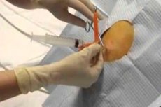
Technique pleural puncture
First of all, it is necessary to give the patient a comfortable position. The doctor may ask the patient to raise his arm, which will allow the intercostal space to be expanded. If the patient is in a serious condition, the procedure is performed in a lying position.
Algorithm for performing pleural puncture
Not only a doctor but also a nurse takes part in the procedure, since the patient needs support and special preparation for the procedure. The doctor also needs help, since it is almost impossible for one person to perform such a procedure. The first mandatory step is to disinfect the area that will be punctured. Various antiseptics are used for this. In this case, a regular iodine solution or chlorhexidine has proven itself to be the best. Then the treated area is dried with a napkin.
The puncture site is anesthetized, which can be achieved by introducing novocaine. Then, using a needle designed for pleural puncture, the doctor punctures the pleura. A rubber tube for drainage is always used, which ensures the removal of air and prevents it from entering the pleural cavity. The doctor determines exactly where the puncture will be performed based on the diagnosis. For each disease, the puncture site is individual, in most cases it is determined by the localization of the fluid or inflammatory process. If, during the procedure, a fluid similar to blood or foam begins to appear, you must urgently finish the procedure and pull the needle out.
After all the fluid has been pumped out, the needle is carefully pulled out, the puncture area is pressed with a finger, and then treated with antiseptic solutions. The procedure ends with the application of a sterile tampon and a sterile bandage. If complications arise, resuscitation may be required, but this happens extremely rarely.
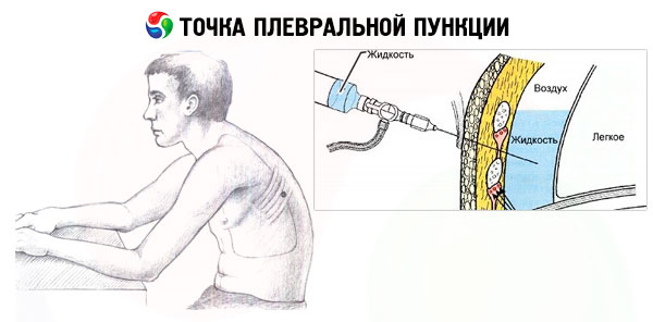
Puncture site for pleural puncture
The choice is determined by the doctor, depends on the disease and the purpose. If air removal is necessary, it is carried out in the area of the 2-3 intercostal space. If fluid removal is planned, it is done in the area of the 7-8 intercostal space. In order to prevent rupture of blood vessels, it is necessarily carried out along the upper edge of the ribs.
Pleural puncture along the edge of the rib
Traditionally, the puncture site is the upper edge of the rib, since it contains a minimum number of vessels and nerves. The puncture is performed with a needle with a diameter of approximately 1 mm. First, an anesthetic is drawn into the needle, a careful puncture is made, and the drug is injected into the puncture site. Then the needle is pulled out. Gradually, a second needle of a larger diameter is inserted, which is attached to a syringe. This syringe is used to puncture and pump out the liquid. After the procedure, the needle is carefully pulled out, and the puncture site is treated with an anesthetic.
Pleural puncture according to Bulau
This is a method used when it is necessary to drain the pleural cavity. The method is named after its discoverer. The patient should be seated with his arms crossed in front of his chest, his head resting on them. The legs are on a special support, the back should be straight. The puncture is made at the bottom, at the base of the lung, and allows fluid to be removed from its diaphragmatic part.
A special kit is used to perform the procedure, which includes a sterile tray, Bobrov's apparatus, clamp, tweezers, scissors, silk thread. Rubber finger cots and gloves are used for the work. Furacilin solution is also used.
Pleural puncture in children
The procedure algorithm and preparation for children are no different from those for adults. The only difference is that a smaller dosage of the drug may be required for pain relief and premedication. Smaller needles are used. The duration of the procedure may be shorter. The puncture depth is significantly less than in adults, which is due to the anatomical features of the child's body. General anesthesia is used. Psychological preparation and support of the little patient are especially important.
Contraindications to the procedure
The procedure is contraindicated in hemophilia - a disorder of the blood clotting process. In an emergency, if the patient's life depends on it, there are no contraindications, it is carried out even in an unconscious state.
Complications after the procedure
It can have serious consequences and complications. But sometimes it is the only way to save the patient's life. It is dangerous because of the development of pneumothorax, air embolism, in which the vessels are blocked by an air clot. The person may start coughing up blood. If it is performed carelessly, there is a high probability of stomach injury. This is indicated by cold sweat appearing on the person's forehead, dizziness. This condition can end with blockage of the vessels. Sometimes there are situations when the procedure cannot be avoided, since a life-threatening condition may arise, in which removal of the lung will be the only way to save life.
Complications are very common, but overall, statistics show that this procedure cannot be cancelled, as there are no alternatives. It makes it possible to save many lives. Due to the close proximity, a lung, diaphragm, or other nearby organ can be accidentally punctured. Intrapleural bleeding or air embolism of blood vessels can occur. Bleeding can occur from the puncture site. If any, even the slightest, complication occurs, the manipulation must be stopped immediately. The needle is urgently removed, the patient must assume a supine position. Urgent surgical assistance is required.
[ 16 ]
Reviews
If you analyze the reviews, they can be both positive and negative. Many have quite serious complications, after which the patient is in the intensive care unit. For others, the procedure goes without any complications, the patient's condition improves dramatically. If the procedure is carried out for diagnostic purposes, it is very informative, often developing into a therapeutic one.
If we analyze the doctors' reviews of the procedure, we can note that they are often forced to resort to this procedure, especially in emergency situations when there is a threat to life. They consider this method radical, but very effective, despite the not very high risk of complications. There is no other alternative to this method.
Experts note that in many cases after such an intervention the pleura thickens. This leads to a sharp decrease in respiratory volume. Sometimes normal breathing can only be restored with a special operation – decortication. During this operation, part of the pleura is removed.
If we analyze the reviews of practicing specialists, we can note the following main complications: fainting and collapse, which occur as a result of the effects of local anesthesia, due to a sharp drop in intrapleural pressure, and changes in vascular tone.
Pneumothorax develops when the tightness of blood vessels is broken, when the tightness of the working system itself is broken, and also as a result of damage to the lung by a needle. There is sharp pain, shortness of breath, pressure and burning in the chest area. Breathing is weakened or not heard at all.
Often, serous pleurisy turns into purulent pleurisy. In this case, the patient's condition deteriorates sharply, intoxication occurs. The exudate becomes turbid, and a purulent sediment appears.
Often there is intrapleural bleeding from the intercostal vessels. If the pleural puncture is performed incorrectly, the liver and spleen can be damaged. Hollow organ injury and diaphragmatic hernia development are often observed. Bleeding into the abdominal cavity occurs. Diagnosis can be made using ultrasound and emergency laparoscopy. In case of severe damage, hemostasis or laparoscopy is performed, depending on the severity of the pathology.