Medical expert of the article

New publications
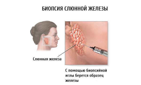
Salivary gland puncture and biopsy
Last reviewed: 04.07.2025

All iLive content is medically reviewed or fact checked to ensure as much factual accuracy as possible.
We have strict sourcing guidelines and only link to reputable media sites, academic research institutions and, whenever possible, medically peer reviewed studies. Note that the numbers in parentheses ([1], [2], etc.) are clickable links to these studies.
If you feel that any of our content is inaccurate, out-of-date, or otherwise questionable, please select it and press Ctrl + Enter.
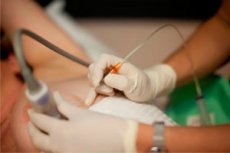
Diagnostic puncture of the salivary gland refers to morphological methods of examination. It is performed with a needle no more than 1 mm in diameter and a 20 ml syringe. After inserting the needle into the examined area of the organ, the contents are aspirated with several movements of the piston. Then the contents of the needle are transferred to glass and a smear is made. Smears are stained with azure II-eosin, according to Romanovsky. In the punctate of intact salivary glands, there are small quantities of cubic and cylindrical epithelial cells. Rarely, cells and narrow dense threads of mature connective tissue are found in the field of vision.
If there are difficulties in interpreting the cytological picture during diagnostic puncture, a puncture biopsy is performed.
 [
[ How is a salivary gland biopsy performed?
For morphological examination, a tissue column is taken using a special hollow needle with a trocar. Puncture is performed using a special thick needle with a core to collect a small piece of gland tissue for subsequent pathomorphological examination. Normally, lobules of the salivary gland or fatty and connective tissue are found in the biopsy. After performing a puncture biopsy, temporary paresis of the facial muscles may occur due to injury to the branches of the facial nerve.
Incisional biopsy of the parotid glands is performed under general or local anesthesia using a bordering incision of the skin and subcutaneous tissue (similar to the incision of G.P. Kovtunovich) in the posterior maxillary fossa. The capsule is dissected, the section of the gland in question is exposed, and a piece of glandular tissue is excised to a depth of no more than 1 cm to avoid injury to the branches of the facial nerve. The wound is carefully sutured layer by layer to prevent the formation of salivary fistulas. Incisional biopsy of the submandibular glands is usually not performed, since, if necessary, an extended biopsy with organ removal is performed.
Minor salivary gland biopsy
A biopsy of the minor salivary gland of the lower lip is used for differential diagnostics of variants of chronic sialadenitis of the major salivary glands, since morphological changes in the minor salivary gland are in many ways similar to those in the major salivary glands. This research method is mainly used to diagnose Sjogren's syndrome or disease. A biopsy of the minor salivary gland of the lower lip is performed under infiltration anesthesia. A 1 cm long incision is made in the mucous membrane of the lower lip. 2-3 minor salivary glands are found and removed. The wound is tightly sutured with interrupted sutures. When analyzing the morphological picture, the degree of lymphoid infiltration is taken into account. Its intensity is assessed by degrees: 1st degree (focal infiltration) - accumulation of more than 50 lymphocytes in focus; 2nd degree (focal-diffuse infiltration) - a preserved lobule may be located next to a lobule partially replaced by lymphoid tissue; 3rd degree (diffuse infiltration) - almost all acinar tissue is replaced by lymphoid tissue.