Medical expert of the article

New publications
Progressive facial atrophy
Last reviewed: 05.07.2025

All iLive content is medically reviewed or fact checked to ensure as much factual accuracy as possible.
We have strict sourcing guidelines and only link to reputable media sites, academic research institutions and, whenever possible, medically peer reviewed studies. Note that the numbers in parentheses ([1], [2], etc.) are clickable links to these studies.
If you feel that any of our content is inaccurate, out-of-date, or otherwise questionable, please select it and press Ctrl + Enter.
In the literature, this disease is known under two terms: hemispheric progressive facial atrophy (hemiatrophia faciei progressiva) and bilateral progressive facial atrophy (atrophia faciei progressiva bilateralis).
In addition, hemispheric and cross atrophy of the face and body may be observed.
Causes progressive facial atrophy
It is assumed that the disease may be caused by trauma to the skull or face, general or local infection, syphilis, syringomyelia, damage to the V or VII pair of cranial nerves, extirpation or injury to the cervical sympathetic trunk, etc. Some authors admit the possibility of hemiatrophy of the face, combined with hemiatrophy of the body due to dystrophy in the diencephalic parts of the autonomic nervous system.
There are cases of hemiatrophy after epidemic encephalitis, as well as with pulmonary tuberculosis that has affected the cervical sympathetic trunk.
According to the available data, progressive facial atrophy in the overwhelming majority of cases is a syndrome of various diseases, in which the autonomic nervous system is involved in the pathological process at its various levels. Obviously, trauma and other factors are only an impetus for the development of these serious neurodystrophic phenomena.
Symptoms progressive facial atrophy
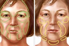
Patients usually complain that the affected half of the face is smaller than the healthy one; the difference in the volume of the facial part of the skull and soft tissues gradually increases; on the affected side the skin is dark-aspen in color, thinned, and gathers into many folds when smiling.
Sometimes patients note a tingling pain in the area of the affected cheek or in the entire half of the face, lacrimation from the eye on the affected side, especially in the cold, in the wind, and a difference in the color of the cheeks, especially noticeable in the cold.
In severe hemiatrophy, it appears as if one half of the face belongs to a person who is emaciated to the limit by starvation or cancer intoxication, and the other half belongs to a healthy person. The skin on the affected side is yellowish-gray or brownish in color and does not blush. The palpebral fissure is widened due to the lower eyelid sinking.
When pressing on the supraorbital, infraorbital and mental foramina, pain occurs.
The corneal reflex is reduced, but the pupils are uniformly dilated and react equally to light.
The thinned skin feels like parchment; atrophy also extends to the subcutaneous tissue, the chewing and temporal muscles, and bone tissue (jaw, zygomatic bone, and zygomatic arch).
The chin is displaced to the affected side, since the size of the body and the branch of the lower jaw are reduced, this is especially pronounced in patients suffering from hemiatrophy of the face since childhood; half of the nose is also reduced, the auricle is wrinkled.
In some cases, hemiatrophy of the face is combined with atrophy of the same half of the body, and sometimes with atrophy of the opposite side of the body (hemiatrophia cruciata), with unilateral scleroderma or excessive pigment deposition in the skin, impaired growth or depigmentation of hair, hemiatrophy of the tongue, soft palate and alveolar processes, caries and tooth loss, and impaired sweating.
Having reached one degree or another, facial hemiatrophy stops, stabilizes and does not progress further.
Clinical and physiological examinations of this group of patients showed that in all forms of progressive facial atrophy, there are, to varying degrees, pronounced disturbances in the function of the autonomic nervous system.
In patients with unilateral facial dystrophy, asymmetry in electrical potentials and skin temperature is usually detected, with a predominance on the affected side.
In most cases, a decrease in the oscillographic index and spasm of the capillaries on the affected side are observed, which indicates the predominance of the tone of the sympathetic nervous system.
Almost all patients show changes in electroencephalograms that are characteristic of damage to the hypothalamic-mesencephalic formations of the brain. Electromyographic studies almost always reveal changes in the electrical activity of muscles on the side of the dystrophy, including where atrophic manifestations in tissues are clinically observed.
Based on a set of clinical and physiological research data, L.A. Shurinok identifies two stages of facial atrophy: progressive and stationary.
Diagnostics progressive facial atrophy
Hemiatrophy of the face should be differentiated from asymmetry in congenital (non-progressive) underdevelopment of the face, hemi-hypertrophy of the face, as well as muscular torticollis, focal scleroderma, tissue atrophy in lipodystrophies and dermatomyositis. The latter diseases are considered in courses on general orthopedics and dermatology.
Treatment progressive facial atrophy
Surgical methods of treatment of progressive facial atrophy are permissible only (!) after suspension or inhibition of the process progression, i.e. in its second completed stage. For this purpose, complex drug and physiotherapeutic treatment in combination with vago-sympathetic blockade, and sometimes - blockade of the cervicothoracic ganglion, is recommended.
To improve tissue metabolism, vitamins (thiamine, pyridoxine, cyanocobalamin, tocopherol acetate), aloe, vitreous body or lidase should be prescribed for 20-30 days. In order to stimulate metabolism in muscle tissue, ATP is administered intramuscularly at 1-2 ml for 30 days. Thiamine helps to normalize carbohydrate metabolism, as a result of which the amount of ATP (formed by oxidative phosphorylation occurring in the mitochondria) increases. Cyanocobalamin, nerobol, retabolil help to normalize protein metabolism.
To influence the central and peripheral parts of the autonomic nervous system (ANS), electrophoresis of the cervical sympathetic ganglia, a galvanic collar, endonasal electrophoresis with a 2% solution of calcium chloride or diphenhydramine (7-10 sessions), UHF on the hypothalamic region (6-7 sessions) and a galvanic half mask with lidase (No. 7-8) are combined.
It is necessary to exclude sources of irritation originating from the liver, stomach, pelvic organs, etc.
In case of increased tone of the sympathetic and simultaneous weakness of the parasympathetic divisions of the nervous system, it is recommended to combine sympatholytic and cholinomimetic drugs, taking into account the level of damage: in case of damage to the central vegetative structures, central adrenolytic agents are prescribed (chlorpromazine, oxazil, reserpine, etc.): ganglia are best treated with ganglioplegics (pachycarpine, hexonium, pentamine, gangleron, etc.). When both peripheral and central divisions of the VNS are involved in the process, antispasmodics such as papaverine, dibazol, euphyllin, platiphylline, khellin, spasmolytin, nicotinic acid are used.
Sympathetic tone is reduced by limiting proteins and fats in the diet; to enhance the parasympathetic effect, acetylcholine, carbachol, as well as anticholinesterase substances (for example, proserin, oxamizine, mestinon) and antihistamines (diphenhydramine, pipolfen, suprastin) are prescribed. In addition, carbohydrate-rich foods, a cool mountain or sea climate, carbon dioxide baths (37°C) and other means and methods prescribed by neurologists are indicated (L. A. Shurinok, 1975).
As a result of conservative preoperative treatment, the process stabilizes, although atrophy, as a rule, remains outwardly expressed.
Myograms of facial muscles show an increase in their bioelectrical activity, a decrease or even disappearance of the asymmetry of the indicators of the state of the autonomic nervous system, a decrease in a number of cases (initial forms of the disease) in the values of electrical potentials of the skin of the face, and the disappearance of disturbances in the thermotopography of the skin.
Methods of surgical treatment of progressive facial atrophy
The main methods of surgical treatment of facial atrophy include the following.
- Injections of paraffin under the skin of the atrophied cheek. Due to cases of thrombosis and embolism of the vessels, surgeons do not currently use this method.
- Subcutaneous tissue grafting (due to its gradual and uneven wrinkling, it also has not found wide application).
- The introduction of plastic explants, which eliminate facial asymmetry at rest, but at the same time immobilizes the affected side and eliminates the symmetry of the smile. Patients are also not satisfied with the rigidity of the plastic, which is located in places that are typically soft and pliable. In this regard, the implantation of porous plastics is more promising, but there are no convincing reports in the literature on the results of their use. It is also recommended to use silicone explants, which have biological inertness and stable elasticity.
- The implantation of crushed cartilage and connective tissue base of the Filatov stem under the skin has almost the same disadvantages: rigidity (cartilage), the ability to immobilize the face (cartilage, stem).
- Replantation of a de-epidermized and subcutaneous tissue-free skin flap or the protein coat of a bull testicle using the methods of Yu. I. Vernadsky.
Correction of facial contours using the method of Yu. I. Vernadsky
An incision is made in the submandibular region, through which the skin, previously “lifted” with a 0.25% solution of novocaine, is peeled off using large curved blunt-ended Cooper scissors or a special raspatory with a long handle.
Having tamped and pressed the resulting pocket from the outside, the contours of the future transplant are outlined on the anterior surface of the abdomen under local anesthesia using a pre-prepared plastic template. In the outlined area (before taking the transplant), the skin is de-epidermized, and then the flap is separated, trying not to capture the subcutaneous tissue.
Having taken the flap on plastic threads (holders), their ends are threaded through the eye of 3-4 straight thick ("gypsy") needles, with the help of which the ends of the holders are pulled into the subcutaneous wound on the face, and then from the upper and lateral arches of the wound they are brought out and tied on small iodoform rollers. In this way, the skin graft appears to be stretched over the entire subcutaneous wound surface. Due to the fact that the graft has a wound surface on both sides, it grows to the skin and subcutaneous tissues inside the wound pocket.
In places of the greatest cheek depression, the flap is doubled or laid in three layers by sewing a kind of "patch"-duplicate to the main flap. The cosmetic effect of this method is quite high: facial asymmetry is eliminated; the mobility of the affected half of the face, although reduced, is not completely paralyzed.
During and after the operation, there are usually no complications (unless an infection occurs, leading to rejection of the transplant or explant). However, over time, some atrophy of the transplanted skin (or other biological material) occurs and a new layer has to be added. In some patients, after transplantation of de-epidermized autoskin, gradually enlarging sebaceous cysts develop. In these cases, it is recommended to puncture the skin above the fat accumulation site (in 2-3 places) with a thick injection needle and squeeze it out through the punctures. Then the empty cavity is washed with 95% ethyl alcohol to cause denaturation of the activated cells of the sebaceous glands; part of the alcohol is left in the cavity under a pressure bandage applied for 3-4 days.
To avoid the formation of sebaceous cysts (atheromas) and additional trauma, it is advisable to use the protein coat of a bull's testicle instead of autoderma, which is perforated with a scalpel in a checkerboard pattern and injected under the skin of the affected area of the face (in the same way as autoderma).
 [ 19 ]
Correction of facial contour using the A.T. method Titova and N.I. Yarchuk
Contour plastic surgery is performed using allogenic preserved broad fascia of the thigh, grafting it in one or two layers or accordion-shaped (corrugating it) if a significant amount of plastic material is required.
A pressure bandage is applied to the face for 2.5-3 weeks.
2-3 days after the operation, fluctuation is determined in the transplant area, caused not by the accumulation of fluid under the skin, but by swelling of the fascial graft and aseptic inflammation of the wound.
To reduce swelling after surgery, apply cold to the transplant area for 3 days, and take diphenhydramine orally at 0.05 g 3 times a day for 5-7 days.
Postoperative graft swelling is dangerous when the incision for forming the bed and introducing the fascia is located directly above the transplant area. This can cause excessive tension at the edges of the wound, causing them to separate and part of the fascia to fall out. To prevent this complication, skin incisions should be located outside the transplant area, and if it does occur, then in the early stages it is possible to limit oneself to removing part of the fascial graft, and secondary sutures should be applied to the wound.
If infection occurs and inflammation develops in the wound, the entire transplant must be removed.
Despite the extensive tissue detachment during fascia transplantation, subcutaneous hematomas and intradermal hemorrhages are extremely rare, which can be explained to some extent by the hemostatic effect of fascial tissue. The greatest risk of hematoma formation exists when eliminating pronounced deformations of the lateral part of the face. Extensive tissue detachment through an incision in front of the auricle creates a prerequisite for blood accumulation in the lower, closed section of the formed bed. If hematoma formation is suspected, it is recommended to create an outflow in the lower part of the wound.
Complications
The most severe complication is suppuration of the surgical wound, which occurs when the graft or the receiving bed becomes infected. To prevent this, it is necessary to strictly observe aseptic requirements when preparing fascial grafts and during their transplantation, trying not to damage the oral mucosa when forming the bed in the cheek and lip area.
The occurrence of a communication between the surgical wound and the oral cavity during surgery is a contraindication for fascia transplantation, protein membrane, etc. Repeated intervention is permissible only after several months.
Considering that the subcutaneous fat tissue of the sole of the human foot (the thickness of which is from (5 to 25 mm), as well as the dermis of the foot, differ sharply from the fat and dermis of other areas, and that they are very strong, dense, elastic, and have low antigenic properties, N.E. Sel'skiy et al. (1991) recommend this allomaterial for contour plastic surgery of the face. Having used it in 21 patients, the authors noted suppuration and rejection of the transplant in 3 people. Obviously, it is necessary to continue studying the immediate and remote results of using this plastic material, since, unlike the de-epithelialized skin of other areas, the plantar skin is devoid of sweat and sebaceous glands, which is very important (in terms of preventing cyst formation).