Medical expert of the article

New publications
Rectal polyps
Last reviewed: 04.07.2025

All iLive content is medically reviewed or fact checked to ensure as much factual accuracy as possible.
We have strict sourcing guidelines and only link to reputable media sites, academic research institutions and, whenever possible, medically peer reviewed studies. Note that the numbers in parentheses ([1], [2], etc.) are clickable links to these studies.
If you feel that any of our content is inaccurate, out-of-date, or otherwise questionable, please select it and press Ctrl + Enter.
Rectal polyps are benign epithelial tumors. They account for approximately 92% of all benign intestinal tumors.
According to clinical classification, polyps are divided into single, multiple (group and scattered in different sections) and diffuse polyposis of the colon. Polyposis is characterized by massiveness of the lesion, can be inherited, i.e. it is a genetically determined disease, and the term "diffuse familial polyposis" is used to describe it.
The size of single and group polyps varies from a millet grain to a walnut. Polyps can have a stalk, sometimes reaching 1.5-2 cm, or be located on a wide base. In diffuse polyposis, they densely cover the entire mucous membrane of the rectum and colon. According to their histological structure, polyps are divided into adenomatous, villous and mixed (adenomatous-villous).
 [ 1 ]
[ 1 ]
Symptoms of rectal polyps
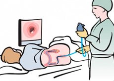
In most cases, rectal polyps are asymptomatic and are an accidental finding during endoscopy performed for some other disease or for the purpose of preventive examination of the colon. However, as the polyps increase in size and their surface ulcerates, such clinical symptoms of rectal polyps as nagging pain in the lower abdomen or lumbosacral region, pathological discharge from the rectum may appear and then progress. Large villous tumors are characterized by metabolic disorders (changes in water-electrolyte balance, significant loss of protein). Anemia may be observed.
Diagnosis of rectal polyps
During the period of appearance of the clinical symptoms described above, all methods of proctological examination are used, starting from digital examination and ending with colonoscopy. Detection of polyps at an earlier (asymptomatic) stage is possible during preventive examinations of people over 40 years of age, which, in the opinion of V.D. Fedorov and Yu.V. Dultsev (1984), will allow diagnosing about 50% of all benign tumors. Since from 50 to 70% of tumors are located in the left sections of the colon, then rectoscopy can be used for preventive examination. At the same time, detection of polyps in the rectum and distal sigmoid colon is a direct indication for colonoscopy in order to exclude multiple lesions.
Adenomatous (glandular) polyps are the most common. They are rounded formations on a stalk or a broad base, rarely bleeding or ulcerating.
Adenomatous-villous (adenopapillomatous, or glandularvillous) polyps are usually larger than adenomatous polyps and exceed 1 cm in diameter. During endoscopy, these polyps are visible as multi-lobular formations. In fact, their multi-lobular appearance is explained by the unevenness of the surface, which can ulcerate, become covered with fibrinous deposits and bleed.
Villous tumors can reach large sizes. During endoscopy, they are determined either as a polypoid formation on a long thick stalk, or as a formation spreading along the intestinal wall over a significant distance. Villous tumors have different surface colors (from whitish to bright red), ulcerate, bleed, and often become malignant.
What do need to examine?
How to examine?
Treatment of rectal polyps
Conservative treatment of rectal polyps with celandine juice was proposed in 1965 by A. M. Aminev. However, it was not widely used due to its insufficient effectiveness. Specialists working on this problem are against the use of celandine for the treatment of polyps, since an attempt at conservative treatment of polyps leads to the postponement of surgical treatment.
The most common methods of surgical treatment of rectal polyps are:
- polypectomy through an endoscope with electrocoagulation of the stalk or base of the polyp;
- transanal excision of the neoplasm;
- tumor removal by colotomy or intestinal resection using the transperitoneal method.
Taking into account the possibility of recurrence and malignancy of polyps, a system of clinical examination of patients after surgical treatment has been developed. It includes endoscopic monitoring of the condition of the rectum and colon, especially in the most dangerous period - the first 2 years after surgery. During these years, the interval between endoscopic examinations does not exceed 6 months, and in patients after removal of villous tumors, which are most prone to recurrence and malignancy in the early stages, this interval does not exceed 3 months.
In case of relapses, repeated surgical treatment of rectal polyps with subsequent systematic endoscopic control is recommended. In cases where the results of the histological examination of the removed polyp indicate malignancy of the process, but there are no signs of malignancy in the base or stalk of the polyp, the first endoscopic control examination with multiple biopsy is carried out 1 month after the operation. If the biopsy result is favorable, patients continue to be examined every 3 months, and then - 2 times a year. If invasive growth extends to the stalk of the polyp or its base, radical oncological surgery is indicated.