Medical expert of the article

New publications
Pathogens of superficial mycoses
Last reviewed: 08.07.2025

All iLive content is medically reviewed or fact checked to ensure as much factual accuracy as possible.
We have strict sourcing guidelines and only link to reputable media sites, academic research institutions and, whenever possible, medically peer reviewed studies. Note that the numbers in parentheses ([1], [2], etc.) are clickable links to these studies.
If you feel that any of our content is inaccurate, out-of-date, or otherwise questionable, please select it and press Ctrl + Enter.
Superficial mycoses (keratomycosis) are caused by keratomycetes - low-contagious fungi that affect the stratum corneum of the epidermis and the surface of the hair. These include:
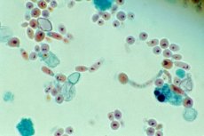
Pityrosporum orbiculare is a widespread yeast-like lipophilic fungi that normally live on human skin. They cause pityriasis versicolor (variegated, multi-colored) lichen, characterized by the appearance of pinkish-yellow non-inflammatory spots on the skin of the trunk, neck, and arms. When scraped off, scales similar to bran appear on the spots. In the scales treated with 20% alkali, short curved hyphae and yeast-like budding fungal cells are detected. They are grown on media containing lipid components. Colonies grow better under a layer of sterile olive oil. Growth is noted after a week in the form of creamy whitish-cream colonies consisting of oval, bottle-shaped budding cells measuring 2x6 μm. Treatment with amphotericin B, ketoconazole, fluconazole.
Exophiala werneckii causes black lichen. Brown or black spots appear on the palms and soles. The fungus is found in the tropics. It grows in the stratum corneum of the epidermis in the form of budding cells and fragments of brown, branched, septate hyphae. It produces melanin and grows on sugar media in the form of brown, black colonies. The colonies consist of yeast-like cells. In old cultures, mycelial forms and conidia predominate. The fungus is detected by microscopy of a smear from clinical material treated with potassium hydroxide. Treatment is with local antifungals.
Piedraia hortae causes mycosis of the scalp - black piedra (piedriasis), found in tropical regions of South America, Africa and Indonesia. Dense black nodules 1 mm in diameter appear on the infected hair, consisting of dark-brown septate, branching threads 4-8 μm thick. Colonization of the hair, up to the penetration of the fungus into the cuticle, occurs as a result of sexual reproduction of the fungus (teleomorph). Cultures growing on Sabouraud's medium reproduce asexually (anamorph). Colonies are small, dark brown with velvety edges. They consist of mycelium and chlamydospores. Antifungal drugs for local use are prescribed.
Richosporon beigelii causes white piedra (trichosporosis) - an infection of the hair shafts of the head, moustache, and beard. The disease is more common in countries with a tropical climate. The causative agent is a yeast-like fungus that forms a greenish-yellow case of hard nodules around the hair and affects the hair cuticle. Septate typhuses of the fungus 4 μm thick are fragmented to form oval arthroconidia. Cream and gray wrinkled colonies consisting of septate mycelium, arthroconidia, chlamydospores, and blastoconidia are formed on a nutrient medium. Treatment with flucytosine, nitrogen series drugs; hair removal with a razor and personal hygiene are also effective.
 [
[