Medical expert of the article
New publications
Parainfluenza viruses
Last reviewed: 06.07.2025

All iLive content is medically reviewed or fact checked to ensure as much factual accuracy as possible.
We have strict sourcing guidelines and only link to reputable media sites, academic research institutions and, whenever possible, medically peer reviewed studies. Note that the numbers in parentheses ([1], [2], etc.) are clickable links to these studies.
If you feel that any of our content is inaccurate, out-of-date, or otherwise questionable, please select it and press Ctrl + Enter.
Parainfluenza is an acute infectious disease characterized by catarrhal manifestations of the upper respiratory tract; laryngotracheobronchitis, bronchiolitis, and pneumonia develop.
Human parainfluenza viruses (HPIV) were discovered in 1956 by R. Chenock.
Structure and antigenic properties of parainfluenza virus
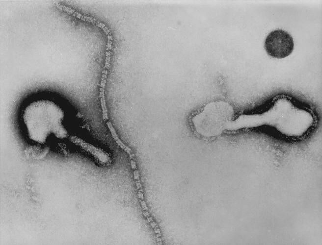
Human parainfluenza viruses are similar to other members of the family. Single-stranded, non-fragmented minus RNA of the virus encodes 7 proteins. The nucleocapsid is an internal antibody. The virus envelope contains glycoprotein spikes (HN and F). According to the antigenic properties of the HN, NP and F proteins, there are 4 main serotypes of parainfluenza viruses (HPHV-1, HPHV-2, HPHV-3, HPHV-4). HPHV-1, HPHV-2 and HPHV-3 have common antigens with the mumps virus. The hemagglutinin of viruses differs in the spectrum of action: HPGV-1 and HPGV-2 agglutinate different erythrocytes (human, chicken, guinea pig, etc.), parainfluenza virus-3 does not agglutinate chicken erythrocytes, parainfluenza virus-4 agglutinates only guinea pig erythrocytes.

Virus cultivation is carried out on primary cell cultures.
Resistance of parainfluenza virus
Human parainfluenza viruses do not differ in resistance from other members of the family.
Pathogenesis and symptoms of parainfluenza
The entry gate of infection is the upper respiratory tract. Parainfluenza viruses are adsorbed on the cells of the columnar epithelium of the mucous membrane of the upper respiratory tract, penetrate into them and multiply, destroying the cells. Edema of the mucous membrane of the larynx develops. The pathological process quickly descends to the lower parts of the respiratory tract. Viremia is short-term. Parainfluenza viruses cause secondary immunodeficiency, contributing to the development of bacterial complications.
After the incubation period (3-6 days), the temperature rises, weakness, runny nose, sore throat, hoarseness and dry, rough cough appear. The fever lasts from 1 to 14 days. HPGV-1 and HPGV-2 are a common cause of croup (acute laryngotracheobronchitis in children). Parainfluenza virus - 3 causes focal pneumonia. Parainfluenza virus - 4 is less aggressive. In adults, the disease usually occurs as laryngitis.
Immunity after the disease is due to the presence of serum IgG and secretory IgA, but it is fragile and short-lived. Reinfections caused by the same types of virus are possible.
Epidemiology of parainfluenza
The source of parainfluenza is a sick person, especially on the 2nd-3rd day of the disease. Infection occurs airborne. The main route of transmission of the virus is airborne. The contact-household route is also possible. The disease parainfluenza is characterized by its wide distribution and contagiousness. Most often, HPGV-1, HPGV-2 and HPGV-3 are isolated from patients.
 [ 9 ], [ 10 ], [ 11 ], [ 12 ], [ 13 ], [ 14 ], [ 15 ], [ 16 ]
Microbiological diagnostics of parainfluenza
Mucus or respiratory tract swabs and sputum are taken from the patient. Using RIF, viral antigens are detected in the epithelial cells of the nasopharynx. Parainfluenza virus is isolated on a Hep-2 cell culture. Indication is carried out according to the cytopathic effect of viruses, RGA and the hemadsorption reaction, which is most pronounced in parainfluenza viruses - 1, 2, 3 (they were previously called hemadsorbing). Identification is carried out using RTGA, RSK, RN. Using the serological method, using RTGA, RSK or RN, it is possible to detect both viral antigens and antibodies in paired sera of the patient.

