Medical expert of the article

New publications
Osteoblastoclastoma
Last reviewed: 29.06.2025

All iLive content is medically reviewed or fact checked to ensure as much factual accuracy as possible.
We have strict sourcing guidelines and only link to reputable media sites, academic research institutions and, whenever possible, medically peer reviewed studies. Note that the numbers in parentheses ([1], [2], etc.) are clickable links to these studies.
If you feel that any of our content is inaccurate, out-of-date, or otherwise questionable, please select it and press Ctrl + Enter.
Osteoblastoclastoma is a tumor process that can be either benign or malignant and damages different skeletal bones. At first, the pathology was called gigantocellular tumor (since 1912), 10 years later Dr. Stewart proposed the name osteoclastoma. And only in 1924, Professor Rusakov introduced the refined term "osteoblastoclastoma", which more fully corresponded to the cellular composition of the neoplasm.
Today, osteoblastoclastoma is considered a true neoplasm, a soft tissue tumor with an extensive vascular network. The only correct treatment option is removal of the tumor within healthy tissues, sometimes simultaneously with bone grafting. [1]
Epidemiology
The incidence of bone tumors worldwide ranges from 0.5 to 2%. According to United States statistics, osteosarcoma (about 34% of cases), chondrosarcoma (27%), and Ewing's tumor (18-19%) are the most common. Chordomas, fibrosarcomas, fibrosarcomas, histiocytomas, giant cell tumors, and angiosarcomas are less common.
The incidence rate is highly correlated with age. Thus, the first surge of tumor growth is detected in adolescence (around 16 years of age), and the second surge in middle age.
Osteoblastoclastoma is a relatively common tumor. It occurs in about 2-30% of all bone neoplasms. Women are more often affected, but men can also be affected, mainly between the ages of 18 and 40. Children under 12 years of age are rarely affected, but even in this age period the incidence is not excluded. There are descriptions of familial and hereditary cases of osteoblastoclastoma.
Most often (about 75%) the tumor is found in long tubular bones, much less often flat and small bones are affected.
In long tubular bones, the epimetaphysis is mainly affected, and in childhood the metaphysis is affected. The neoplasm does not sprout into the area of epiphyseal and articular cartilage. Very rarely the problem is found in the diaphysis (less than 0.5% of cases).
It is noted that with the development of medicine, the incidence of osteoblastoclastoma remains stable, but mortality rates have significantly decreased. The main and most probable cause of pathology is considered to be the impact of ionizing radiation. Thus, the risks are increased in people who have received high doses of radiation therapy, as well as in those patients who have been injected with radioisotopes (for diagnosis or therapeutic purposes). Other common etiologic factors include unfavorable ecology and heredity. [2]
Causes of the osteoblastoclastomas
Osteoblastoclastoma is a focus of pathologically altered cells that can appear in almost any part of the skeleton. Despite the abnormalities of structure, pathological cells continue to divide, as in healthy tissues. Their structure differs to a large extent from the norm, which entails the replacement of the properties of the directly affected bone and its typical function. Pathologically altered malignant cells acquire a propensity for uncontrolled, often rapid multiplication, as a result of which the tumor volume increases. Previously normal bone tissue can be displaced by the structures of the neoplasm, and individual pathological cells can be separated and transported with blood or lymph to other, distant anatomical zones. In this way, metastases are formed.
It is known that the source of malignant osteoblastoclastoma can be any malignant neoplasm located in any part of the body (including tumors of internal organs). The way of spread of the process is metastasis. But most osteoblastoclastomas (both benign and malignant) are primary neoplasms that appear and develop first and in the same place.
In general, osteoblastoclastomas are multifactorial tumors whose exact causes have not been established at this time. Conditions for neoplasm occurrence include such things as:
- An immunodeficiency state;
- Congenital tissue changes;
- Mutagenic environmental influences;
- Hormonal changes;
- Concomitant pathologies and injuries (trauma is often present in the anamnesis).
Risk factors
There is a lack of precise data regarding the causes of osteoblastoclastoma formation. However, experts suggest the involvement of a number of factors associated with an increased risk of bone oncopathologies:
- Heredity. In many cases, the tendency to tumor processes is genetically determined. In particular, this may be the case with Leigh Fraumeni syndrome, which predisposes to the development of various neoplasms, including cancerous tumors and sarcomas.
- Paget's disease. The disease can affect one or more bones and belongs to pre-tumor pathologies. In patients with this disorder, the bones thicken and become brittle at the same time, resulting in frequent pathologic fractures. Osteosarcomas occur in about 8% of cases of severe Paget's disease.
- Multiple bony overgrowths, exostoses.
- Multiple osteochondromas (including hereditary).
- Multiple enchondromas (risk is small but still present).
- Radiation exposure (including intense radiation used to treat other tumor processes and the effects of radioactive radium and strontium).
A special category of risk includes radiation treatment in childhood and young age, receiving doses over 60 Gray.
Experts draw attention to the fact that non-ionizing rays - in particular, microwave and electromagnetic radiation, which are formed from power lines, cell phones and household appliances - do not carry risks of osteoblastoclastoma.
Pathogenesis
The pathogenetic features of the appearance and development of osteoblastoclastoma are not fully understood, which is due to the complexity of the pathology. The basic cause of tumor formation is a failure in cell differentiation due to the improper functioning of the immune system. This gives rise to the growth of a tumor consisting of "wrong", undifferentiated cells, which determine the properties of the neoplasm and structurally resemble immature cells. If the cellular structure is close to normal, but is not, it is said to be a benign osteoblastoclastoma. With pronounced changes in the structure of cells, the tumor is attributed to malignant processes. For such a neoplasm, a change in the antigenic cellular fold, uncontrolled growth and cell division are typical. Together with the loss of specificity of the cellular structure, the functionality also suffers. Among other things, malignant osteoblastoclastoma differs from benign osteoblastoclastoma by the process of invasion into nearby healthy tissues. In benign bone neoplasm there is no sprouting into healthy structures, there is no tendency to rapid growth and spread throughout the body, there is no tendency to arbitrary self-destruction and intoxication by products of tumor decomposition.
The bone structure is destroyed in all cases, regardless of the benignity of the pathology. As a result, the affected bone segment becomes fragile, brittle. Often the reason for turning to doctors is a pathological fracture that occurs even under minimal load.
It is important to note: the benignity of the process is always a conditional state, because there are risks of malignization, and the benign focus is transformed, malignant osteoblastoclastoma occurs.
Symptoms of the osteoblastoclastomas
The clinical picture in osteoblastoclastoma mainly depends on the localization and stage of the pathological process. As a rule, the tumor is characterized by the following features:
- The neoplasm is solitary;
- Affects mainly the tubular bones of the lower or upper limbs;
- Is less commonly found in flat bones;
- There is a nagging pain in the affected segment;
- The skin and vascular pattern over the pathologic focus increases;
- The diseased limb is deformed (localized volume increase);
- The work of the joint nearest to the osteoblastoclastoma or the limb as a whole is disturbed;
- Palpatorily determined compacted focus with a characteristic "parchment crunch".
In general, symptoms can be divided into local and general symptoms. Local symptoms are detected visually - in particular, you can see the presence of curvature or bulging of the bone fragment. Attention is also drawn to the change in the skin over the pathological focus: a vascular pattern is clearly manifested, the tissues are swollen or flattened. The tumor can be palpated - often it is painless, but has a characteristic structure. Malignant tumors are typically lumpy and irregular in configuration.
The adjacent joint may be limited in movement, persistently painful. Due to compression of vessels and nerve trunks, sensitivity is often impaired, and persistent swelling appears. The lymphatic system also reacts: nearby lymph nodes become enlarged.
General symptomatology is more typical for malignant osteoblastoclastomas and is due to the processes of intoxication of the body. Patients may have:
- Fever, febrile conditions;
- Gauntness;
- Constant weakness;
- Drowsiness or insomnia, appetite disturbances;
- Nighttime excessive sweating;
- Collapse.
There is also a small percentage of osteoblastoclastomas, which are usually small and not clinically apparent. They become an incidental finding during radiologic or imaging studies for other reasons.
First signs of osteoblastoclastoma ossification
- Accelerating the growth of the neoplasm.
- Increased pain syndrome.
- Expansion of the destructive focus in diameter, or transformation of the cellular-trabecular form into a lytic form.
- Disintegration of the cortical layer over a relatively long area.
- Loss of clarity of configurations of the destructive focus.
- Disintegration of the closure plate that used to block the medullary canal.
- Periosteal reaction.
Osteoblastoclastoma malignancy is based on clinical and radiological indicators and is necessarily confirmed by morphological diagnosis of tumor tissues.
In addition to osloplasticization of an initially benign neoplasm, there is also a primary malignant osteoblastoclastoma. In fact, such a tumor is a type of sarcoma of osteogenic etiology.
The location of malignant osteoblastoclastoma is the same as in the benign process. Radiography reveals a destructive focus in the bone tissue without clear contours. The destruction of the cortical layer is extended, often sprouting into soft tissue structures is observed.
Signs to distinguish malignant osteoblastoclastoma from the osteogenic form of osteoclastic sarcoma:
- The predominantly elderly age of the patients;
- Less vivid symptomatology;
- A more favorable long-term prognosis.
Osteoblastoclastoma in children
Osteoblastoclastoma in childhood is rare: there are only two or three cases per one million children. It should be noted that among all pediatric patients, those older than 10-15 years of age predominate.
Scientists cannot name the exact cause of osteoblastoclastoma in children. Presumably, the pathology is associated with intensive growth of the child's body, as well as with a genetic factor.
There are also indications of such possible causes as radioactive exposure (in particular, radiation therapy), chemotherapy (taking cytostatics). Many chemotherapy drugs can destroy the genetic material of bone cells, which leads to the development of tumorigenesis.
In addition, the risk of osteoblastoclastoma is higher in children with certain congenital conditions, such as bilateral retinoblastoma or Li-Fraumeni syndrome. A causal link also exists with Paget's disease.
It is also known that in the vast majority of children (about 90%), doctors are unable to detect any of the risk factors mentioned above.
It is difficult to predict the course of osteoblastoclastoma in childhood, as it depends on the characteristics of a particular tumor, its localization, the degree of spread at the time of diagnosis, the timeliness of treatment and completeness of removal of the neoplasm.
The quality of osteoblastoclastoma treatment has made great progress in the last 2-3 decades. The therapeutic protocol has become combined and the cure rate has increased to more than 70-80%. A favorable outcome can be said if the tumor process is radically surgically removed and the effect is consolidated with a sufficient course of chemotherapy. Children with benign osteoblastoclastoma have the best chance of recovery.
When specific figures of cured patients are announced, we see only general figures: no statistics can accurately predict and determine the chances for a particular child. The term "recovery" is understood primarily as "absence of tumor process in the body", as modern therapeutic approaches are able to ensure long-term absence of recurrence. However, one should not forget about the possibility of undesirable side effects and late complications. Therefore, any treatment, regardless of its complexity, should flow into high-quality rehabilitation measures. In addition, children still need orthopedic care for a long time.
Forms
The classification of bone tissue neoplasms is quite broad. Attention is mainly paid to variations in cellular structure, morphological characteristics of the tumor process. Thus, tumors are divided into two categories:
- Osteogenic (formed on the basis of bone cells);
- Neosteogenic (formed in bone under the influence of other cell types - for example, vascular or connective tissue structures).
Osteoblastoclastoma of bone is predominantly a benign neoplasm. However, despite this, it often has aggressive growth, contributes to the destruction and thinning of bone tissues, which makes surgical intervention mandatory. At the same time, giant cell osteoblastoclastoma can also be malignant.
Depending on clinical and radiologic parameters and morphologic picture, three basic forms of osteoblastoclastomas are distinguished:
- The cellular form is found mainly in elderly people, it is characterized by slow development. Diagnosis reveals a thickened, lumpy swelling, without the possibility of clinical delineation of the tumor focus from healthy bone zones.
- Cystic form, first of all, manifests itself with pain. Palpatorily, the symptom of "parchment crunch" is determined. Visually, a bony tumor of smoothly convex, dome-shaped configuration is noted.
- The lytic form is considered a rare variant of pathology, it is detected mainly in adolescence. The tumor process develops quickly enough, the patient begins to be bothered by pain, including at palpation.
A giant cell tumor can form on almost any bone of the skeleton, although the tubular bones of the limbs, ribs, and spine are affected somewhat more often. Osteoblastoclastoma of the lower jaw occurs twice as often as on the upper jaw. Palpatorily, a dense neoplasm with softened zones is noted. The most common complaints of patients: the presence of a bulge that bleeds and creates discomfort when chewing food. As the problem progresses, it is complemented by impaired function of the temporomandibular joint. Among the tubular bones, the tumor more often affects the femur and tibia. Osteoblastoclastoma of the femur is found predominantly in middle-aged people. The disease is accompanied by impaired function of the corresponding joint, lameness occurs, and the skin over the neoplasm is covered with a pronounced vascular pattern.
In addition to the above classification, there are central and peripheral forms of pathology, although there are no morphological differences between them. Peripheral osteoblastoclastoma has a gingival localization, and the central form develops in the bone and is distinguished by the presence of multiple hemorrhages in it (therefore, the second name of the central osteoblastoclastoma is a brown tumor). The appearance of a brown color is due to the deposition of erythrocytes, which disintegrate with the formation of hemosiderin.
Malignant bone neoplasms go through the following stages in their development:
- A T1 foci measuring 3-5 cm is located within the bone and one musculofascial segment.
- The T2 foci extend no more than 10 cm along the course of the bone but do not extend beyond one fascial case.
- The T3 foci leave the confines of one musculofascial case and sprout into a nearby one.
- The T4 foci sprout from the skin or neurovascular trunks.
In a similar way, the degree of lymph node involvement and the spread of metastases are categorized.
Complications and consequences
Among the complications of osteoblastoclastoma is any increase in the activity of the neoplasm, which especially often occurs against the background of a long quiet period. In some such cases, we are talking about malignant degeneration of the tumor process, or its sprouting into sensitive nearby anatomical structures:
- Spread to the nerve trunk provokes the occurrence of neuropathic pain syndrome due to the effect on the large-caliber nerve. Such pain is practically not eliminated after taking conventional analgesics, so it literally exhausts the patient.
- Spread to the blood vessels can be complicated by sudden massive bleeding and hematoma formation.
Complications are not excluded, which are accompanied by a violation of the function of nearby articulations: the growth of osteoblastoclastoma in such a situation blocks the adequate functioning of the musculoskeletal mechanism, which leads to a limited range of motion and the appearance of pain syndrome.
The most common complications of osteoblastoclastoma are considered pathological fractures in the affected area. The problem occurs even with a minor traumatic impact, as the bone tissue becomes extremely fragile and unstable.
In addition, specialists also talk about specific general and local adverse effects characteristic of malignant osteoblastoclastoma:
- The formation of distant and near metastases;
- Intoxication of the body with decay products.
If metastases are detected some time after the initial diagnostic measures, it indicates the ineffectiveness of ongoing treatment and progression of the neoplasm.
A separate line of complications is the emergence of new tumor or general pathology due to chemotherapy or irradiation of the osteoblastoclastoma bone focus.
Diagnostics of the osteoblastoclastomas
Diagnostic methods used to detect osteoblastoclastoma include:
- Clinical, which includes external examination and palpation of the pathologically altered area;
- X-ray (anteroposterior and lateral radiography, if indicated - targeted and oblique radiography);
- Tomographic (using computerized or magnetic resonance imaging);
- Radioisotope;
- Morphological, which includes histologic, histochemical, cytologic analysis of biomaterial obtained during puncture or trepanobiopsy;
- Laboratory.
The doctor carefully studies the history of the disease, determines the first signs, specifies the location and type of pain syndrome, its features, takes into account the results of previous examinations and treatment procedures, assesses the dynamics of the patient's general condition. If the pathology of long tubular bones is suspected, the specialist pays attention to the presence of swelling, motor restriction in the closer articulation, as well as the presence of neurological symptoms, muscle weakness and hypotrophy. It is important to carefully examine internal organs for possible spread of metastases to them.
All patients take general blood and urine tests with determination of protein and protein fractions, phosphorus and calcium, sialic acids. It is also necessary to determine the enzymatic activity of phosphatases, conduct a definil test, study the index of C-reactive protein. If it is necessary to differentiate osteoblastoclastoma from myloma, the patient passes a urine test for the presence of pathologic Bence-Jones protein.
Radiological diagnosis is fundamental for the diagnosis of osteoblastoclastoma. Obligatory appointed review and targeted X-ray, high-quality tomography, allowing to clarify the location, type of pathological focus, its spread to other tissues and organs. Thanks to CT, it is possible to clarify the state of soft tissue and the thinnest bone structures in the necessary plane, to identify deep foci of pathological destruction, to describe their parameters within the bone limits, to determine the degree of damage to the surrounding tissues.
At the same time, MRI is considered the most informative diagnostic procedure, which has a number of advantages over both radiography and CT. The method allows you to examine even the thinnest tissue layers, form a picture of pathological chag using a spatial three-dimensional image.
Mandatory instrumental diagnostics is represented by morphologic studies. Biomaterial is evaluated, which is obtained during aspiration and trepanobiopsy, or during resection of bone segments together with the neoplasm. Puncture biopsy is performed using special needles and radiologic control.
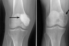
The following x-ray signs are considered typical of osteoblastoclastoma:
- Porosity limitation;
- Homogeneity of bone lysis in the type of thin trabeculization;
- The presence of pseudocystic lucencies that have the structure of peculiar "soap bubbles".
This radiologic picture is accompanied by the absence of primary or secondary reactive osteoformative periostosis. Thinning and atrophy of the cortical layer is detected.
Malignant type of osteoblastoclastoma as a result of intensive vascular sprouting entails an increase in venous stasis. Vascular changes have the appearance of a neoplasm with abundant vascularization.
Differential diagnosis
It is sometimes very difficult to identify osteoblastoclastoma. Problems arise during differential diagnosis of the disease with osteogenic sarcoma and bone cysts in patients of different ages. According to statistics, in more than 3% of cases osteoblastoclastoma was mistaken for osteogenic sarcoma, and in almost 14% of cases - for bone cyst.
The table summarizes the main signs of these pathologies:
|
Indicators |
Osteoblastoclastoma |
Osteogenic osteoplastic sarcoma |
Bone cyst |
|
Most common age of incidence |
20 to 30 years old |
20 to 26 years old |
Children under 14 years of age |
|
Location |
Epimetaphyseal region |
Epimetaphyseal region |
Metadiaphysis area |
|
Bone reconfiguration |
Severe asymmetrical bulge. |
Small transverse expansion |
A spindle-shaped bulge. |
|
Configuration of the destructive focus |
The contours are clear |
The contours are blurry, there's no clarity |
The contours are clear |
|
The condition of the spinal canal |
Covered by a closure plate |
Open at the border with the neoplasm |
No change. |
|
Condition of the cortical layer |
Thin, fibrous, discontinuous. |
Thinning, ruined |
Thin, flat |
|
Sclerosis phenomena |
Atypical |
Present |
Atypical |
|
Periosteal reaction |
Absent |
Present in a "periosteal visor" type of way |
Absent |
|
The condition of the epiphysis |
The lamina is thin, wavy. |
In the initial stage, part of the epiphysis remains intact |
No change. |
|
Nearby bone section |
No change. |
Signs of osteoporosis |
No change. |
Obligatory attention requires such indicators as the age of the patient, the duration of pathology, the location of the affected focus, other anamnestic information indicated in the table.
The following diagnostic errors are the most common, when osteoblastoclastoma is confused with such pathologic processes:
- Aneurysmal cyst (localized in the diaphysis or metaphysis of long tubular bones);
- Monoaxial type of fibrous osteodysplasia (manifested mainly in childhood, accompanied by bone curvature without bone ballooning);
- Hyperparathyroid osteodystrophy (no clear delineation of the focus from the healthy bone area, no clear bone bulge);
- Solitary cancerous bone metastasis (characterized by destructive foci with curved "eaten" contours).
It is important to keep in mind that benign osteoblastoclastoma can always transform and become malignant. The causes of malignancy have not yet been precisely determined, but scientists believe that trauma and hormonal changes (e.g., during pregnancy) contribute to it. According to some observations, malignization has also occurred with repeated series of remote radiation treatments.
Symptoms of ossification:
- The neoplasm starts to grow rapidly;
- The pain's getting worse;
- The size of the destructive focus increases, and the cellular-trabecular phase transitions to the lytic phase;
- The cortical layer is breaking down;
- The contours of the destructive focus become indistinct;
- The locking plate is collapsing;
- There's a periosteal reaction.
In the process of differentiation of primary malignant neoplasm (osteogenic osteoclastic sarcoma) and malignant osteoblastoclastoma, special attention is paid to the duration of pathology, assessment of the radiological picture in dynamics. On the X-ray image of the primary malignant neoplasm there is no bone protrusion typical for osteoblastoclastoma, there are no bone bridges, sclerosed area with indistinct contours can be detected. In malignization, however, there is often a small area of the closure plate, which used to serve as a barrier to the healthy bone segment.
Who to contact?
Treatment of the osteoblastoclastomas
The only correct treatment for patients with osteoblastoclastoma is surgery. The most gentle intervention takes place at the initial stages of tumor process development and represents excision of the affected tissues with further filling of the cavity with a graft. The graft is taken from another healthy bone of the patient. Such intervention is the most favorable and less traumatic, however, in some cases it is also less radical. Excision of the affected bone fragment together with the neoplasm is considered a more reliable method, which reduces the probability of tumor re-growth to a minimum.
If it is a neglected osteoblastoclastoma of large size, especially prone to malignization or already malignant, partial or complete amputation of the limb is often considered.
In general, the tactics of surgical treatment for osteoblastoclastoma is selected depending on the location, spread and aggressiveness of the pathologic focus.
If the tumor affects the long tubular bones, then it is recommended to pay attention to these types of surgical interventions:
- Edge resection with alloplasty or autoplasty for benign, delayed process, foci with a cellular structure and in the periphery of the epimetaphysis. Fixation with metal screws.
- When cellular osteoblastoclastoma spreads to the middle of the bone diameter, two thirds of the condyle, partially of the diaphysis and the articular surface are resected. The defect is filled with articular cartilage allograft. It is fixed firmly with tie bolts and screws.
- In the case of epimetaphysis decay along the entire length or pathological fracture, such tactics as segmental resection with articular excision and filling the defect with allograft are used. It is fixed with a cemented rod.
- In case of pathological fracture and malignization of osteoblastoclastoma in the proximal femur region, total hip arthroplasty is performed.
- In case of resection of the ends in the joint zone of the knee, the technique of allopolysubstance transplantation with fixation is used. Total endoprosthesis with an extended titanium stem is often preferred to ensure subsequent radiation treatment.
- If the pathologic focus is located at the distal end of the tibia, resection with bone-plastic ankle arthrodesis is performed. If the talus bone is affected, it is extirpated with extension arthrodesis.
- In cervical spine lesions, an anterior access to the C1And C2Vertebrae is performed. An anterolateral access is preferred. At the Th1-Th2Level, an anterior access with oblique sternotomy to the third intercostal space is used (vessels are carefully shifted downward). If the tumor affects the bodies of 3-5 thoracic vertebrae, an anterolateral access with resection of the third rib is performed. The scapula is shifted backwards without cutting off the musculature. If the osteoblastoclastoma is found in the thoracolumbar region between Th11And L2, the operation of choice is right-sided thoracofrenolumbotomy. Access to the anterior part of the upper 3 vertebrae of the sacrum is more difficult. An anterolateral retroperitoneal right-sided access with careful drainage of the vascular trunks and ureter is recommended.
- If the vertebral bodies are destroyed severely, or the pathology has spread to the arch region in the thoracic and lumbosacral spine, then in this case, transpedicular-translaminar fixation of the spine is performed, after which the destroyed vertebrae are removed with further autoplasty.
- If a benign form of osteoblastoclastoma is detected in the brow and sciatic bone, the pathologically altered segment is removed within healthy tissues, without bone grafting. If the floor and roof of the acetabulum are affected, resection is performed with further bone grafting to replace the defect, with fixation with spongiosis fasteners.
- If the iliac, bosom or sciatic bone is affected, alloplasty with a structural allograft, transplant osteosynthesis, cement-based plastic insertion, and repositioning of the prosthetic head into an artificial cavity are performed.
- If the sacrum and L2AreAffected, a two-stage intervention is performed, including posterior access resection of the pathologically altered lower sacral fragment (up to S2), transpedicular fixation and removal of the neoplasm from the anterior side by retroperitoneal method with bone grafting.
In each specific situation, the doctor determines the most appropriate method of surgical intervention, including considering the possibility of applying the latest technology to improve the results of treatment and ensure the normal quality of life of the patient.
Prevention
There is no specific prevention of osteoblastoclastoma. First of all, this is due to the insufficient study of the causes of the development of such tumors. Many experts emphasize the prevention of trauma to the bone system among the main preventive points. However, there is no evidence of the direct influence of trauma on the formation of bone neoplasms, and trauma in many cases only draws attention to the existing tumor process and has no obvious significance in the origin of the pathological focus, but at the same time, it can contribute to its growth.
It should not be forgotten that osteoblastoclastoma often forms in bones that have previously been exposed to ionizing radiation - for example, for the purpose of therapy of other tumor processes. Radioinduced neoplasms usually occur no earlier than 3 years after the radiation exposure.
Non-specific preventive measures include:
- Elimination of bad habits;
- Leading a healthy lifestyle;
- Quality and sustainable nutrition;
- Moderate regular physical activity;
- Prevention of injuries, timely treatment of any pathological processes in the body, stabilization of immunity.
Forecast
Pathologic fractures often occur in the affected area of bone tissue. In this case, benign neoplasms, provided that a radical method of treatment is used, have a favorable prognosis, although the possibility of recurrences and malignancy of the pathological focus is not excluded. An unfavorable outcome of benign osteoblastoclastoma is not excluded if the focus is characterized by active growth and pronounced bone destruction. Such a tumor can quickly destroy an entire bone segment, which entails the development of a pathological fracture and significant impairment of bone function. Such patients often have problems with surgical replacement of the bone tissue defect, and complications associated with non-healing of the fracture develop.
The average five-year survival rate for all variants of malignant osteoblastoclastomas in both children and adults is 70%, which can be considered quite good. Hence, we can conclude that in many cases such neoplasms are cured quite successfully. Of course, such points as the type of tumor process, its stage, the degree of lesion and malignancy are also of great importance.
Obviously, it is malignant osteoblastoclastoma that poses the greatest threat. In this situation, we can speak about a favorable prognosis only in cases of early detection, accessible surgical localization, sensitivity of the focus to chemopreventive agents and radiation therapy.