Medical expert of the article

New publications
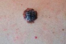
Nodular melanoma: what it looks like, prognosis
Last reviewed: 04.07.2025

All iLive content is medically reviewed or fact checked to ensure as much factual accuracy as possible.
We have strict sourcing guidelines and only link to reputable media sites, academic research institutions and, whenever possible, medically peer reviewed studies. Note that the numbers in parentheses ([1], [2], etc.) are clickable links to these studies.
If you feel that any of our content is inaccurate, out-of-date, or otherwise questionable, please select it and press Ctrl + Enter.
Today, various neoplasms affecting the skin are increasingly common. At the same time, approximately 4-10% of them are malignant tumors. They affect people of both sexes with equal frequency. In most cases, the tumor does not form spontaneously. Its formation is preceded by a number of conditions, and it is formed gradually, passing through a number of stages. The cancerous process develops evenly, creating an unfavorable background. In the presence of certain factors, it turns into an independent disease - cancer. Nodular melanoma is one of such tumors. A characteristic feature of this tumor is the ability to grow unlimitedly
Epidemiology
As statistics show, melanoma is detected approximately 2-3 times more often in mature individuals, which can be explained by the fact that immunity is significantly reduced and loses the ability to eliminate genetically foreign material, which transformed cells become for the body. The risk of genetic mutations also increases significantly, as a result of which the normal functioning of the gene responsible for cell apoptosis may be disrupted. A certain contribution is made by changes in hormonal levels, increased viral load, an increase in the number of carcinogenic, potentially oncogenic viruses, which trigger the processes of cell degeneration.
Most often, melanoma affects fair-haired women, as well as people with light skin and blue eyes. There is no exact explanation for this phenomenon, but it may be due to genetic predisposition and greater susceptibility of light skin to solar radiation and other types of radiation. Light skin is more susceptible to damage and is least protected from the effects of external environmental factors.
Causes nodular melanoma
To date, the causes of nodular melanoma have not yet been established. Presumably, melanoma develops from an ordinary mole (nevus), which undergoes malignant transformations. A number of factors can trigger the process of malignancy, including trauma, mechanical, chemical damage. Self-medication, cauterization, cuts, rupture of spots can lead to the degeneration of any growths into a malignant neoplasm. This also includes excessive insolation, exposure to a number of irritants, hormonal disorders, and a decrease in the immune system.
Risk factors
The risk group includes people who are exposed to adverse factors, such as physical and mechanical impact on the skin, the influence of toxic vapors, work with chemicals, reagents, vapors settling on the skin. Risk factors also include prolonged exposure to sunlight, exposure to various types of radiation (X-ray, ultraviolet, infrared radiation). Some chemicals and even low-quality cosmetics can lead to malignancy (malignant transformation of cells). This also includes people with reduced immunity, various hormonal imbalances, hidden pathologies, chronic diseases. An important factor is genetic predisposition.
Pathogenesis
Pathogenesis is based on malignant degeneration of cells. They undergo malignization – transformation. As the results of recent studies show, cell death is determined by genes that program apoptosis – cell death. In malignant tumors, the cell loses the ability to die, and, in fact, is a cell that has achieved immortality. Thus, a characteristic feature of a malignant tumor is unlimited growth.
 [ 22 ], [ 23 ], [ 24 ], [ 25 ], [ 26 ], [ 27 ], [ 28 ], [ 29 ], [ 30 ]
Symptoms nodular melanoma
The main symptom is skin tumors of various sizes that begin to grow rapidly. At the initial stages, the size ranges from a pinhead to the size of a large coin. They are mainly localized on the upper layer of the skin - the epidermis. But some are also found in other layers - the dermis, subcutaneous tissue (keratoma, dermatoepithelioma). They can be flat or elevated. But a characteristic feature and an unfavorable prognostic sign is the moment when they begin to grow and multiply quite quickly. Often, growths are the only form of manifestation of this disease. As the condition progresses, regional lymph nodes are affected, then internal organs (metastases are formed).
The first sign of melanoma is the formation of moles (nevi), their sharp increase in size, as well as multiple manifestations. Also, concern should be caused by the fact that the tumor spreads and affects other areas. In particular, pain and swelling of the lymph nodes is an unfavorable prognostic factor that may indicate the development of a malignant process.
Nodular amelanotic melanoma on the eyelid
The appearance of nodular amelanotic melanoma on the eyelid is most often associated with the dissemination of the primary lesion. It is usually quite easy to recognize visually, but to confirm the diagnosis, it may be necessary to use a radiometric method that accurately recognizes the malignancy of the process.
Nodular melanoma of the skin
To identify a pathological process and establish a differential diagnosis, cytological studies are performed. But there is one nuance - cytology can only be performed if there is an ulcer on the skin surface, or an affected surface from which a smear-print is taken. Subsequently, the features of the structure and growth of cells are studied.
How fast does melanoma grow?
It is impossible to give a clear answer to the question of how fast melanoma will grow. This process is individual for everyone, since it depends on a number of factors, including genetic characteristics, the person's immunological status, viral and bacterial load, hormonal background, and the person's age. Histological studies, in which a piece of tissue (melanoma) is taken and seeded on nutrient media, will help answer this question. The nature and rate of cell growth, and then the tissues on the medium, can be used to predict the growth rate.
Stages
There are three stages of melanoma growth. At the initial stage, skin malignancy occurs, that is, cells degenerate, transform and give rise to malignant growth. At this stage, treatment will be most effective. This manifests itself as the initial stage of growth, when the mole acquires an increased size, begins to grow and multiply.
At the second stage, there is a progressive rapid growth of the tumor. It increases sharply in size. At these stages, the condition can deteriorate sharply. The size of the tumor increases, an increase in lymph nodes is also observed, their pain appears. It is worth noting that at this stage, treatment can be effective, but action is needed.
The third stage is the most severe, advanced stage.
At this stage, a person usually experiences pain, the condition noticeably worsens. Cancer is reflected in the biochemical parameters of the blood. The prognosis is serious. A fatal outcome is not excluded.
At the initial stage of melanoma development, it is a standard birthmark (nevus), which gradually increases in size. But pathological processes are already occurring in it. In particular, cells undergo malignization and undergo malignant transformations.
The most well-known scale used to determine the severity of a condition is the Clark scale, which distinguishes 3 degrees of severity of the pathological process.
Forms
There are several types of melanoma, depending on the classification features. Thus, nodular melanoma can be pigmented and non-pigmented. Separately, there is a horizontal form of melanoma, as well as an epithelial cell form.
- Nodular amelanotic melanoma
It occurs in approximately 30% of patients with malignant skin neoplasms. It is quite common in patients with AIDS and other immunodeficiency conditions. It is worth noting that decreased immunity and hormonal imbalance are the main predisposing factors that contribute to the development of a malignant process.
- Nodular pigmented melanoma
Nodular pigment melanoma is a malignant process in which malignant degeneration of cells occurs. The pathological process involves melanocytes synthesizing pigment. If the function of melanocytes is not impaired and they do not stop synthesizing pigments, melanoma retains pigmentation.
- Horizontal nodular melanoma
The distinctive feature of the horizontal form of nodular melanoma is that it spreads quite quickly and has a tendency to expand.
- Nodular amelanotic epithelial cell melanoma
First of all, melanocytes are involved in the pathological process - cells that normally produce the pigment melanin. When the function of melanocytes is impaired, they stop synthesizing pigment, which contributes to the development of nodular apigmented melanoma.
Complications and consequences
First of all, it is worth noting such complications as the formation of metastases, relapses, and death.
- Relapse
When nodular melanoma is surgically removed, a relapse may develop after some time.
- Ulcerations
Melanoma has the ability to spread (disseminate): first to neighboring areas in the form of satellite nodules, then to regional lymph nodes, and in later periods it metastasizes to internal organs. Early ulceration of nodular melanoma is considered an unfavorable prognostic factor. The malignancy of the process increases sharply with tumor trauma.
Diagnostics nodular melanoma
Differential diagnostics are important, as they allow one to distinguish one type of wart from another, as well as to identify the exact species and generic name of the virus that caused the development of the pathology.
[ 42 ], [ 43 ], [ 44 ], [ 45 ]
Tests
The main method of confirming the diagnosis is to confirm the presence of malignant degeneration (malignancy), which can only be achieved after passing the appropriate tests. The only accurate method is histological examination, the essence of which is that a piece of tissue (biopsy) is taken for analysis. Then, a seed is made on special nutrient media, and the nature of the growth is used to determine whether the tumor is benign or malignant. Also, an analysis for tumor markers is a direct confirmation of the presence or absence of a malignant neoplasm. There are a number of factors in human blood that appear only if a cancerous tumor develops in the body, and which are not normally diagnosed. The nature and quantity of these markers are used to judge the localization, severity of the tumor, stage, etc. A biochemical blood test can also bring some clarity to the diagnosis.
Standard clinical methods are uninformative, however, they are used because they can show the general picture of the pathology. Based on the results, one can indirectly judge the nature of the neoplasm (the malignant process is reflected in the blood parameters).
If a viral infection is suspected, serological and virological methods of research are used. Also often used are methods such as DNA probing, hybridization, genome sequencing, PCR analysis. These methods allow to detect not only the virus itself in the blood, but also the products of its vital activity and even DNA, or its individual fragments.
Additional methods may include microscopic examination and scraping. Microscopy will help to identify the virus itself or its waste products in a smear. In case of tumor ulceration (melanoma), a smear is taken from its surface. Cytological examination is important, allowing to examine cells and identify transformed cells, characteristic of a malignant tumor.
[ 46 ], [ 47 ], [ 48 ], [ 49 ]
Instrumental diagnostics
The essence of instrumental diagnostics is that the study is carried out using special equipment, tools and devices. They allow visualizing the picture of pathology, identifying structural and functional changes, predicting their consequences, the rate of progression.
Differential diagnosis
Differential diagnostics is one of the main stages of making a final diagnosis. It allows differentiating the signs of several pathologies that have similar external manifestations. Most often, it is necessary to differentiate malignant tumors from benign ones, since they are very similar in appearance, but differ in many parameters detected during the study. In particular, histological examination is one of such methods of making a differential diagnosis. During this analysis, the characteristics of the tumor are determined by the nature of growth. It is often necessary to differentiate various types and forms of warts, nevi, melanomas, keratomas, for example, from papillomas, fibromas, tumors, traumatic scars and other pathologies. It is also important to determine what exactly is the cause of malignant degeneration. This will prevent relapse in the future, and will also prevent metastasis. For example, if the cause was a virus, then it is necessary to accurately determine the species and generic name of this virus, and conduct appropriate treatment against this virus.
Lentiginous melanoma
They occur several years after the start of chemotherapy for patients with psoriasis. First, lentiginous spots appear, then their malignant transformation occurs, melanomas are formed. It is considered a complication of chemotherapy, developing against the background of reduced immunity.
Acral lentiginous melanoma
It is a tumor that is formed as a result of malignant transformation of lentiginous spots. There can be many reasons for such transformation, but first of all, it is trauma to an existing skin growth. In second place is a viral infection (carcinogenic viruses), in third place is decreased immunity. Often these reasons act in combination.
There are many viruses that can trigger the development of melanoma. Basically, the triggers (starting mechanisms) are HPV (type 16, 33, 58), herpes virus, chickenpox, cytomegalovirus, retroviruses. A special role is given to HIV infection. Malignant degeneration of skin neoplasms is observed in 56% of AIDS patients. This occurs against the background of severe immunodeficiency and the inability of the body to resist foreign agents. There are forms that promote the transition of a flat wart or nevus from a passive, flat state to a hanging position and trigger further growth. With age, growths, papillomas and hanging warts on the skin may appear, with a tendency to grow.
Often the cause is a decrease in immunity. Normally, the immune system suppresses the activity of viruses and destroys all foreign agents, including its own cells that have undergone malignant degeneration. With reduced immunity, this does not happen. This is also due to the persistence of viruses that are activated against the background of reduced immunity. Immunity decreases sharply after an illness, in the postoperative period, during pregnancy, during menopause, in adolescence, after a course of antibiotic therapy, with AIDS. Often, increased growth and a change in the shape of growths occurs during pregnancy, or some time after childbirth, which is also associated with a decrease in immunity. A similar phenomenon is observed during menopause, against the background of some diseases of the immune and endocrine systems, with metabolic disorders, biochemistry.
[ 50 ]
Basalioma
It is a tumor of the basal layer of the epidermis. It develops under certain conditions (predisposing factors): decreased immunity, increased reactivity and sensitization, weakened body, disruption of the biochemical and menstrual cycle, hormonal background.
[ 51 ], [ 52 ], [ 53 ], [ 54 ], [ 55 ]
Warts
Warts can be localized on any part of the body. They are represented by connective tissue. They are covered with multilayered epithelium on top. As a rule, they are flat at first, but over time they can grow, become hanging, multiple. They are formed everywhere. In fact, there is no such area where a wart could not form. They are formed even on mucous membranes.
The armpit area is an area that is quite prone to the formation of hanging warts (the skin is thin, a fairly large number of sweat glands are formed in its surface layer, there is almost always high humidity due to abundant sweating).
Another place where warts often form is the groin area. Often in the groin area, hanging warts associated with a viral infection that is sexually transmitted are formed. They are transmitted during sexual contact. These warts are potentially oncogenic, that is, they provoke the development of malignant neoplasms under certain conditions.
At first glance, it may seem that warts are not dangerous, but simply unattractive in appearance, spoil the aesthetic appearance. But this is only part of the consequences, so to speak, the "tip of the iceberg." One of the most dangerous consequences is the possibility of malignant degeneration of the wart and the risk of tumor development. Tumors located on internal organs are especially dangerous: they can be damaged, cause bleeding. Also, a great danger develops during pregnancy, since warts can lead to pregnancy pathologies. There is an increased risk of infection of the child during childbirth.
Various methods are used to remove hanging warts. These can be both traditional medicinal and radical methods. Radical methods include surgical excision of the wart. Such methods are the most effective.
Dysplastic nevus
A nevus is a common birthmark (pigmented spot). They can be congenital or acquired. A dysplastic nevus is a growing, malignantly transformed spot. This is facilitated by a decrease in immunity, as well as hormonal changes, viral infections, and dysbacteriosis.
[ 56 ], [ 57 ], [ 58 ], [ 59 ], [ 60 ], [ 61 ], [ 62 ]
Keratoma
This is a malignant tumor localized in the deep layers of the skin. Most often it develops in people with reduced immunity, with senile, age-related changes in the body. The risk group includes people who are often ill, those who are in contact with people suffering from warts, pigment spots. People who are subject to frequent stress, chronic diseases, malnutrition, with a disrupted work and rest regime are more at risk.
Angiokeratoma
They are vascular tumors localized in epithelial tissue. They are formed mainly on the neck. They can rise quite strongly above the surface (on a stalk).
[ 63 ], [ 64 ], [ 65 ], [ 66 ], [ 67 ], [ 68 ], [ 69 ]
Dermatofibroma
It is a benign skin tumor with a high risk of malignant transformation. People with such a diagnosis should constantly monitor their condition, be observed by an oncologist (to prevent malignant transformation of the tumor). The risk group includes people who are carriers of carcinogenic viruses. These are, first of all, the herpes virus, papillomas, retroviruses, and others. This also includes people with reduced immunity, with disrupted or altered hormonal background, frequently ill people, patients with chronic pathologies, immunodeficiencies, AIDS. There are certain stages of life, during which the risk of malignant transformation increases sharply - adolescence, adolescence, pregnancy, lactation, menopause, old age. Elderly people are especially at risk, since their body often has disrupted metabolic processes and hormonal background.
[ 70 ], [ 71 ], [ 72 ], [ 73 ], [ 74 ]
Lentigo
This is a malignantly degenerated pigment spot. It requires removal. Medication is rarely used. But it is often ineffective. High immunity will help improve the condition and prevent malignant degeneration. For this, it is recommended to take immunostimulants, vitamins. You should regularly consult with an immunologist, oncologist. You can also try some folk remedies that have immunostimulant, antiviral properties.
Recipe No. 1.
Add a tablespoon of dandelion roots, orchis tuber roots, greater celandine herb, comfrey roots, and parsnip herb to regular alcohol (500 ml). Drink a tablespoon twice a day.
Recipe No. 2.
To prepare, take a tablespoon of peony roots, buds and needles of Siberian fir, club moss, blueberry leaves, bird cherry flowers and fruits. Infuse all this for at least 3-4 days, drink a tablespoon 2-3 times a day.
Recipe No. 3.
Take equal parts of plantain leaves, wormwood leaves, male fern rhizomes, three-part Bidens herb, horseradish roots, and pour 500 ml of alcohol over them. Drink a third of a glass per day.
Recipe No. 4.
Vodka or pure alcohol is used as a base. Then about a tablespoon of the following components is added: motherwort herb, chamomile baskets, pine needles, marsh cudweed herb, creeping thyme herb. Mix, then set aside and allow to brew. Drink a tablespoon 3-4 times a day.
Recipe No. 5.
Add a tablespoon of wild pansy and horsetail to regular alcohol (500 ml). Drink a tablespoon twice a day.
Who to contact?
Treatment nodular melanoma
You should not attempt to remove melanoma on your own, as this may result in dissemination and development of a malignant process (metastasis). In addition, special equipment and tools are required, sterile conditions must be created, and it is important to know the exact technique of removal. Incorrect removal, damage can lead to multiple metastases, including in internal organs. It is worth noting that incomplete excision of tissue is no less dangerous, as new tumors then develop from it, and metastases occur.
In drug therapy, mainly drugs for internal use are used (antibiotics, antitumor, antiviral agents, immunomodulators). Various antitumor ointments for local use have proven themselves quite well, but they have serious side effects.
Radical methods include surgical excision, laser removal, and cauterization using various methods.
Surgical treatment
Surgical treatment is resorted to if the neoplasm begins to grow, and the analysis confirms a malignant process. It is necessary that the operation be performed by an experienced oncologist, since the tumor must not be damaged in any way, and it is also impossible for even a small piece of tissue to remain. Otherwise, metastases will begin to form, and relapses will begin. First, metastases affect the nearest lymph nodes, and then they can move to the internal organs. Removal is mandatory if the neoplasm reaches a sufficiently large size, or if it is located in the lumen of internal organs, and there is a risk of their blockage. The main method is mechanical excision. Laser removal and cryodestruction are also used.
Prevention
Prevention is based primarily on increasing immunity, eliminating the viral load, and normalizing the microflora. It is important to monitor the condition of all skin neoplasms - nevi, moles, warts, papillomas. You should periodically consult an immunologist and oncologist. If necessary, you should take tests for viruses, latent infections, and tumor markers. At the slightest suspicion of malignant degeneration of a neoplasm, it is necessary to conduct a histological examination of the sample, which will help determine the nature of the tumor. Make a prognosis, and select adequate treatment. A prerequisite is proper nutrition, vitaminization of the body, and a healthy lifestyle. You should follow a daily routine, avoid hypothermia, and avoid stress. You should not allow injury or damage to skin growths or spots. You should avoid exposure to ultraviolet light, chemical reagents, and radiation.
Forecast
In most cases, with proper and timely treatment, the prognosis will be favorable. Nodular melanoma is successfully removed by surgical methods. If everything is done correctly, in a timely manner, and in the future all the doctor's recommendations are followed, and an oncologist is observed, everything will end well. Otherwise, relapses, metastases may occur, and everything will end in death.