Medical expert of the article
New publications
Neuron
Last reviewed: 04.07.2025

All iLive content is medically reviewed or fact checked to ensure as much factual accuracy as possible.
We have strict sourcing guidelines and only link to reputable media sites, academic research institutions and, whenever possible, medically peer reviewed studies. Note that the numbers in parentheses ([1], [2], etc.) are clickable links to these studies.
If you feel that any of our content is inaccurate, out-of-date, or otherwise questionable, please select it and press Ctrl + Enter.
A neuron is a morphologically and functionally independent unit. With the help of processes (axon and dendrites) it makes contacts with other neurons, forming reflex arcs - links from which the nervous system is built.
Depending on the functions in the reflex arc, a distinction is made between afferent (sensory), associative and efferent (effector) neurons. Afferent neurons perceive impulses, efferent neurons transmit them to the tissues of the working organs, prompting them to act, and associative neurons provide interneuronal connections. The reflex arc is a chain of neurons connected to each other by synapses and providing for the conduction of a nerve impulse from the receptor of a sensory neuron to the efferent ending in the working organ.
Neurons are distinguished by a great variety of shapes and sizes. The diameter of the bodies of granular cells of the cerebellar cortex is about 10 µm, and giant pyramidal neurons of the motor zone of the cerebral cortex are 130-150 µm.
The main difference between nerve cells and other cells in the body is that they have a long axon and several shorter dendrites. The terms "dendrite" and "axon" are used to refer to the processes on which the incoming fibers form contacts that receive information about excitation or inhibition. The long process of the cell, along which the impulse is transmitted from the cell body and forms contact with the target cell, is called the axon.
The axon and its collaterals branch into several branches called telodendrons, the latter ending in terminal thickenings. The axon contains mitochondria, neurotubules and neurofilaments, as well as agranular endoplasmic reticulum.
The three-dimensional area in which the dendrites of a single neuron branch is called the dendritic field. Dendrites are true protrusions of the cell body. They contain the same organelles as the cell body: chromophilic substance (granular endoplasmic reticulum and polysomes), mitochondria, a large number of microtubules (neurotubules) and neurofilaments. Due to the dendrites, the receptor surface of a neuron increases by 1000 times or more. Thus, the dendrites of pear-shaped neurons (Purkinje cells) of the cerebellar cortex increase the receptor surface area from 250 to 27,000 μm2; up to 200,000 synaptic endings are found on the surface of these cells.

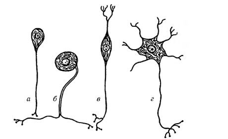
Types of nerve cells: a - unipolar neuron; b - pseudounipolar neuron; c - bipolar neuron; d - multipolar neuron
Neuron structure
Not all neurons conform to the simple cell structure shown in the figure. Some neurons lack axons. Others have cells whose dendrites can conduct impulses and form connections with target cells. The retinal ganglion cell conforms to the standard neuron diagram with dendrites, a cell body, and an axon, while photoreceptor cells have no obvious dendrites or axon because they are activated not by other neurons but by external stimuli (light quanta).
The neuron body contains a nucleus and other intracellular organelles common to all cells. The vast majority of human neurons have one nucleus, usually located in the center, less often eccentrically. Binuclear and especially multinuclear neurons are extremely rare. The exception is the neurons of some ganglia of the autonomic nervous system. The nuclei of neurons are rounded. In accordance with the high metabolic activity of neurons, the chromatin in their nuclei is dispersed. The nucleus contains one, sometimes two or three large nucleoli. Increased functional activity of neurons is usually accompanied by an increase in the volume (and number) of nucleoli.
The plasma membrane of a neuron has the ability to generate and conduct an impulse; its structural components are proteins that function as selective ion channels, as well as receptor proteins that provide neuronal responses to specific stimuli. In a resting neuron, the transmembrane potential is 60-80 mV.
When staining the nervous tissue with aniline dyes, a chromophilic substance is detected in the cytoplasm of neurons, which is found in the form of basophilic granules of various sizes and shapes. Basophilic granules are localized in the perikaryon and dendrites of neurons, but are never found in axons and their cone-shaped bases - axonal hillocks. Their color is explained by the high content of ribonucleotides. Electron microscopy showed that the chromophilic substance includes cisterns of the eudoplasmic reticulum, free ribosomes and polysomes. The granular eudoplasmic reticulum synthesizes neurosecretory and lysosomal proteins, as well as integral proteins of the plasma membrane. Free ribosomes and polysomes synthesize proteins of the cytosol (hyaloplasm) and non-integral membrane proteins.
Neurons require a variety of proteins to maintain their integrity and perform specific functions. Axons that do not have protein-synthesizing organelles are characterized by a constant flow of cytoplasm from the perikaryon to the terminals at a rate of 1-3 mm per day. The Golgi apparatus is well developed in neurons. It is revealed by light microscopy as variously shaped granules, twisted threads, and rings. Its ultrastructure is normal. Vesicles budding from the Golgi apparatus transport proteins synthesized in the granular endoplasmic reticulum either to the plasma membrane (integral membrane proteins), or to the terminals (neuropeptides, neurosecretions), or to lysosomes (lysosomal hydrolases).
Mitochondria provide energy for a variety of cellular functions, including processes such as ion transport and protein synthesis. Neurons require a constant supply of glucose and oxygen in the blood, and cutting off blood flow to the brain is detrimental to nerve cells.
Lysosomes are involved in the enzymatic breakdown of various cellular components, including receptor proteins.
Of the cytoskeleton elements, neurofilaments (12 nm in diameter) and neurotubules (24-27 nm in diameter) are present in the cytoplasm of neurons. Bundles of neurofilaments (neurofibrils) form a network in the body of a neuron, and they are located in parallel in its processes. Neurotubules and neurofilaments participate in maintaining the shape of neuronal cells, in the growth of processes, and in the implementation of axonal transport.
The ability to synthesize and secrete biologically active substances, in particular mediators (acetylcholine, norepinephrine, serotonin, etc.), is inherent in all neurons. There are neurons that specialize primarily in performing this function, for example, cells of the neurosecretory nuclei of the hypothalamic region of the brain.
Secretory neurons have a number of specific morphological features. They are large; the chromophilic substance is located mainly on the periphery of the body of such neurons. In the cytoplasm of the nerve cells themselves and in the axons there are granules of neurosecretion of various sizes containing proteins, and in some cases lipids and polysaccharides. Granules of neurosecretion are excreted into the blood or cerebrospinal fluid. Many secretory neurons have nuclei of irregular shape, which indicates their high functional activity. Secretory granules contain neuroregulators that ensure the interaction of the nervous and humoral systems of the body.
Neurons are highly specialized cells that exist and function in a strictly defined environment. Such an environment is provided to them by neuroglia, which performs the following functions: supporting, trophic, delimiting, protective, secretory, and also maintains the constancy of the environment around neurons. A distinction is made between glial cells of the central and peripheral nervous systems.


 [
[