Medical expert of the article

New publications
Nasal cavity
Last reviewed: 06.07.2025

All iLive content is medically reviewed or fact checked to ensure as much factual accuracy as possible.
We have strict sourcing guidelines and only link to reputable media sites, academic research institutions and, whenever possible, medically peer reviewed studies. Note that the numbers in parentheses ([1], [2], etc.) are clickable links to these studies.
If you feel that any of our content is inaccurate, out-of-date, or otherwise questionable, please select it and press Ctrl + Enter.
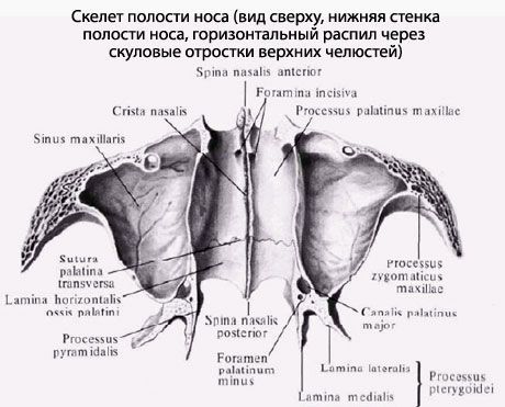
Nasal cavity(cavum nasi) occupies a central position in the facial skull. The bony nasal septum (septum nasi osseum), consisting of the perpendicular plate of the ethmoid bone and the vomer, connected below with the nasal crest, divides the bony nasal cavity into two halves. In front there is a piriform aperture (apertura piriformis), limited by the nasal notches (right and left) of the maxillary bones and the lower edges of the nasal bones. In the lower part of the piriform aperture, the anterior nasal spine (spina nasalis anterior) protrudes forward. Through the posterior openings of the nasal cavity, or choanae (choanae), the nasal cavity communicates with the pharyngeal cavity. Each choana is limited on the laterally by the medial plate of the pterygoid process, on the medial side by the vomer, above by the body of the sphenoid bone, below by the horizontal plate of the palatine bone. The nasal cavity has three walls: superior, inferior and lateral.
The superior wall is formed by the nasal bones, the nasal part of the frontal bone, the cribriform plate of the ethmoid bone and the inferior surface of the body of the sphenoid bone.
The inferior wall consists of the palatine processes of the maxillary bones and the horizontal plates of the palatine bones. Along the midline of this wall, these bones form the nasal crest, to which the bony nasal septum is attached, which is the medial wall for the right and left halves of the nasal cavity.
The lateral wall is formed by the nasal surface of the body and the frontal process of the maxilla, the nasal bone, the lacrimal bone, the ethmoid labyrinth of the ethmoid bone, the perpendicular plate of the palatine bone, and the medial plate of the pterygoid process of the sphenoid bone (in the posterior section).
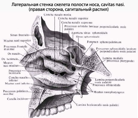
On the lateral wall of the nasal cavity, three nasal conchae are visible, located one above the other. The superior and middle conchae are parts of the ethmoid labyrinth, and the inferior nasal concha is an independent bone. The nasal conchae divide the lateral part of the nasal cavity into three nasal passages: superior, middle, and inferior.
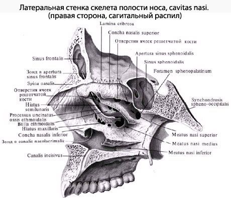
The superior nasal meatus (meatus nasi superior) is bounded above and medially by the superior nasal concha and below by the middle nasal concha. This meatus is located in the posterior part of the nasal cavity. The posterior cells of the ethmoid bone open into it. Above the posterior part of the superior nasal concha is the sphenoid-ethmoidal recess (recessus sphenoethmoidalis), into which the aperture of the sphenoid sinus opens. Through this aperture, the sinus communicates with the nasal cavity.
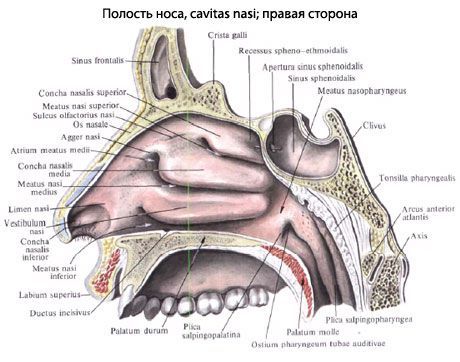
The middle nasal meatus (meatus nasi medius) is located between the middle and lower nasal conchae. The anterior and middle cells of the ethmoid bone, the aperture of the frontal sinus via the ethmoid funnel, and the semilunar cleft leading to the maxillary sinus open into it. The sphenopalatine foramen (foramen sphenopalatinum), located behind the middle nasal concha, connects the middle nasal meatus with the pterygopalatine fossa.
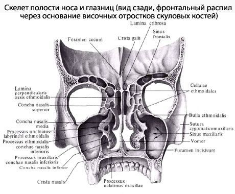
The inferior nasal meatus (meatus nasi inferior) is limited above by the inferior nasal concha, and below by the nasal surfaces of the palatine process of the maxilla and the horizontal plate of the palatine bone. In the anterior part of the inferior nasal meatus, the nasolacrimal canal (canalis nasolacrimal) opens, beginning in the orbit.
A narrow sagittal slit, bounded by the nasal septum on the medial side and the nasal conchae, is the common nasal meatus.
What's bothering you?
How to examine?
What tests are needed?