Medical expert of the article

New publications
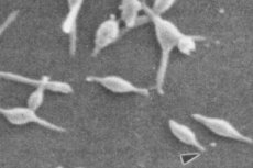
Mycoplasma pneumoniae test
Last reviewed: 04.07.2025

All iLive content is medically reviewed or fact checked to ensure as much factual accuracy as possible.
We have strict sourcing guidelines and only link to reputable media sites, academic research institutions and, whenever possible, medically peer reviewed studies. Note that the numbers in parentheses ([1], [2], etc.) are clickable links to these studies.
If you feel that any of our content is inaccurate, out-of-date, or otherwise questionable, please select it and press Ctrl + Enter.
To date, there are no clinical, epidemiological or laboratory symptoms that would allow early detection of Mycoplasma pneumoniae lung damage. Diagnosis is made only after the appearance of symptoms characteristic of the pathology. There are certain signs that allow one to suspect atypical pneumonia:
- A sharp increase in body temperature from the first sign of the disease to 38 °C.
- Productive cough with the separation of viscous purulent sputum.
- Difficulty breathing, shortness of breath and cyanosis of the nasolabial triangle.
- Increased number of leukocytes in the blood.
Diagnosis of mycoplasma pneumonia consists of the following stages:
- Collecting anamnesis and analyzing the patient's complaints - the doctor will find out how long ago the first painful symptoms appeared, whether there are chronic diseases and other features of the onset and course of the disease.
- Visual inspection and percussion – the doctor examines the patient's chest. If there is a depression in the intercostal spaces or one side lags behind the other during breathing, this indicates pneumonia. Percussion is also performed, that is, tapping the chest with the fingers. Based on the sound obtained, the doctor draws conclusions about the condition of the lungs.
- Auscultation - the lungs are listened to using a stethoscope. Normally, the sound should be clear, and breathing calm and measured. If breathing is difficult, there is gurgling or wheezing, then this is a sign of exudate accumulation, which interferes with the normal functioning of the lungs.
- Laboratory diagnostics – the patient must undergo a general blood and urine test, sputum analysis, PCR, and ELISA.
- Instrumental diagnostics is a set of studies to establish the exact cause of a painful condition. Patients are prescribed X-ray diagnostics, tomography, ultrasound, bronchoscopy, CT and other examinations.
Tests
Laboratory diagnostics of pulmonary mycoplasmosis consists of a set of the following tests:
- Complete blood count
- Erythrocytes are above normal.
- Leukocytes are significantly elevated in the bacterial form of the disease.
- Leukocyte formula - neutrophils with toxic granularity predominate, formula shift to the left.
- Lymphocytes - decreased due to increased neutrophils.
- ESR is above normal.
- Platelets are within normal limits.
The more severe the patient's condition, the more pronounced the changes in the blood.
- Biochemical blood test
- Total protein is normal.
- C-reactive protein is elevated.
- LDH and fibrinogens are elevated.
- Alpha and gamma globulins are elevated.
- Analysis of sputum secreted - increased number of neutrophils, fibrin, elastic fibers, erythrocytes.
- Blood and sputum analysis for immunoglobulins IgM, G to mycoplasmas.
- Blood test for bacterial DNA.
- Blood gas analysis.
The attending physician is responsible for deciphering the results of the tests. Based on their results, the doctor makes a treatment plan or prescribes additional tests.
 [ 1 ], [ 2 ], [ 3 ], [ 4 ], [ 5 ]
PCR
An experimental diagnostic method of molecular biology for determining the state of DNA fragments in biological material is the polymerase chain reaction. PCR in case of suspected mycoplasma pneumonia is a study of blood, sputum, pleural fluid and other types of biomaterial for pathogenic microorganisms.
Advantages of PCR:
- Increased detection rate of pathogen DNA in clinical samples compared to standard diagnostic microbiological methods.
- High sensitivity in cases of suspected generalized processes in the body.
- Identification of difficult to cultivate microorganisms and uncultivated forms of bacteria in persistent infections.
Detection of pathogens in biomaterial does not always have diagnostic value. This is due to the fact that many microorganisms normally live in the respiratory tract, but under certain conditions they realize their pathogenic potential, causing infectious processes.
IFA
Laboratory immunological method of qualitative/quantitative determination of viruses and other pathogenic microorganisms is ELISA. Enzyme immunoassay is performed in the following cases:
- Search for specific antibodies to infectious pathologies.
- Determination of antigens for various diseases.
- Study of hormonal status.
- Screening for autoimmune diseases and tumor markers.
The advantages of ELISA are high sensitivity and specificity, the ability to identify the disease and track the dynamics of the pathological process. The main disadvantage of the method is the detection of antibodies, i.e. the immune response, and not the pathogen itself.
To detect Mycoplasma pneumoniae, blood is taken for ELISA. The analysis is considered confirmed if immunoglobulins IgM, G are detected in the blood. If the increase in antibody titer is increased by 3-4 or more times, then the enzyme immunoassay confirms atypical pneumonia.
Antibodies to mycoplasma pneumonia iG
Specific antibodies produced by the immune system in response to infection with various pathogens are immunoglobulins. Antibodies to mycoplasma pneumoniae igg are serological markers indicating a pathological process in the body.
Mycoplasma pneumoniae occupies an intermediate position between bacteria, protozoa and viruses. It causes damage to the respiratory system and accounts for about 20% of all cases of community-acquired pneumonia. After infection, the immune system begins to actively produce immunoglobulins of class A, M and G.
IgG against mycoplasma infection appears after 2-4 weeks and continues to be produced for a long period of time, usually more than a year. Blood analysis for these immunoglobulins is included in the complex of mandatory laboratory tests if atypical pneumonia is suspected. To reduce the risk of diagnostic errors, simultaneous analysis for IgM and IgG is indicated.
[ 6 ]
Antibodies to mycoplasma pneumonia igM
To confirm acute mycoplasma damage to the respiratory system, patients are prescribed an enzyme immunoassay. Antibodies to mycoplasma pneumonia IgM allow differentiating atypical inflammation from other pathologies of the respiratory tract, for example, an infectious process caused by streptococci or staphylococci.
The following symptoms are a reason for conducting a laboratory test:
- A non-productive cough that lasts for a long period of time.
- Severe pain in the throat and chest.
- Muscle pain.
- Deterioration of general well-being.
The positivity coefficient indicating infection is 0-0.84. A negative result is possible not only in the absence of the disease, but also in chronic mycoplasma infection, early infection, when the body has not yet developed an immune response. It should also be taken into account that IgM is usually not released during repeated initiation.
Cold antibodies in mycoplasma pneumonia
Antibodies that cause red blood cell aggregation when exposed to low temperatures are cold antibodies. In mycoplasma pneumoniae, they most often belong to the IgM class. Normally, they can be found in healthy people, but they increase significantly 7-10 days after the onset of the disease. Cold exposure causes acute transient hemolytic anemia. A persistent increase in the titer of agglutinins leads to the development of a chronic form of pathology.
There are several types of cold agglutinins:
- The disease is caused by primary intravascular hemodialysis with monoclonal antibodies to the I-antigen of erythrocytes. In this case, cold antibodies are formed in lymphoproliferative disorders.
- The disease is caused by secondary intravascular hemolysis. It is characterized by polyclonal antibodies in a low titer and active in a narrow temperature range. It manifests itself in various infections. For example, in mycoplasma pneumonia, cold agglutinins to the I-antigen of erythrocytes occur.
Cold antibodies in atypical pneumonia can act as a mixture of various immunoglobulins. Activation of agglutinins begins already at 37 °C and causes such pathological reactions: acrocyanosis and hemolysis due to activation of complement.
[ 11 ], [ 12 ], [ 13 ], [ 14 ]
Instrumental diagnostics
To determine the localization of the inflammatory focus in the lungs, its size and other features, instrumental diagnostics are indicated. The complex of studies consists of the following procedures:
- Radiography.
- Fiberoptic bronchoscopy.
- CT.
- Function of external respiration.
- Electrocardiography.
The main diagnostic method is radiography. It allows to detect foci of inflammation, which in the image appear darker than the rest of the lung. Also observed is a change in the pulmonary pattern and proliferation of connective tissue. In pneumonia, changes in the pulmonary roots, pleural damage and even the presence of an abscess in the organ are possible. Radiography is performed in two projections - direct and lateral.
Tomography gives the same result as X-ray, so it is rarely performed if atypical pneumonia is suspected. Ultrasound diagnostics is also rarely performed, since it only reveals exudate in the lungs, which is also visible on X-ray. As for bronchoscopy, it is necessary to obtain more accurate research results.
Differential diagnostics
Successful treatment of any disease requires a comprehensive examination. Differential diagnostics of atypical pneumonia is aimed at excluding pathologies with similar symptoms. This allows for an accurate diagnosis and therapy.
Differentiation is carried out in several stages:
- Collection of primary data and formation of a list of possible diseases.
- Study of symptoms, changes in the dynamics of well-being and other factors of the disease.
- Comparative analysis of the obtained data, assessment of similar and different values.
- Identification of extraneous symptoms that are not related to the suspected pathology.
- Exclusion of diseases whose clinical signs are not included in the overall picture.
- Making a final diagnosis and drawing up a treatment plan.
The data collected and analyzed during the diagnostic process provide a reliable picture of the disease state. Differentiation of atypical pneumonia is carried out with the most common harmful microorganisms:
- Mycoplasma - acute onset, catarrh of the upper respiratory tract, cough with poorly separated sputum. As a rule, it develops in young patients.
- Pneumococci - acute onset of the disease, severe fever, severe course, but good response to penicillin antibacterial drugs.
- Staphylococci – acute onset and severe course, limited infiltrates, resistance to penicillins.
- Hemophilus influenzae – severe course, extensive infiltrates, thick sputum with blood impurities, abscess formation. Most often occurs in patients with chronic bronchopulmonary pathologies and alcoholism.
- Legionellosis - severe course, diarrhea and liver dysfunction, neurological disorders. People who are in air-conditioned rooms for a long time are susceptible to the disease
- Aspiration – putrid sputum, multiple and confluent foci of inflammation, reflex cough and increased salivation.
- Pneumocystis - increasing shortness of breath with frequent coughing fits. Severe symptoms with weak radiographic signs.
- Fungi - rapid development of a feverish state, cough with poor expectoration, severe feverish state, chest pain.
Most pathogens have a similar symptom complex, so much attention is paid to bacterial culture. Atypical pneumonia is differentiated from other diseases. During the examination, the doctor determines extrapulmonary pathologies with signs from the respiratory system and limits pulmonary inflammation from other possible disorders from the respiratory system:
- Tuberculosis is most often mistaken for pneumonia. It occurs with a dry cough, subfebrile body temperature and pale skin. If positive tuberculin tests are detected, then the diagnosis is complicated. The main differences from pneumonia: heterogeneous and dense shadows, areas of enlightenment similar to seeded foci. Massive spread of mycobacteria is observed in sputum. Leukocytes are elevated in the blood.
- Bronchitis - occurs after ARVI or against their background. In the early stages, it is accompanied by a dry cough, which gradually becomes productive. The elevated temperature lasts 2-3 days, and then remains in the subfebrile range. There is no infiltration, the pulmonary pattern is enhanced. Very often, pneumonia is diagnosed as an exacerbation of bronchitis.
- Flu - in the epidemiological period it is very difficult to differentiate between pulmonary inflammation and influenza infection. The clinical picture of the disease is taken into account.
- Pleurisy is an inflammatory pathology in the respiratory system, similar to pleural changes. It occurs with pain in the chest and during coughing. The main diagnostic sign of pleurisy is wheezing, that is, the sounds of friction of the pleura during breathing. Particular attention is paid to the results of biochemical analysis.
- Atelectasis is a pulmonary pathology with tissue collapse and impaired gas exchange. Its symptoms resemble pneumonia: respiratory failure, shortness of breath, cyanosis of the skin. Chest pain in this disease is caused by impaired gas exchange. An infection gradually develops in the collapsed area of the organ. Atelectasis is associated with injuries, blockage and compression of the lungs, destructive changes in tissues.
- Oncological processes - the initial stages of the disease are no different from atypical pneumonia. Differentiation is based on a comprehensive diagnostic approach with a thorough study of cancer signs.
In addition to the above-mentioned diseases, mycoplasma pneumonia is differentiated from dysfunctions of the cardiovascular system, hepatitis, rheumatoid arthritis, collagenoses, pulmonary infarction and other disorders of the body.
[ 15 ], [ 16 ], [ 17 ], [ 18 ], [ 19 ], [ 20 ], [ 21 ], [ 22 ], [ 23 ]