Medical expert of the article

New publications
Malignant moles on the body: how to distinguish, what to do, removal
Last reviewed: 04.07.2025

All iLive content is medically reviewed or fact checked to ensure as much factual accuracy as possible.
We have strict sourcing guidelines and only link to reputable media sites, academic research institutions and, whenever possible, medically peer reviewed studies. Note that the numbers in parentheses ([1], [2], etc.) are clickable links to these studies.
If you feel that any of our content is inaccurate, out-of-date, or otherwise questionable, please select it and press Ctrl + Enter.
Malignant moles - in medicine they are called melanomas - are onco-altered neoplasms on the skin that develop from the cells of the birthmark that form pigment (melanocytes). If the mole accelerates its growth, changes color or bleeds - these are symptoms that require mandatory consultation with a doctor. After all, timely diagnosis of melanoma significantly improves the prognosis of the disease.
 [
[ Causes malignant mole
An ordinary harmless mole can become malignant if a person often and for a long time likes to sunbathe. And not only under the sun's rays, but also in a solarium. Exposure to ultraviolet rays leads to the degeneration of pigment cells, which accelerate their growth and reproduction, involving nearby healthy tissues in the process.
A malignant mole can also appear in a hereditary chain. So, if one of the relatives has previously been diagnosed with melanoma, then other family members are also at risk of developing a pigment tumor. In addition, those who have a large number of moles on their body, or birthmarks of significant size, are at risk.
An additional impetus for malignancy can be trauma and damage to the skin of an ordinary mole, friction against clothing, etc.
Why are malignant moles dangerous?
A malignant mole is one of the most unfavorable neoplasms that can affect a person of any age and gender. This is a tumor with a high mortality rate, which begins its development with melanocytes of the epidermal layer of the skin. Melanoma is one of the most aggressive forms of oncopathology, because even an insignificantly small malignant birthmark can give a large number of metastases to various organs in a short time: the respiratory system, the skeletal system, the brain.
If the disease is detected in time, the patient has a chance of recovery. The ill-fated mole is removed. If the tumor has managed to send its daughter cells (metastases) to other organs, the prognosis for the disease becomes extremely unfavorable.
Malignant moles are found less frequently than skin cancer. However, in recent decades, this pathology has been occurring more and more often.
Pathogenesis
Malignancy of a birthmark occurs against the background of rapid growth of melanocytes, which penetrate into nearby tissues and also spread through blood and lymph. The tumor grows both on the surface of the skin and deep into the tissues, gradually penetrating into new adjacent and underlying layers.
Doctors classify the depth of the lesion by the degree of invasion. The greater the degree of germination (VI-V degree), the more unfavorable the prognosis.
A malignant mole is characterized by early and rapid spread of metastases. The nearest lymph nodes are affected first, they enlarge and become dense and elastic, without signs of pain.
After the lymph nodes, metastases often enter the skin, near the main focus. They look like small dark spots localized around the melanoma. Sometimes the malignant area swells and becomes bluish-red.
Metastases can reach almost any organ via the circulatory system. They are most often found in the lungs, adrenal glands, liver, and brain.
Symptoms malignant mole
A malignant mole at the beginning of its development looks like a common nevus. Its growth rate increases, and ulcers, peeling, and bleeding may appear later. The size of the formation can vary from a barely noticeable pea to large-caliber nodes.
Melanoma has an elastic consistency, its density is moderate. The mole cover is mostly smooth, in rare cases with small bumps and growths resembling cauliflower.
Oncologists identify three signs that allow one to suspect a malignant mole:
- dark color;
- glossy surface;
- the presence of decomposition processes in the tumor.
The listed symptoms are explained by the fact that malignant changes occur inside the birthmark: excessive accumulation of pigment, damage to the structure of the epidermis, damage to blood vessels and disruption of tissue trophism.
Sometimes the pigment accumulation occurs only in one part of the tumor. In this case, the mole itself is light, but has dark inclusions or a center.
The decomposition processes are not immediately noticeable. Over time, the birthmark becomes easily vulnerable, often bleeds, and ulcers and crusts form on the surface.
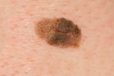
What do malignant moles look like? How to distinguish a malignant mole from a benign one? There are several distinctive features:
- a malignant mole is asymmetrical or blurred (with a benign mole, the borders and shape are clear);
- the edges of a malignant mole are uneven, jagged, or cloudy;
- the color of melanoma is dark or with inclusions (a benign mole is light or brown, uniform);
- a malignant birthmark is large in size and grows rapidly;
- Malignant degeneration is characterized by crusts, peeling, bleeding, and ulcers on the surface.
The clinical picture may vary, as there are different types of malignant moles:
- Superficial spreading melanoma looks like a black or brown spot, up to 3 mm in circumference. It gradually increases in size and becomes round - oval or irregular in shape. The surface acquires a smooth, glossy appearance and becomes dense.
- Malignant lentigo is an uneven plaque with slow growth and uneven coloring. Both light and dark inclusions, up to black, can be observed on the surface. A characteristic sign is the presence of nodules and papillomas with significant hyperkeratosis or elements of atrophy.
- The nodular appearance of a malignant mole often arises from a normal pigment spot. When a mole becomes malignant, it darkens, the surface becomes bumpy, compacted, and perfectly smooth. Sometimes small black nodules appear nearby – the so-called “screenings” of melanoma. Ulcers or crusts may form on top of the mole.
Complications and consequences
What consequences can be expected from a malignant mole? The main complication of melanoma is the active spread of the tumor throughout the body. Metastases form relatively quickly, and they pose the greatest danger to the patient's health and life.
Complications such as secondary malignant neoplasms are very common with melanoma. Tumor elements can spread with the blood or lymph flow, stopping in other organs and growing into them. Most often, such objects are the lungs, liver, bones, brain, and skin.
Some expectant mothers are interested in the question: can a malignant mole affect the fetus if it was diagnosed during pregnancy? Scientists studied this issue at the end of the last century and came to the conclusion that metastases can penetrate the placenta, but this happens extremely rarely. Isolated cases have been described only in the disseminated form of malignant pigment tumor (with chaotic and massive spread of metastases).
No less of a problem is the treatment of a malignant mole during pregnancy, because chemotherapy and radiation therapy can have an extremely negative impact on the development of the fetus. In such a situation, the decision on treatment measures is made by the doctor, taking into account all the pros and cons.
Diagnostics malignant mole
Patients with suspected melanoma often complain of changes occurring in the birthmark. These are mainly the following symptoms:
- bleeding;
- itching, discomfort;
- mole growth;
- change in color and appearance.
In this case, the doctor asks the following questions:
- When did the suspicious mole appear?
- Over what period of time did the changes occur?
- Was there trauma to the mole or exposure to other factors?
- Have you treated the mole and how?
After questioning and examining the birthmark, the doctor prescribes other necessary tests.
- Blood and urine tests for diagnostic purposes in case of malignant moles are not informative. Such studies are relevant only for determining the general condition of the body, which is especially important in case of metastasis of tumor elements.
- Instrumental diagnostics are used to monitor the effectiveness of the selected therapy, or to detect a possible relapse of the tumor:
- X-ray of the lungs – helps to diagnose metastases;
- computed tomography method – detects metastases in the lungs, lymph nodes, etc.;
- Dermatoscopy is a method that allows you to accurately examine a skin problem, which is especially important in the early stages of the development of a malignant mole.
- A melanoma biopsy is prescribed in cases where it is impossible to establish a diagnosis in any other way, as well as after surgical removal of a mole, to clarify its structure. Conducting a biopsy is directly related to a radical operation to remove a malignant mole.

What do need to examine?
How to examine?
Differential diagnosis
Differential diagnostics are applied in relation to pigment basalioma, seborrheic keratosis, hemangioma, granuloma, angiofibroma, histiocytoma.
Who to contact?
Treatment malignant mole
Treatment should be carried out immediately after diagnosis, as melanomas tend to spread quickly and actively throughout the body.
The first and main method of treatment is surgical removal of a malignant mole. This method is indicated for malignant pigmented lesions of stage I and II development. To avoid recurrence of the tumor, the surgeon removes not only the mole, but also the subcutaneous tissue and underlying fascia. The operation ends with skin grafting. The material removed during the procedure is sent for histological and cytological examination.
Are there any consequences after removing a malignant mole? Consequences occur with incomplete or delayed removal of the tumor, which leads to its repeated growth or the appearance of metastases. Therefore, removal should only be performed in a medical institution by a qualified specialist.
In any case, the lack of adequate treatment is guaranteed to lead to a worsening of the situation and, over time, to premature death.
Chemotherapy is also effective for malignant moles. Medicines are actively used for common forms of melanoma, as well as in combination with surgery.
For widespread moles, the following treatment regimens are considered the most effective:
- Imidazolecarboxamide 250 mg per m², once a day for 5 days;
- Lomustine 100 mg/m² + Vincristine 1.2 mg/m² on the first, eighth and fifteenth days, as well as in combination with Dactinomycin 500 mcg three times a week, in the amount of six doses;
- Vinblastine 6 mg per m² by intravenous administration. On the first day together with Cisplatin 120 mg per m², and also with Bleomycetin 10 mg on the first and fifth days.
The time intervals between chemotherapy courses are 1 month.
Radiation therapy is rarely used for malignant moles due to their low sensitivity to ionizing rays.
Folk treatment of malignant moles
Unfortunately, many patients are in no hurry to see a doctor, but treat themselves with all sorts of folk methods. Folk treatment of melanoma is officially not welcomed, since treatment with herbs and other means can take away precious time when the disease is still treatable. Lost time can cost a person not only health, but also life.
However, prescriptions for malignant pigment tumors do exist. However, reliable information on their effectiveness has not been provided.
- Mix equal parts of nettle leaves, angelica, coriander and hyssop. Pour 1 tbsp of the mixture into 200 ml of boiling water and leave to infuse until cool. Take 400-600 ml of the drink per day.
- Take aconite rhizome tincture three times a day, 60 minutes before meals. Treatment regimen: first day - 1 drop, daily increasing the dose by 1 drop, bringing it to 20 drops. Then the amount of the drug is reduced, again bringing it to 1 drop.
- Take 100 ml of a decoction of sweet clover, elderberry, wintergreen, centaury, meadowsweet, duckweed, and agrimony, taken in equal parts, half an hour before meals.
- Prepare a tar ointment: mix tar with Vaseline in equal parts. Lubricate the affected area several times a day.
- Squeeze fresh celandine juice, mix with Vaseline 1:4. Use for compresses.
It is not recommended to resort to folk remedies without consulting an oncologist.
Homeopathy for malignant moles
Homeopathy is often used as an adjunct treatment for malignant moles. Many experts believe that the correct use of such drugs can improve the effectiveness of treatment and reduce the risk of relapse in the future.
The choice of the optimal homeopathic remedy is made individually, depending on the characteristics of the tumor and the patient's condition. Since homeopathic treatment requires precise dosages, self-medication is not encouraged.
- Homeopathic preparations with antihomotoxic action:
- Lymphomyosot;
- Galium-heel;
- Engystol.
- Preparations that catalyze metabolic processes:
- Ubiquinone compositum;
- Coenzyme compositum.
- Homeopathic preparations with organotropic action:
- Cutis compositum;
- Psorinocheel.
- Products that accelerate detoxification of the body:
- Hepar compositum;
- Hepel.
- Preparations whose action is aimed at activating immune forces and stimulating connective tissue processes:
- Echinacea compositum;
- Tonsilla compositum.
Prevention
In order to prevent the degeneration of a common mole into a malignant melanoma, it is necessary to exclude as much as possible the impact of factors stimulating malignancy. To do this, it is necessary to follow these recommendations:
- monitor the growth and appearance of moles on the body, and at the slightest suspicion, consult a doctor;
- avoid injury to moles, chemical or mechanical damage;
- do not overuse tanning, use appropriate cosmetic protective products before and after sunbathing;
- Do not try to remove birthmarks yourself, do not scratch or damage moles.
Most experts agree that the best way to prevent a mole from degenerating is to remove it. One important point to consider is that removal should be performed by a competent qualified specialist in a medical facility, but not in beauty salons or other similar establishments.
By turning to incompetent doctors, you can lose not only your health, but also your life.
Forecast
More than half of the patients can be observed to have a 5-year recovery period. Such positive results are explained by the timely and early detection of the tumor.
If malignancy is detected at later stages, the prognosis worsens, especially with the spread of metastases.
If treatment is started on time and no metastases are found, then the size and depth of tumor penetration play a decisive role in the prognosis. It has been noted that treatment is more effective in female patients than in male patients.
All patients who have undergone treatment are subject to mandatory medical examination. Skin examinations, remaining benign moles, and lymph nodes are carried out regularly.
With proper and adequate treatment, malignant moles do not recur.