Medical expert of the article

New publications
Malignization of gastric ulcer
Last reviewed: 07.07.2025

All iLive content is medically reviewed or fact checked to ensure as much factual accuracy as possible.
We have strict sourcing guidelines and only link to reputable media sites, academic research institutions and, whenever possible, medically peer reviewed studies. Note that the numbers in parentheses ([1], [2], etc.) are clickable links to these studies.
If you feel that any of our content is inaccurate, out-of-date, or otherwise questionable, please select it and press Ctrl + Enter.
According to modern data, the frequency of malignancy of gastric ulcer does not exceed 2%. Data from previous years were overstated. This is explained by the fact that malignancy of gastric ulcer was taken to be primary ulcerative form of gastric cancer, which in clinical course is almost no different from chronic gastric ulcer. In addition, primary ulcerative form of gastric cancer can proceed for quite a long time without generalization of the process and give periods of remission with ulcer healing. At the same time, good appetite and satisfactory condition of the patient are maintained for a long time.
Symptoms gastric ulcer malignization
Malignancy of the stomach can be determined based on the following signs:
- the pain in the epigastric region becomes constant, radiates to the back, the pain is especially intense at night;
- the symptom of localized pain during palpation disappears, the pain in the epigastrium becomes diffuse;
- a progressive decrease in the patient's body weight is noted;
- appetite disappears;
- an unmotivated, increasing weakness appears.
Diagnostics gastric ulcer malignization
- characterized by progressive anemia, a constantly positive Gregersen reaction (a reaction to hidden blood in the stool) and a persistent decrease in the acidity of gastric juice, detection of lactic acid in it; a persistent increase in ESR;
- X-ray examination of the stomach reveals signs of ulcer malignancy: a wide entrance to the ulcer crater, atypical relief of the mucosa around the "niche", disappearance of folds and peristalsis in the affected segment, the infiltration shaft around the ulcer exceeds the diameter of the ulcer crater, the symptom of a sunken niche, the appearance of a filling defect;
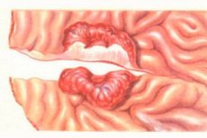
- During FGDS, characteristic signs of a "malignant ulcer" appear. Such ulcers often have an irregular shape, uneven, unclear, bumpy edges. The ulcer bottom is also uneven, bumpy, can be flat, shallow, covered with a grayish coating. In some areas of the ulcer, the edges may be undermined. Diffuse infiltration and deformation of the stomach wall in the ulcer area are characteristic. A common sign is the rigidity of the ulcer edges during targeted biopsy and increased bleeding in the ulcer lesion area. There are erosions on the mucous membrane surrounding the ulcer. To make a final judgment about the nature of the ulcer, it is necessary to perform a targeted biopsy from the edges and bottom of the ulcer in several areas (at least 5-6 biopsies) with subsequent histological and cytological examination of the material.
 [ 8 ], [ 9 ], [ 10 ], [ 11 ], [ 12 ], [ 13 ], [ 14 ], [ 15 ], [ 16 ], [ 17 ]