Medical expert of the article

New publications
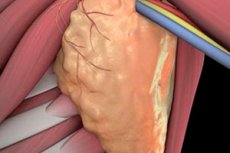
Lymphadenectomy
Last reviewed: 04.07.2025

All iLive content is medically reviewed or fact checked to ensure as much factual accuracy as possible.
We have strict sourcing guidelines and only link to reputable media sites, academic research institutions and, whenever possible, medically peer reviewed studies. Note that the numbers in parentheses ([1], [2], etc.) are clickable links to these studies.
If you feel that any of our content is inaccurate, out-of-date, or otherwise questionable, please select it and press Ctrl + Enter.
Lymph node dissection, or lymphadenectomy, is a surgical procedure that involves removing lymph nodes and then examining them for the presence of atypical cells. Lymph node dissection can be limited or complete, depending on the scale of the operation. The likelihood of complications after such a procedure is quite high. However, the intervention often helps stop the further spread of cancerous structures, thereby saving the patient's life.
Indications for the procedure
The main purpose of the lymphatic system is to retransport fluid from tissues to the circulatory system and to provide immunity, which includes protection against bacteria, viruses and atypical cells.
The lymphatic system consists of nodes, vessels, and small vascular capillaries. Lymph flows through the vessels, and the nodes are bean-shaped formations located along the entire system and act as filters that trap any foreign objects.
The largest clusters of nodes are observed in the neck, armpits, pelvis and groin area.
The lymphatic system is the first to accept the spread of tumor cells from the focus to other points in the body: sometimes such cells are retained in the lymph nodes and continue to grow there. This process is called metastasis. By removing several lymph nodes, the doctor can determine whether the patient has metastasis.
Lymph node dissection is used not only for diagnosis but also to block further spread of cancerous structures throughout the body.
In addition, indications include severe pain in the lymph nodes, as well as the ineffectiveness of conservative therapy.
Lymph node dissection in cancer is an integral part of a qualified and comprehensive approach to the treatment of oncopathology. Even before the operation, the surgeon clarifies the probability of damage to the "sentinel" lymph nodes and their groups that are directly involved in the outflow of lymph from the area affected by the tumor process. Suspicion of the presence of metastases in a certain lymphatic collector is a direct indication for lymph node dissection. As a rule, the lymphatic capillaries, outgoing vessels, lymph flow directions, including regional and distant lymph nodes, as well as the surrounding tissue are subject to removal. Such an operation can significantly improve the quality of life of postoperative patients and speed up their recovery.
Preparation
The preparatory stage is simple, but mandatory. It includes the following sequential activities:
- Consultation with an oncosurgeon who will perform the lymphadenectomy, as well as with an anesthesiologist.
- Agreeing on the main points and date of the intervention.
- Preoperative diagnostics, including general urine analysis, general and biochemical blood analysis, ultrasound examination, and sometimes fine-needle biopsy of the lymph nodes.
- Examination by a therapist, with an assessment of the likelihood of contraindications to surgery.
- Discontinuation of drugs that may negatively affect the course of the operation and the postoperative period (for example, non-steroidal anti-inflammatory drugs, barbiturates, heparin, etc.).
- The day before the lymphadenectomy, the patient should limit their diet, avoid overeating, and avoid heavy, fatty, and sweet foods. On the day of the operation, they should not drink or eat.
Technique lymphadenectomies
Most often, in the presence of oncology, surgeons resort to such types of surgical lymphadenectomy as dissection of the axillary lymph nodes (in case of breast cancer), cervical dissection (in case of thyroid cancer or cancer of the neck and head organs), D2 lymphadenectomy with removal of nodes located in the stomach, liver and spleen area (in case of stomach cancer). [ 1 ]
A qualified surgeon can in most cases predict to which lymph nodes the atypical cells will move during the spread of metastases. The nodes that will be affected first are called sentinel nodes. That is why the doctor first removes exactly these nodes, which are immediately sent for examination - biopsy of sentinel lymph nodes.
To determine the priority nodes for lymphadenectomy, a mapping procedure is performed: a radioisotope substance (indicator) is introduced into the affected area, indicating the direction of lymph flow.
The average duration of lymphadenectomy surgery is one hour. However, the duration may vary depending on the specifics of the surgical intervention.
- Pelvic lymphadenectomy can be performed using laparoscopic and laparotomic access. During laparoscopy, pneumoperitoneum conditions are maintained (10 to 15 mm Hg), laparoscopes and trocars are used. The parietal peritoneum is dissected above the iliac vessel zone, in a parallel direction with the external iliac vessels. The ureters are necessarily examined. Using clamps, the periadventitial tissue with lymph nodes and vessels located in the proximal area of the incision is grasped. With a closed clamp, the tissue is peeled off from the frontal part of the external iliac vessels. After this, the obturator nerve is isolated and all the tissue localized around the internal iliac vessels is removed, along with the lymph nodes. The lymph nodes located near the external iliac vein are removed especially carefully. It is preferable if the entire chain is excised. Finally, the fatty tissue with the nodes localized in it is removed from the space between the external iliac arterial and venous vessels. The biomaterial is sent for histological analysis, the damaged vessels are coagulated to prevent bleeding - for this, electrosurgery is used. [ 2 ]
- Inguinal lymphadenectomy in its classic version is performed according to the description of the French oncologist Duquesne. The essence of the operation is the excision of the lymph nodes of the femoral-inguinal zone together with the tissue, fascia and an element of the great femoral saphenous vein. First, the surgeon makes a vertical incision above the middle of the inguinal ligament and below, cutting to the subcutaneous fat layer. The skin tissue is separated at the level of the superficial subcutaneous fascia. The subcutaneous fat is excised, achieving exposure of the iliac part of the abdominal wall and the entire femoral triangle. Next, the incision is extended to the underlying muscles, after which the great saphenous vein is isolated, ligated and crossed at the apex of the femoral triangle. The tissue with the lymph nodes is moved inward, the sartorius muscle is taken outward using hooks: this helps to examine the femoral vascular bed. The tissue area to be removed and the outer wall of the vascular sheath are separated from the femoral vessels and lifted upwards to the area where the great saphenous vein joins directly to the femoral vein. The biomaterial is removed and sent for further examination. [ 3 ]
- Axillary lymphadenectomy rarely lasts more than 60 minutes. Usually, the surgeon makes an incision in the armpit area of about 50-60 mm. The intervention is performed under general anesthesia, sometimes in combination with radical mastectomy. During a lumpectomy, the nodes can be removed later or during the operation. In the classic version, mainly the lymph nodes of the first row and the lower part of the second row are removed, after which they are sent for histological examination. In general, about ten nodes are excised (complete lymphadenectomy involves the excision of about two dozen nodes). In the complete version, lymph nodes belonging to all rows of the axillary chain are excised, but such operations are currently not performed very often. Preserving intervention involves dissecting tissues by five and seven centimeters in the axillary fossa. The removed tissues are sent for examination, the results of which can be obtained in a few days. Such diagnostics are necessary for the appointment of further postoperative treatment, which may include chemotherapy, radiation, etc. [ 4 ]
- Cervical lymphadenectomy is due to the fact that metastases of cancer foci from the neck and head area quite often end up in the regional cervical lymph nodes. In this case, the classic option is considered to be the intervention according to Crail, named after the American surgeon. The operation involves the complex removal of the suprahyoid, cervical and supraclavicular nodes on one side, simultaneously with the submandibular salivary gland, internal jugular vein, omohyoid and sternocleidomastoid muscles. Cervical lymphadenectomy is indicated for cancerous lesions of the laryngopharyngeal region, thyroid gland, salivary glands, tongue, oral cavity or nasopharynx. The most commonly performed surgical options are radical removal of all cervical lymph nodes (levels 1-5), modified or selective excision, or an extended radical method. Another common method is considered to be a sparing intervention, which involves the removal of lymph nodes and tissue. This method is called functional cervical dissection: during the operation, the sternocleidomastoid muscle, internal jugular vein and accessory nerve are preserved. [ 5 ]
- Inguinofemoral lymphadenectomy is used to remove cancer metastases in the inguinal and femoral lymph nodes. The surgeon makes two semi-oval incisions in a direction parallel to the inguinal fold. After dissecting the skin and subcutaneous fat layer, tissue flaps are separated upward to the aponeurosis of the external oblique abdominal muscle and downward to the middle of the femoral triangle. The inguinal ligament is divided, removing the fascia of the external oblique muscle. The prepubic tissue is moved back, exposing the base of the femoral triangle. Then the tissue is cut, starting from the point of the anterior superior iliac spine to the middle of the femoral triangle, and also from the pubic tubercle to the apex. The block of tissue and lymph nodes is removed, after which they proceed to iliac lymphadenectomy. This surgical technique helps to reduce the duration of scarring, reduce the likelihood of infection in the wound, and optimize the aesthetic appearance of the postoperative area. [ 6 ]
- Retroperitoneal lymphadenectomy involves the removal of the retroperitoneal lymph nodes. Abdominal surgery involves radical excision of adipose tissue and lymph nodes in the retroperitoneal space. Possible postoperative complications may include infertility and retrograde ejaculation into the bladder. This is due to the fact that during the procedure, the postganglionic efferent sympathetic fibers responsible for ejaculation and located para-aortically under the level of the inferior mesenteric artery are crossed. Minimal metastatic foci are those whose size does not exceed 20 mm: after removal of such metastases, the likelihood of developing postoperative complications is reduced to a minimum. [ 7 ]
- Iliac lymphadenectomy is performed as part of the iliopsoas-inguinofemoral surgery for verified metastases in the inguinal lymph nodes. Bilateral lymphadenectomy is appropriate for cancer of the penis or vulva. The classic Duquesne technique, described in the last century, is used. A long longitudinal incision is made through the middle of the inguinal ligament (with its intersection). The upper point of the incision is localized in the area 7 cm above the inguinal ligament, and the lower point coincides with the apex of the femoral triangle. Tissue flaps are separated at the level of the superficial subcutaneous fascia, the subcutaneous fat layer is excised, exposing the iliac part of the abdominal wall with the femoral triangle. Next, a large subcutaneous venous vessel is isolated, ligated and crossed in the lower wound corner, the block of lymph nodes with tissue is retracted inward, and the sartorius muscles are retracted outward. The tissues to be removed are gradually separated from the femoral vessels, lifting them to the confluence of the great saphenous venous vessel of the thigh and the femoral vein. The nervous and external oblique muscles are dissected, the peritoneum is shifted medially, and the cellular tissue and lymph nodes are separated along the iliac vessels. The iliac cellular tissue is removed together with the femoroinguinal cellular tissue. The tissues are sutured layer by layer. If necessary, plastic surgery of the inguinal region is performed. Ilioinguinal-femoral lymphadenectomy usually involves the removal of an average of eight to eleven nodes. [ 8 ]
- Para-aortic lymphadenectomy involves radical excision of the periaortic lymph nodes. The procedure is performed under general anesthesia using endovideosurgical methods. The scope of such an operation includes removal of the tissue containing the lymph nodes above and below the level of the inferior mesenteric artery, to the upper line in the area of the upper edge of the left renal vein. Para-aortic lymphadenectomy is successfully used to treat endometrial cancer. Midline laparotomy is performed above the umbilical opening and completed under the pubic symphysis. Extraperitoneal access may be used. The round ligament of the uterus is crossed, avoiding damage to the inferior epigastric vessels. The parietal peritoneum is dissected, the ureter area is visualized. The infundibulopelvic ligament is crossed and ligated. The peritoneum is dissected downwards to the round ligament of the uterus along the external iliac artery. The ligament is clamped, transected and ligated. Lymph node dissection is performed near the branch of the internal iliac artery. The separated block of tissue located lateral to the vascular network is clamped and transected, and the proximal end is ligated to block the lymph flow. Next, the perivascular tissue and lymph nodes are removed along the lateral walls of the vessels to the level of the obturator nerve. The nodes located medial to the external iliac artery and at the entrance to the femoral canal are also subject to excision. The fatty layer with the lymph nodes along the external iliac vein to the obturator fossa are also separated. After the obturator nerve is detected, the obturator fossa is visualized and the tissue between the obturator nerve and the superior vesical arterial vessel is retracted. The tissue is clamped, transected and ligated. The manipulations are performed very carefully, avoiding damage to the veins. The uterine artery is then transected and ligated, and the lymph nodes along the internal iliac vessels are removed. The removed nodes are sent for histological examination. [ 9 ], [ 10 ]
- Lymph node dissection for breast cancer is performed in relation to the nodes located in the armpit area on the affected side. Excision can also extend to the cervical, supraclavicular and subclavian nodes. The operation is performed in combination with the removal of the mammary gland, completely or partially. The surgeon makes an incision in the armpit area up to 6 cm long. Lymph node dissection itself is performed at several levels of the relative location of the nodes to the pectoralis minor muscle. The first level includes the lymph nodes located below this muscle, the second level includes those located immediately under the muscle, and the third level includes those located above the pectoralis minor muscle. In the lumpectomy tract, the nodes of the first and second levels are removed. If a mastectomy is performed - a radical resection of the mammary gland with regional lymph node dissection, then the nodes belonging to the first, second and third levels are excised, with subsequent plastic reconstruction of the breast. Such an operation lasts on average about an hour and a half. [ 11 ]
To date, experts have not come to a consensus on the advisability of removing all regional lymph nodes in any oncological processes in the mammary glands. Most surgeons and mammologists believe that such radical intervention is required only in extreme cases, when there is a clear risk of metastasis. The presence of such an indication is verified by conducting a sentinel biopsy, or sentinel lymph node biopsy. Sentinel nodes are those that are closest to the tumor focus - it is in them that atypical cells first get and metastases form. Therefore, an intervention that involves removing the sentinel lymph node is always a sure way to determine the likelihood of metastasis of the tumor. If the biopsy shows a negative result (atypical cells are not detected), then there is no need for a large-scale lymphadenectomy operation with the removal of all levels of the lymph nodes. [ 12 ], [ 13 ]
- Thyroidectomy with lymphadenectomy is a standard type of surgery for thyroid cancer. Most often, this cancer metastasizes to the sixth (central) group of cervical lymph nodes. Specialists recommend and practice thyroidectomy with one-stage central removal of lymph nodes for oncological formations with dimensions exceeding 10 mm. This approach reduces the likelihood of relapse and eliminates the need for repeated surgical intervention in this area. Central lymphadenectomy in this case involves excision of the prelaryngeal, para- and pretracheal nodes, as well as those located along the inner surface of the carotid artery and internal jugular vein. [ 14 ]
- Rectal resection with extended lymphadenectomy can be performed using different techniques, which depends mainly on the intestinal segment in which the tumor develops. If the upper third of the rectum is affected, an operation called Anterior resection is performed. If the middle third is affected, a Low anterior operation is performed. Both the first and second interventions are performed through the abdominal cavity. The doctor makes an incision in the abdominal wall to the left of the navel. After detecting and removing the tumor, he connects the remaining segments of the intestine, removes nearby lymph nodes, carefully examines all tissues, and sutures. If necessary, drainage is installed (for several days). The most difficult and traumatic for the patient is surgical removal of the lower rectal third. This intervention is called Abdominal Perineal Resection, or Miles' operation: it involves removing the tumor together with the anus. In order to provide the patient with the ability to eliminate feces, the surgeon forms a permanent colostomy. The procedure usually proceeds as follows: the doctor makes an incision in the lower abdominal cavity and perineum, removes the sigmoid colon and rectum, as well as the anus and nearby lymph nodes. In most cases, the patient must additionally undergo chemotherapy. This type of intervention can last several hours (on average, 2.5 hours). [ 15 ],
- Pancreaticoduodenal lymphadenectomy is a common type of surgery for adenocarcinoma of the head of the pancreas, which has two rows of regional lymph nodes. These nodes surround the organ or are located around large nearby vessels (abdominal aorta with branches, including the celiac trunk, superior renal and mesenteric artery). To clarify the oncological stage of pancreatic cancer, it is recommended to remove and subject to histological diagnosis at least ten lymph nodes. After crossing the gastrocolic ligament, the surgeon performs adhesioviscerolysis in the omental bursa, mobilizes the lower edge of the gland with exposure of the superior mesenteric vein. Then he crosses the right gastroepiploic vessels. The duodenum is mobilized using the Kocher method and crossed in the proximal segment. Next, parts of the hepatoduodenal ligament are mobilized, the gastroduodenal artery and small intestine are transected. After mobilization of the uncinate process, lymphadenectomy is performed along the superior mesenteric arterial vessel. [ 16 ]
- Lymph node dissection for gastric cancer can be performed in three variants. The first variant is a classic gastrectomy, during which D1 lymph node dissection is performed, including removal of paragastric lymph nodes - 1-6 rows of regional nodes according to the Japanese classification. The second variant is a radical gastrectomy with D2 lymph node dissection, including lymph basins localized in the direction of the branches of the celiac trunk - 7-11 rows of lymph nodes. The third variant is an extended radical gastrectomy with removal of retroperitoneal lymph nodes (12-16 rows). The choice of one or another type of operation with lymph node dissection is directly related to the stage of gastric cancer. For example, at the first "A" stage, radical surgical intervention may involve endoscopic resection of the gastric mucosa or the use of other techniques up to classic gastrectomy. [ 17 ]
Lymph node dissection during colon resection
Colon surgery can be performed using several techniques, depending on which part of the intestine contains the tumor. Usually, the affected segment of the intestine is removed, as well as the lymph nodes into which lymph flows from the tumor. This is due to the fact that lymphadenectomy can reduce the risk of recurrence of cancer. In addition, specialists will be able to carefully examine the removed structures, which will directly affect the nature of subsequent treatment. [ 18 ]
Surgical removal of an intestinal element is called colectomy. If the cancerous lesion is removed and is located in the right half of the colon, then we speak of a right-sided hemicolectomy, and if in the left half, then a left-sided hemicolectomy. Standard resection involves removing up to 40 cm of the colon, although this figure largely depends on the patient's weight and height.
Distal resection is when the distal two-thirds of the sigmoid colon and the upper third of the rectum are removed, and the upper rectal and sigmoid vessels are ligated. An anastomosis is applied to restore rectal function.
Left hemilectomy with extended lymphadenectomy involves removal of the left colon, which includes the sigmoid, descending, and distal half of the transverse colon. The inferior mesenteric vessels are ligated and transected, and a transverse rectal anastomosis is formed.
Right-sided hemilectomy with extended lymphadenectomy includes resection of the cecum and the distal element of the ileum – about 100-150 mm. The ascending colon and proximal third of the transverse colon are also removed, the ileocolic vessels, right colic artery and right branch of the middle colic artery are ligated and transected. Additionally, an ileotransverse anastomosis is formed.
There is another variant of the operation: subtotal removal with resection of the entire colon without the distal element of the sigmoid colon. In this case, all the basic vessels that supply the colon with nutrition are separated.
Classification of lymphadenectomy
Different variations of cancer require different amounts of lymphadenectomy. A more complete resection is referred to as extended lymphadenectomy, which in turn is further subdivided into a number of subtypes, depending on the location of the lymph nodes being removed, such as:
- aortoiliac lymphadenectomy;
- pancreatoduodenal;
- iliopelvic, etc.
Unlike extended lymphadenectomy, regional lymphadenectomy involves the removal of only certain lymph nodes that are in close proximity to the tumor site.
An auxiliary term is radical lymphadenectomy, which involves the removal of all or the dominant number of lymph nodes located near the neoplasm (along the lymph flow).
Depending on the method of operation, removal of lymph nodes can be open or laparoscopic.
Laparoscopic lymphadenectomy is performed by access through punctures in the skin, through which the surgeon inserts a special laparoscopic device and instruments. This method is less traumatic and is less likely to cause complications. Cavity lymphadenectomies are performed less frequently today: we are talking about the classic method, when tissues are dissected by an incision, and direct access is carried out. After a laparoscopic operation, healing occurs much faster, the risk of bleeding and wound infection is reduced.
Lymph node dissection and lymphadenectomy
Classic radical interventions in the treatment of oncological diseases consisted of monoblock removal of regional lymph nodes. As for preventive extended lymph node dissection, this term is used to describe surgical operations to remove the affected organ and areas with regional metastasis. It turns out that the name lymph node dissection implies a more extensive intervention, in contrast to the term lymphadenectomy, since it involves excision of not only the lymph nodes, but also the entire lymph flow section, along with the surrounding subcutaneous fat layer within the fascia sheaths. Thus, it is appropriate to talk about lymphadenectomy if regional removal of lymph nodes is performed, and about lymph node dissection - if lymph nodes, vessels and fatty tissue are removed.
Contraindications to the procedure
Lymph node dissection is not prescribed if there is no possibility of complete removal of the primary tumor. This happens if the tumor process is detected at a late stage of development. In this case, we are talking not so much about a contraindication as about the inappropriateness of lymph node dissection, since the tumor focus has already managed to spread its cells not only to the nearest lymph nodes, but also to distant tissues and organs. Even after excision of the lymph nodes, atypical structures will remain in the body, provoking the development of new cancerous (secondary) foci.
Lymph node dissection is not performed if the patient is in a serious condition, for example, if he or she suffers from serious cardiovascular, liver, or kidney diseases, or if he or she develops an acute cerebrovascular accident. Such pathologies may prevent both the operation as a whole and the anesthesia.
Consequences after the procedure
The most common adverse effect after lymphadenectomy is lymphedema, a complication that manifests itself as difficulty in lymph drainage from the area where the operation is performed. This disorder develops in approximately every tenth patient. The main symptom is severe tissue swelling. The disease has several stages of development:
- The swelling occurs throughout the day, but disappears when the damaged area is in a vertical position. If you press with your finger, a kind of "pit" is formed, which slowly disappears.
- Edema is present regardless of the location of the damaged area. The skin becomes denser, and no "pit" appears when pressed.
- The swelling is pronounced, like elephantiasis.
When the first stage of lymphedema is detected, it is necessary to urgently consult a doctor. This will stop the further development of the pathology, and in some cases – reduce its manifestation.
In addition to swelling, bleeding that is detected in the early postoperative period can become a dangerous condition.
Complications after the procedure
In general, doctors sometimes encounter the following possible complications associated with lymphadenectomy:
- Loss or deterioration of sensitivity in the area of the operation, which is caused by damage (cutting) of nerve fibers. In most patients, sensitivity is restored after a certain period of time.
- A feeling of weakness, numbness, “crawling sensations”, contractures, which requires the prescription of special therapeutic exercises to reduce the feeling of discomfort.
- Lymphedema is lymphatic swelling.
- Phlebitis in the area of the operation, with possible transition to thrombophlebitis. With timely administration of blood thinners and anti-inflammatory drugs, such phenomena quickly disappear.
- The addition of an infection, which is accompanied by pain, redness and swelling in the area of the intervention. This condition requires antibiotic therapy.
Most often, complications associated with lymphadenectomy develop in elderly patients and individuals suffering from diabetes and obesity.
Care after the procedure
Immediately after lymphadenectomy, the patient is transferred to the postoperative ward: there he is observed until the anesthesia wears off. If everything is in order, the patient is transported to a regular ward.
If necessary, the affected area is elevated. For example, after axillary lymphadenectomy, the arm on the side of the intervention is raised upward, and after removal of the inguinal lymph nodes, the patient's legs are raised.
Sometimes, during the first day after surgery, the patient is attached to a catheter to collect urine, and in some cases, a temporary or permanent colostomy bag (depending on where exactly and to what extent the lymphadenectomy was performed).
If drainage was installed during the operation, it is removed as the condition improves (usually after a few days).
If the patient cannot eat independently, then he is given nutrients intravenously. If the surgical intervention affected the digestive system, then the patient will be told about changes in the diet.
The duration of hospitalization is negotiated individually.
After discharge, the patient is advised not to lift or carry heavy objects, and to avoid wearing tight or constricting clothing or accessories.
Rehabilitation measures are indicated for all patients who have undergone lymphadenectomy. Such measures allow:
- prevent the emergence of psychological problems;
- eliminate pain;
- prevent the development of complications;
- return to your normal lifestyle faster.
Standard rehabilitation methods include exercise therapy (a special set of exercises), physiotherapy, vitamin therapy, and additional conservative treatment. The main procedures are aimed at restoring tissue nutrition and lymph flow, accelerating blood circulation and healing.
If after lymphadenectomy the patient's temperature rises, or suspicious symptoms suddenly appear in the form of chills, nausea, paroxysmal vomiting, difficulties with urine and feces, bleeding or severe pain, then it is necessary to immediately inform the surgeon who performed the operation.
Lymphatic massage after axillary lymphadenectomy surgery
Lymphatic drainage massage is a physiotherapeutic procedure, the main purpose of which is to accelerate lymph flow. The person performing the lymphatic massage should have an idea of the location of the lymphatic system and the direction of lymph flow. The procedure should not cause pain, so stroking and light pressure are recommended as basic effects. It is better to conduct sessions 1-2 times a week. [ 19 ]
Under the influence of such massage, the flow of lymph is stimulated, which contributes to:
- reduction of tissue swelling;
- increasing skin turgor;
- optimization of metabolic processes;
- improving local immunity;
- activation of blood circulation.
Contraindications to lymphatic massage after lymphadenectomy may include:
- acute thrombophlebitis;
- skin diseases;
- cardiovascular diseases;
- infectious pathologies.
After the session, the patient should lie quietly for ten minutes. You can drink a glass of warm water. The result becomes noticeable, as a rule, after the first or second procedure.
Reviews
Lymph node dissection is often a mandatory procedure that determines the effectiveness of the treatment. The procedure involves removing affected or suspicious lymph nodes and sending them to a laboratory for histological examination. Reviews of the operation are mostly positive, since it helps prevent further spread of the pathology, reduce or completely eliminate the manifestations of the disease. Complications after the procedure are rare if you follow the doctor's strict recommendations:
- limit activity and do not put pressure on the operated part of the body;
- do not squeeze or tighten the affected side with clothing or accessories;
- Avoid crossing your legs (for patients who have had inguinal lymphadenectomy).
The frequency of complications also depends on the affected area where lymphadenectomy is performed. For example, removal of axillary lymph nodes in about 10% of cases leads to the development of lymphedema and skin discomfort. Removal of pelvic lymph nodes is complicated by lymphedema in only 6% of cases, and inguinal lymph nodes - in 15% of cases. However, much also depends on the general health of the patient and the qualifications of the operating physician.