Medical expert of the article

New publications
Keratoglobus
Last reviewed: 29.06.2025

All iLive content is medically reviewed or fact checked to ensure as much factual accuracy as possible.
We have strict sourcing guidelines and only link to reputable media sites, academic research institutions and, whenever possible, medically peer reviewed studies. Note that the numbers in parentheses ([1], [2], etc.) are clickable links to these studies.
If you feel that any of our content is inaccurate, out-of-date, or otherwise questionable, please select it and press Ctrl + Enter.
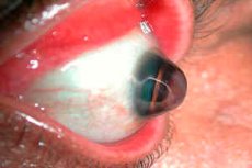
Keratoglobus is a rare condition characterized by curvature and thinning of the cornea of the eye. This condition belongs to the group of corneal dystrophies and is often associated with a progressive bulging (protrusion) of the cornea.
Keratoglobus is a variant of keratoconus, a more common condition in which the cornea is also thin and bulging but cone-shaped. In the case of keratoglobus, the protrusion is more diffuse and usually involves the entire cornea, which becomes spherical in shape. [1]
Epidemiology
Keratoglobus is a relatively rare corneal disease and therefore its exact epidemiology is not fully understood. Unlike keratoconus, which is relatively common and has a well-defined epidemiologic pattern, data on the prevalence of keratoglobus are limited.
Frequency of occurrence
Due to the rarity of the condition, there are no accurate statistics on the incidence of keratoglobus, but the general consensus is that the condition is diagnosed much less frequently than keratoconus. Keratoglobus can occur at any age, but is most often diagnosed in childhood or adolescence.
Distribution by gender and age
There is little reliable data on the distribution of keratoglobus by sex and age, but some sources indicate that the condition may be evenly distributed between males and females. The condition can manifest as early as childhood and is often diagnosed in adolescents.
Geographical distribution
Information on the geographic distribution of keratoglobus is also limited. There is no clear evidence that keratoglobus is more common in certain regions of the world or among certain ethnic groups.
Association with other diseases
Keratoglobus is sometimes associated with rare systemic connective tissue diseases such as Marfan syndrome and Ehlers-Danlos syndrome. In such cases, the distribution and epidemiology of keratoglobus may reflect the prevalence of these underlying conditions. [2]
Causes of the keratoglobus
The exact causes of keratoglobus are not fully understood. However, several theories about possible causes and risk factors have been proposed based on observation and clinical studies.
Genetic factors
A genetic component is considered one of the possible causes of keratoglobus. The condition is sometimes associated with inherited systemic connective tissue diseases such as:
- Ehlers-Danlos syndrome.
- Marfan syndrome
- Down syndrome
Patients with these diseases may have abnormalities in the structure of collagen and elastin fibers, which can affect the structural integrity of the cornea.
Disorder of corneal biomechanics
Corneal thinning and bulging in keratoglobus may be due to abnormalities in the biomechanical stability of the cornea. This may be due to abnormalities in intercellular interactions and the composition of the corneal extracellular matrix.
Inflammatory processes
Some researchers consider the possibility of inflammation in the cornea as one of the mechanisms leading to the development of keratoglobus. However, the presence of inflammation may be a consequence of other diseases or conditions rather than the underlying cause of keratoglobus.
Environmental Exposure
Some environmental and lifestyle factors, such as ultraviolet radiation or mechanical exposure of the eye, may contribute to the development or progression of keratoglobus, although their role is not precisely established.
Infections and injuries
Eye trauma or surgery may also be associated with the development of keratoglobus. In some cases, the development of keratoglobus is associated with eye infections, which can cause thinning and weakness of the cornea.
Other diseases
Rarely, but keratoglobus may be associated with other ophthalmologic diseases that affect corneal structure and function.
Because keratoglobus is a rare disease and research is limited, many aspects of this condition remain poorly understood. Future research may uncover new factors contributing to keratoglobus and help develop new treatment and prevention strategies. [3]
Pathogenesis
The pathogenesis of keratoglobus is not fully understood, but there are theories based on anatomic and biochemical changes in the cornea that may play a role in the development of this condition.
Biomechanical instability of the cornea
One of the key features of keratoglobus is the biomechanical instability of the cornea, which can be caused by a variety of factors:
- Collagen fiber weakness: The quantity and quality of collagen fibers in the cornea decreases, which reduces its strength and elasticity.
- Extracellular matrix abnormalities: The cornea contains an extracellular matrix that provides structural support and regulation of cellular functions. Changes in the composition and organization of the extracellular matrix can lead to thinning and bulging of the cornea.
Genetic factors
The presence of familial cases of keratoglobus implies that genetic factors may play a role in the development of the disease. Mutations in certain genes that regulate the synthesis and structure of collagen and other connective tissue components can lead to the development of keratoglobus.
Associated systemic diseases
Keratoglobus may be associated with systemic connective tissue diseases such as Marfan syndrome or Ehlers-Danlos syndrome. These diseases affect collagen and can lead to structural abnormalities in the cornea.
Enzymatic disorders
Some studies suggest that the activity of certain enzymes that break down components of the extracellular matrix may be increased in the cornea of patients with keratoglobus. This leads to degradation of collagen fibers and other structural components of the cornea.
Inflammatory processes
Although inflammation is not always present in the pathogenesis of keratoglobus, its role is being studied as a possible contributing factor that may enhance corneal degenerative processes.
Oxidative stress
Increased oxidative stress in the cornea can lead to cellular and matrix damage, which can also contribute to the development and progression of keratoglobus.
A common theme in the pathogenesis of keratoglobus is thinning and weakness of the cornea, resulting in its abnormal bulging. Pathogenetic mechanisms may include structural and biochemical abnormalities in the cornea that arise from congenital or acquired causes. However, further research is required to fully understand the pathogenesis of keratoglobus. [4]
Symptoms of the keratoglobus
Keratoglobus is characterized by a number of clinical signs and symptoms that can range from mild to severe. The main symptoms of this disease are:
Visual symptoms:
- Decreased visual acuity: Vision may become blurred or distorted due to distortion of the shape of the cornea.
- Myopia and Astigmatism: Pathologic change in corneal curvature often leads to the onset or worsening ofmyopia and irregular astigmatism.
- Photophobia: Sensitivity to light due to the thinness and transparency of the cornea.
Physical Symptoms:
- Corneal bulge: The appearance of the eye may change due to the cornea bulging forward.
- Thin cornea: Examination of the patient may show thinning of the central and peripheral parts of the cornea.
- Scleral indication: The periphery of the cornea may become so thin that the sclera (the white of the eye) can be seen through the cornea.
Other symptoms:
- Eye irritation: Patients may experience constant irritation or a foreign body sensation in the eye.
- Frequent conjunctivitis: Inflammatory processes may occur due to constant irritation and mechanical trauma to the cornea.
- Risk of corneal rupture: In rare cases, a very thin cornea can lead to spontaneous or traumatic rupture.
Diagnostic techniques such as ophthalmoscopy, pachymetry (measurement of corneal thickness) and corneal topography can reveal the degree of corneal thinning and the extent of corneal deformity.
Keratoglobus symptoms can worsen over time, and patients with this condition often require vision correction (through special contact lenses or surgery) and ongoing medical monitoring. [5]
Stages
The stages of keratoglobus may not be as well-defined as in other, better-studied eye diseases, such as keratoconus. However, certain stages of disease progression can be distinguished based on the degree of corneal thinning and severity of symptoms.
Initial stage:
- Mild corneal bulge: A slight distortion of vision may be noticeable, which patients often ignore or compensate for with glasses or soft contact lenses.
- Myopia and mild astigmatism: Appearance or worsening of myopia and mild astigmatism.
Intermediate stage:
- Moderate thinning and bulging of the cornea: Changes in the shape of the eye become more noticeable and visual acuity deteriorates even with correction.
- Increased astigmatism: Irregular astigmatism becomes more pronounced and difficult to correct.
Late stage:
- Severe bulging and thinning of the cornea: Severe thinning can cause the sclera to show through the cornea (scleral indication).
- High myopia and severe astigmatism: Significant vision problems that are not amenable to conventional correction.
- Photophobia, irritation and eye pain: These symptoms may worsen.
Critical Stage:
- Risk of corneal tearing: The thinnest parts of the cornea may be at risk of tearing even with minor trauma.
- Abrupt visual impairment and pain syndrome: Significant decrease in visual acuity and increase in pain.
Complications and consequences
Keratoglobus can lead to a number of complications that impair a patient's vision and quality of life. Here are some of the potential complications associated with keratoglobus:
- Corneal hydrops: A sudden intraocular intrusion of moisture causing swelling and clouding of the cornea. This can cause sudden decreased vision and pain.
- Spontaneous corneal tears: Due to thinning and bulging of the cornea, spontaneous corneal tears can occur, which can cause serious damage to vision and require urgent surgical intervention.
- Corneal scleralization: Corneal thinning can cause the white sclera to show through the cornea.
- High irregular astigmatism: Distortion of the curvature of the cornea can lead to complex astigmatism that is difficult to correct with regular glasses or contact lenses.
- Severe myopia: Progression of corneal thinning may increase myopia.
- Chronic conjunctivitis: Constant eye irritation can lead to recurrent inflammatory eye disease.
- Pain and discomfort: Patients may develop chronic pain due to constant irritation and eye strain.
- Contact lens problems: Because of the unusual shape of the cornea, it can be difficult to fit and wear contact lenses.
- Psychological problems: Visual impairment and visible deformities of the eye can lead to emotional and psychological problems, including decreased self-esteem and depression.
- Need for surgery: In severe cases, keratoplasty (corneal transplantation) or other surgical procedures may be required to restore corneal function.
Diagnostics of the keratoglobus
Diagnosing keratoglobus involves several steps and examination methods that help eye doctors identify specific changes in the structure and shape of the cornea that are characteristic of this condition. Here are some of the key methods for diagnosing keratoglobus:
- History: Collect a complete medical and family history, including any complaints of vision changes, eye pain, photophobia, or prior eye disease.
- External eye examination: Examination of the eyeball for abnormalities of shape and structure.
- Ophthalmoscopy: Used to evaluate the back of the eye and the condition of the retina and optic disc.
- Refractometry: Measurement of optical abnormalities of the eye, such as myopia and astigmatism, which are often associated with keratoglobus.
- Sleet-lamp biomicroscopy: Detailed examination of the front of the eye using a specialized microscope to detect corneal thinning and other abnormalities.
- Keratometry: A measurement of the curvature of the cornea that can detect abnormally high values indicating corneal bulging.
- Corneal Topography: An advanced evaluation method that builds a map of corneal curvature and shape, identifying unusual areas of thinning and bulging.
- Pachymetry: A corneal thickness measurement that helps to assess the degree of corneal thinning, which is an important parameter in the diagnosis of keratoglobus.
- Anterior Segment Optical Coherence Tomography (OCT): A state-of-the-art, non-invasive imaging technique that provides detailed slices of the front of the eye and cornea.
Combining these methods allows doctors to make an accurate diagnosis and distinguish keratoglobus from other similar conditions such as keratoconus or other corneal dystrophies. In cases where standard examination methods do not provide a complete picture, additional tests may be used to assess the structural integrity of the cornea and the risk of corneal rupture. [6]
What do need to examine?
Differential diagnosis
Differential diagnosis of keratoglobus is the process of ruling out other conditions that may mimic or look similar to keratoglobus in order to establish an accurate diagnosis. Key diseases and conditions to consider are:
- Keratoconus: This is the most common condition in which the cornea thins and bulges forward into a cone-like shape. The difference from keratoglobus is the distribution of thinning and the shape of the bulge, and the fact that keratoconus progresses more slowly and is more common in younger people.
- Globus megalocornea: A rare condition in which the cornea is enlarged in size but not thinned, and its structure is more stable than in keratoglobus.
- Pterygium: A growth of connective tissue film that can deform the cornea but has a different nature and treatment.
- Acanthamoeba keratitis: An infectious disease of the cornea that can cause thinning and reshaping of the cornea, but is accompanied by inflammation and more specific symptoms.
- Dilated (post-LASIK) ectasia: Thinning and bulging of the cornea following vision correction surgery, such as post-LASIK, which may resemble keratoglobus in symptomatology.
- Corneal Dystrophies: Various hereditary corneal dystrophies can cause changes in corneal structure and transparency that need to be distinguished from keratoglobus.
- Corneal inflammatory diseases: For example, keratitis of various etiologies can lead to thinning and reshaping of the cornea.
- Ocular trauma: The effects of trauma resulting in thinning or reshaping of the cornea should also be considered in the differential diagnosis.
Instrumental studies such as corneal topography, pachymetry, and optical coherence tomography, which can accurately measure corneal thickness and the shape of the bulge, play an important role in the differential diagnosis. A careful history should also be taken, especially considering age of onset, family history, and previous eye surgery or trauma. [7]
Who to contact?
Treatment of the keratoglobus
Treatment of keratoglobus depends on the stage and severity of the disease. Treatment options include:
- Glasses or contact lenses: Glasses or soft contact lenses can be used to correct minor vision changes caused by keratoglobus. More severe corneal curvatures may require rigid gas permeable contact lenses that help shape the front surface of the eye, improving vision.
- Scleral lenses: These are a special type of contact lens that do not touch the cornea but rest on the sclera (the white of the eye). They can be helpful for patients with keratoglobus as they provide more stable vision and comfort.
- Corneal collagen cross-linking: This procedure strengthens the collagen fibers in the cornea, which helps prevent further thinning and bulging of the cornea. This method can be effective in the early stages of keratoglobus.
- Intrastromal Corneal Rings (ICR or INTACS): Can be implanted to improve corneal shape and correct refractive errors.
- Corneal transplantation: In cases of severe thinning and bulging of the cornea, when other treatments are ineffective or when there is a risk of corneal rupture, partial (lamellar) or full (penetrating) corneal transplantation may be indicated.
- Surgical treatment: In rare cases where there is a threat of corneal perforation, surgery may be indicated.
- Drug therapy: Medications may be prescribed to relieve symptoms such as pain or inflammation. These may be moisturizing drops, antibiotics to prevent infection, or anti-inflammatory drugs.
- Regular follow-up: Patients with keratoglobus are recommended to have regular follow-up with an ophthalmologist to monitor changes in the cornea and adjust treatment if necessary.
All treatments should be individualized and discussed with an ophthalmologist. Since keratoglobus may progress, it is important to keep a constant monitoring of the condition and adjust the treatment according to changes in the corneal structure.
Prevention
Prevention of keratoglobus is limited to measures aimed at preventing its progression and minimizing the risk of complications, as the exact causes of this disease are unknown and there are no ways to prevent its occurrence. Here are some general recommendations for patients with keratoglobus or at high risk of developing it:
- Regular medical follow-up: It is important to see an ophthalmologist regularly to monitor the condition of the cornea and vision.
- Avoiding eye injuries: Protecting your eyes from injury, especially during sports and other potentially dangerous activities, can help prevent your condition from worsening.
- Control of inflammatory eye disease: Timely treatment of inflammatory eye conditions such as conjunctivitis and keratitis can help reduce the risk of keratoglobus-related complications.
- Use of moisturizing drops: The use of artificial tears is recommended to relieve symptoms of dryness and discomfort.
- Controlling allergic reactions: Managing allergic conditions can help avoid excessive eye rubbing, which is important to prevent progression of keratoglobus.
- UV protection: Wearing sunglasses to protect the cornea from UV radiation can prevent additional damage.
- Avoiding active eye friction: Eye friction can contribute to further thinning and deformation of the cornea and should be avoided.
- Adequate nutrition: Some studies suggest that deficiencies in certain nutrients may contribute to corneal disease, so a balanced diet may be important.
- Informing your ophthalmologist of any changes: At the first sign of vision changes, discomfort or any other changes in your eyes, you should contact your doctor immediately.
Although keratoglobus is rarely preventable, these measures can help reduce the risk of disease progression and improve patients' quality of life.
Forecast
The prognosis for keratoglobus can vary and depends on several factors, including the degree of corneal thinning and bulging, the rate of disease progression, the presence of complications, and the timeliness and effectiveness of treatment.
In mild cases, when the disease proceeds without rapid progression and serious complications, the prognosis is usually favorable. Vision correction with glasses or contact lenses may be sufficient to lead a normal life.
However, if the disease progresses, there may be worsening of vision that may not be fully corrected by standard methods. In such cases, more complex treatments may be required, including surgery such as intrastromal corneal ring implantation, collagen corneal cross-linking or corneal transplantation.
Corneal transplantation can have a high risk of rejection and other complications, so it is usually considered a last resort for severe forms of keratoglobus.
In cases where keratoglobus is associated with other systemic diseases or syndromes, such as Marfan syndrome or Ehlers-Danlos syndrome, the prognosis may be more complex and require an integrated treatment approach.
It is important to note that modern methods of diagnosis and treatment have significantly improved the prognosis for most patients with keratoglobus. Regular follow-up with a specialist and compliance with recommendations help to control the disease and maintain the quality of vision.
Literature used
"Keratoconus and Keratoglobus" is part of the book Cornea, third edition, edited by Krachmer JH, Mannis MJ, Holland EJ
"Keratoconus and Keratoglobus" in Cornea (third edition) - Margaret S. MacDonald, Michael Belenky, Charles Sheffield
"Ophthalmology" - Author: Myron Yanoff, Jay S. Duker, Year of latest edition: 2018.
"Vaughan & Asbury's General Ophthalmology" - Authors: Paul Riordan-Eva, Emmett T. Cunningham, year of latest edition: 2017.
"Clinical Ophthalmology: A Systematic Approach" - Author: Jack J. Kanski, Year of last publication: 2019.
"Ophthalmology: Expert Consult: Online and Print" - Author: Myron Yanoff, Jay S. Duker, Year of latest edition: 2018.