Medical expert of the article

New publications
Keller's osteochondropathy.
Last reviewed: 04.07.2025

All iLive content is medically reviewed or fact checked to ensure as much factual accuracy as possible.
We have strict sourcing guidelines and only link to reputable media sites, academic research institutions and, whenever possible, medically peer reviewed studies. Note that the numbers in parentheses ([1], [2], etc.) are clickable links to these studies.
If you feel that any of our content is inaccurate, out-of-date, or otherwise questionable, please select it and press Ctrl + Enter.
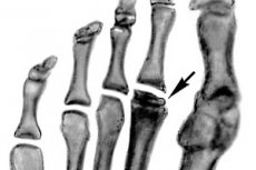
One of the varieties of aseptic necrosis is Keller's disease. It occurs in two forms, affects the bones of the foot and is age-related. Most often it occurs in children and adolescents.
Causes osteochondropathies
The main causes of necrosis of spongy bone tissue are associated with persistent disruption of its blood supply:
- Regular foot injury.
- Endocrine diseases and metabolic disorders: diabetes mellitus, thyroid disease, obesity.
- Wearing tight or ill-fitting shoes.
- Congenital and acquired defects of the arch of the foot.
- Genetic predisposition.
In Keller's osteochondropathy, there is an insufficient supply of oxygen and other useful substances to bone tissue. Because of this, degenerative processes begin, bone structures die off and aseptic necrosis develops.
 [ 1 ]
[ 1 ]
Symptoms osteochondropathies
The pathological condition occurs in two forms:
- Keller's disease I
Characterized by changes in the navicular bone. Most often occurs in boys aged 3-7 years. Manifested by swelling near the inner edge of the dorsum of the foot. Discomfort occurs during palpation and walking. The patient begins to limp, since the entire load is transferred to the healthy foot.
Constant pain leads to progression of the pathology. There is no inflammatory process. The disease does not spread to the second leg. The duration of this form is about a year, after which the painful symptoms completely disappear.
- Keller's disease II
It is bilateral in nature and causes damage to the heads of the II and III metatarsal bones of the feet. The onset of the pathological process occurs with mild pain at the base of the 2 and 3 toes. Discomfort increases with palpation, walking and other loads on the toes, but the pain subsides at rest.
As it progresses, the pain becomes severe and constant, not stopping even at rest. Visual examination reveals limited movement in the finger joints and shortening of the phalanges. This form is bilateral. It lasts for about 2-3 years.
The destruction and slow restoration of spongy bone tissue occurs in stages with the following pathological changes:
- Aseptic necrosis – bone beams die, that is, one of the bone structures. Bone density decreases, so it cannot withstand the previous loads.
- Compression fracture – new but not strong enough beams are formed, which, under normal loads, burst and wedge into each other.
- Fragmentation – osteoclasts resorb broken and dead parts of bone beams.
- Reparation is a gradual restoration of the structure and shape of the bone. Full regeneration is possible with the provision of normal blood supply to the affected bone area.
The symptoms of all forms of the disease negatively affect the patient's motor activity. Painful sensations and swelling of the foot cause changes in gait, lameness, and the inability to move quickly and run. The pathological condition is complicated by regular microfractures in the affected area.
Treatment osteochondropathies
Treatment is the same for both types of pathology and consists of a set of the following measures:
- Immobilization of the affected limb with a plaster cast for 1 month or longer.
- Drug therapy – non-narcotic analgesics for pain relief. Drugs for improving peripheral circulation and activating calcium metabolism, vitamin and mineral complexes.
- Physiotherapy – after the plaster cast is removed, the patient is prescribed foot massage, foot baths, electrophoresis, mud therapy, and magnetic therapy.
- Therapeutic exercise complex – the doctor selects special exercises that allow you to develop the foot after prolonged immobilization and restore its functionality.
- Surgical treatment – revascularizing osteoperforation is performed as an operation, i.e. holes are created in the bone to improve arterial blood flow. Thanks to this, the bone tissue is supplied with blood bypassing the affected vessels.
Prevention
Particular attention is paid to preventive measures. To prevent Keller's disease, it is necessary to choose the right shoes with an orthopedic insole. Also, increased physical activity should be avoided for preschool children. In case of any injuries or painful symptoms, it is necessary to consult a doctor.