Medical expert of the article

New publications
Ischemic optic neuropathy: anterior, posterior
Last reviewed: 04.07.2025

All iLive content is medically reviewed or fact checked to ensure as much factual accuracy as possible.
We have strict sourcing guidelines and only link to reputable media sites, academic research institutions and, whenever possible, medically peer reviewed studies. Note that the numbers in parentheses ([1], [2], etc.) are clickable links to these studies.
If you feel that any of our content is inaccurate, out-of-date, or otherwise questionable, please select it and press Ctrl + Enter.
Causes ischemic optic neuropathy.
The following three factors play a major role in the development of this pathology: disruption of general hemodynamics, local changes in the vascular wall, coagulation and lipoprotein shifts in the blood.
General hemodynamic disorders are most often caused by hypertension, hypotension, atherosclerosis, diabetes, stressful situations and profuse bleeding, atheromatosis of the carotid arteries, occlusive diseases of the brachiocephalic arteries, blood diseases, and the development of giant cell arteritis.
Local factors. Currently, great importance is attached to local factors that cause thrombus formation. Among them are changes in the endothelium of the vessel wall, the presence of atheromatous plaques and areas of stenosis with the formation of blood flow turbulence. The presented factors determine the pathogenetically oriented therapy of this severe disease.
Symptoms ischemic optic neuropathy.
There are two forms of ischemic neuropathy - anterior and posterior. They can manifest as partial (limited) or complete (total) damage.
Anterior ischemic neuropathy
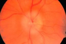
Acute circulatory disorder in the intrabulbar part of the optic nerve. Changes occurring in the head of the optic nerve are detected by ophthalmoscopy.
In case of total damage of the optic nerve, vision decreases to hundredths and even to blindness, in case of partial damage - remains high, but characteristic wedge-shaped scotomas are noted, and the apex of the wedge is always directed to the point of fixation of the gaze. Wedge-shaped losses are explained by the sectoral nature of the blood supply to the optic nerve. Wedge-shaped defects, merging, cause quadrant or half-loss in the field of vision. Defects of the field of vision are most often localized in its lower half. Vision decreases within a few minutes or hours. Usually, patients accurately indicate the day and hour when vision sharply decreased. Sometimes precursors in the form of headache or transient blindness can be noted, but more often the disease develops without precursors. Ophthalmoscopy shows a pale edematous optic disc. The vessels of the retina, primarily the veins, change secondarily. They are wide, dark, tortuous. There may be hemorrhages on the disc and in the parapapillary zone.
The acute period of the disease lasts 4-5 weeks. Then the swelling gradually decreases, hemorrhages are absorbed and optic nerve atrophy of varying severity appears. Visual field defects remain, although they can be significantly reduced.
 [ 13 ], [ 14 ], [ 15 ], [ 16 ]
Posterior ischemic neuropathy
Acute ischemic disorders develop along the optic nerve behind the eyeball - in the intraorbital region. These are posterior manifestations of ischemic neuropathy. The pathogenesis and clinical course of the disease are identical to those of anterior ischemic neuropathy, but in the acute period there are no changes in the fundus. The optic disc is of a natural color with clear boundaries. Only after 4-5 weeks does disc decolorization appear, partial or complete atrophy begins to develop. With total damage to the optic nerve, central vision can decrease to hundredths or to blindness, as with anterior ischemic neuropathy, with partial visual acuity can remain high, but characteristic wedge-shaped losses are detected in the visual field, more often in the lower or lower nasal sections. Diagnostics at an early stage is more difficult than with ischemia of the optic nerve head. Differential diagnostics are carried out with retrobulbar neuritis, space-occupying lesions of the orbit and central nervous system.
In 1/3 of patients with ischemic neuropathy, the second eye is affected, on average after 1-3 years, but this interval can vary from several days to 10-15 years.
What's bothering you?
What do need to examine?
How to examine?
Treatment ischemic optic neuropathy.
Treatment of ischemic neuropathy should be comprehensive, pathogenetically determined, taking into account the general vascular pathology of the patient. First of all, it is envisaged to use:
- antispasmodics (sermion, nicergoline, trental, xanthinol, nicotinic acid, etc.);
- thrombolytic drugs - plasmin (fibrinolysin) and its activators (urokinase, hemase, cavikinase);
- anticoagulants;
- symptomatic agents;
- B vitamins.
Magnetic therapy, electrical and laser stimulation of the optic nerve are also performed.
Patients who have suffered ischemic neuropathy of one eye should be under dispensary observation and receive appropriate preventive therapy.