Medical expert of the article

New publications
Hemodynamic study of the eye
Last reviewed: 07.07.2025

All iLive content is medically reviewed or fact checked to ensure as much factual accuracy as possible.
We have strict sourcing guidelines and only link to reputable media sites, academic research institutions and, whenever possible, medically peer reviewed studies. Note that the numbers in parentheses ([1], [2], etc.) are clickable links to these studies.
If you feel that any of our content is inaccurate, out-of-date, or otherwise questionable, please select it and press Ctrl + Enter.
The study of eye hemodynamics is important in the diagnosis of various local and general vascular pathological conditions. The following main methods are used to conduct the study: ophthalmodynamometry, ophthalmoplethysmography, ophthalmosphygmography, rheoophthalmography, ultrasound Dopplerography.
Ophthalmodynamometry (tonoscopy)
This method allows determining the level of blood pressure in the central artery (CAS) and central vein (CV) of the retina using a special device - a spring ophthalmodynamometer. In practical terms, more important is the measurement of systolic and diastolic pressure in the CAS and the calculation of the ratio between these indicators and the blood pressure in the brachial artery. The method is used to diagnose the cerebral form of hypertension, stenosis and thrombosis of the carotid arteries.
The study is based on the following principle: if intraocular pressure is artificially increased and ophthalmoscopy is performed, the appearance of a pulse in the CAS can initially be observed, which corresponds to the moment of equalization of intraocular and arterial pressure (diastolic pressure phase). With a further increase in intraocular pressure, the arterial pulse disappears (systolic pressure phase). Intraocular pressure is increased by pressing the device sensor onto the patient's anesthetized sclera. The device readings, expressed in grams, are then converted into millimeters of mercury using the Bayard-Majito nomogram. Normally, systolic pressure in the ophthalmic artery is 65-70 mm Hg, diastolic 45-50 mm Hg.
For normal nutrition of the retina, it is necessary to maintain a certain ratio between the amount of blood pressure in its vessels and the level of intraocular pressure.
Ophthalmoplethysmography
A method for recording and measuring eye volume fluctuations that occur in connection with cardiac contractions. The method is used to diagnose occlusion in the carotid artery system, and to assess the condition of the walls of intraocular vessels in glaucoma, atherosclerosis, and hypertension.
Ophthalmosphygmography
A research method that allows recording and measuring pulse fluctuations in intraocular pressure during four-minute Grant tonography.
Rheophthalmography
Allows quantitative assessment of changes in volumetric blood flow velocity in eye tissues based on their resistance (impedance) to high-frequency alternating electric current: with an increase in volumetric blood flow velocity, tissue impedance decreases. This method can be used to determine the dynamics of the pathological process in the vascular tract of the eye, the degree of effectiveness of therapeutic, laser and surgical treatment, and to study the mechanisms of development of diseases of the organ of vision.
Ultrasound Dopplerography
Allows to determine the linear velocity and direction of blood flow in the internal carotid and ophthalmic arteries. The method is used for diagnostic purposes in case of eye injuries and diseases caused by stenotic or occlusive processes in the said arteries.
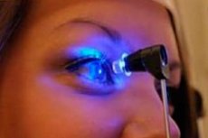
Transillumination and diaphanoscopy of the eyeball
Intraocular structures can be examined not only by sending a beam of light through the pupil with an ophthalmoscope, but also by directing light into the eye through the sclera - diascleral transillumination (diaphanoscopy). Transillumination of the eye through the cornea is called transillumination. These examinations can be performed using transillumination lamp-powered diaphanoscopes or fiber-optic light guides, which are preferred because they do not have an adverse thermal effect on the eye tissue.
The examination is carried out after careful anesthesia of the eyeball in a well-darkened room. Weakening or disappearance of the glow may be noted in the presence of a dense formation (tumor) inside the eye at the moment when the illuminator is above it, or in the case of massive hemorrhage into the vitreous body. In the area opposite the illuminated area of the sclera, during such an examination, it is possible to see a shadow from a parietal foreign body, if it is not too small and holds the light well.
With transillumination, the ciliary body "belt" can be clearly seen, as well as post-contusion subconjunctival ruptures of the sclera.
Fluorescein angiography of the retina
This method of studying retinal vessels is based on objective recording of the passage of a 5-10% solution of sodium fluorescein through the bloodstream by serial photography. The method is based on the ability of fluorescein to produce a bright glow when irradiated with poly- or monochromatic light.
Fluorescein angiography can be performed only in the presence of transparent optical media of the eyeball. In order to contrast the retinal vessels, a sterile, apyrogenic 5-10% solution of sodium fluorescein is injected into the cubital vein. Special devices are used for dynamic observation of the passage of fluorescein through the retinal vessels: retinophotes and fundus cameras of various models.
When the dye passes through the retinal vessels, the following stages are distinguished: choroidal, arterial, early and late venous. Normally, the duration of the period from the introduction of the dye to its appearance in the retinal arteries is 8-13 sec.
The results of this study are of great importance in the differential diagnosis of various diseases and injuries of the retina and optic nerve.
 [ 1 ], [ 2 ], [ 3 ], [ 4 ], [ 5 ]
Echophthalmography
Echo-ophthalmography is an ultrasound method of examining the structures of the eyeball, used in ophthalmology for diagnostic purposes. The method is based on the principle of ultrasound location, which consists in the ability of ultrasound to reflect from the interface of two media with different densities. The source and receiver of ultrasound vibrations is a piezoelectric plate placed in a special probe, which is applied to the eyeball. Reflected and perceived echo signals are reproduced on the screen of the cathode-ray tube in the form of vertical pulses.
The method is used to measure normal anatomical and topographic relationships of intraocular structures, to diagnose various pathological conditions inside the eye: retinal and choroidal detachment, tumors and foreign bodies. The value of ultrasound location is especially increased in the presence of opacities in the optical media of the eye, when the use of the main research methods - ophthalmoscopy and biomicroscopy - is impossible.
To conduct the study, special devices are used - echo-ophthalmoscopes, some of which operate in one-dimensional A-mode (ECHO-21, EOM-24, etc.), while others operate in two-dimensional B-mode.
When working in A-mode (obtaining a one-dimensional image), it is possible to measure the anterior-posterior axis of the eye and obtain echo signals from normal structures of the eyeball, as well as to identify some pathological formations inside the eye (blood clots, foreign bodies, tumors).
B-mode examination has a significant advantage, since it recreates a clear two-dimensional picture, i.e. an image of a “section” of the eyeball, which significantly increases the accuracy and information content of the examination.
Entoptometry
Since the most frequently used methods of assessing the state of the organ of vision in clinical practice (visometry, perimetry ) do not always provide an accurate and complete picture of the functional state of the retina and the entire visual analyzer, there is a need to use not more complex, but more informative functional ophthalmological tests. These include entoptic phenomena (Greek ento - inside, ortho - I see). This term denotes the patient's subjective visual sensations that arise as a result of the impact of adequate and inadequate stimuli on the receptor field of the retina, and they can have different natures: mechanical, electrical, light, etc.
Mechanophosphene is a phenomenon in the form of a glow in the eye when pressing on the eyeball. The study is conducted in a dark room, isolated from external sound and light stimuli, and pressure on the eye can be exerted either using a glass ophthalmological rod or by pressing a finger through the skin of the eyelids.
Pressure on the eyeball is applied in four quadrants at a distance of 12-14 mm from the limbus with the patient looking in the direction opposite to the location of the quadrant in which stimulation is performed. The results of the study are considered positive if the patient sees a dark spot with a bright luminous rim on the opposite side from the quadrant where stimulation is performed. This indicates the preservation of the retinal function in this particular quadrant.
[ 6 ]
Autoophthalmoscopy
A method that allows assessing the preservation of the functional state of the central sections of the retina even with opaque optical media of the eyeball. The results of the study are considered positive if, with rhythmic movements of the tip of the diaphanoscope on the surface of the sclera (after drop anesthesia), the patient notes the appearance of a picture of a "spider web", "tree branches without leaves" or "cracked earth", which corresponds to the picture of branching of the retinal vessels.
The light strip test is designed to assess the functional integrity of the retina in opaque optical media (corneal opacity, cataract ). The study is conducted by illuminating a Maddox cylinder with an ophthalmoscope, which is placed on the patient's eye being examined. If the central sections of the retina are functionally intact, the subject sees a strip of light directed perpendicular to the long axis of the Maddox cylinder prisms, regardless of its orientation in space.