Medical expert of the article
New publications
Invasive breast carcinoma
Last reviewed: 05.07.2025

All iLive content is medically reviewed or fact checked to ensure as much factual accuracy as possible.
We have strict sourcing guidelines and only link to reputable media sites, academic research institutions and, whenever possible, medically peer reviewed studies. Note that the numbers in parentheses ([1], [2], etc.) are clickable links to these studies.
If you feel that any of our content is inaccurate, out-of-date, or otherwise questionable, please select it and press Ctrl + Enter.

Invasive breast carcinoma is a pathology that can affect absolutely anyone - at any age, both men and women. However, the disease is most often detected in women of reproductive age.
Unfortunately, patients with carcinoma can live for a long time without suspecting that they have this dangerous pathology.
But for successful treatment it is very important to seek medical help as early as possible: to do this, it is necessary to understand and distinguish the typical signs of carcinoma.
ICD 10 code
- D 00-D 09 – tumors in situ;
- D 05 – non-invasive breast carcinoma;
- D 05.0 – non-invasive lobular carcinoma;
- D 05.1 – non-invasive intraductal carcinoma;
- D 05.7 – non-invasive breast carcinoma of other locations;
- D 05.9 – non-invasive carcinoma of the mammary gland, unspecified;
- C 50 – malignant tumor of the mammary gland.
Causes of Invasive Breast Carcinoma
The causes of the appearance of invasive neoplasms in the mammary gland have not yet been fully established. Specialists only identify risk factors that can trigger the development of malignant pathology.
- Hereditary predisposition. If close relatives suffered from oncology, then the probability that other family members will also get sick increases.
- Malignant tumor in one breast. If a patient has had a cancerous tumor in one gland, then the risk of developing cancer in the other gland increases.
- Peculiarities of sexual development and reproduction of the patient. The risk of developing carcinoma increases if a woman has premature puberty, delayed menopause, late first pregnancy or primary infertility, etc.
- Benign neoplasm in the mammary gland. A benign process (cysts, fibroadenomas) can sometimes degenerate or serve as a trigger for the development of a malignant neoplasm.
- Exposure to radiation. Radiation, whether environmental or used for medical purposes, significantly increases the risk of developing cancer.
- Endocrine disorders, metabolic disorders. Diseases such as diabetes, thyroid dysfunction, hypertension, and obesity contribute to the growth of atypical cells.
- Hormonal therapy, taking oral contraceptives. Hormonal imbalance can also be an indirect cause of breast tumors.
 [ 1 ], [ 2 ], [ 3 ], [ 4 ], [ 5 ]
Pathogenesis
Such stages of carcinoma progression as initiation, promotion and progression are not fully understood. It is known that pathogenesis is provoked by mutational processes of proto-oncogenes, which are transformed into oncogenes and activate cell growth. Also, proto-oncogenes increase the synthesis of mutational growth factors or affect external cell receptors.

When the integrity of the cell is violated by estrogen hormones, replication of the destroyed cell is activated even before the process of its regeneration. The intervention of estrogen is one of the mandatory conditions for the occurrence of a cancerous tumor in the breast. In this way, such a stage as promotion is launched. Distant metastasis occurs in the latent period (clinical symptoms are not yet expressed) - this usually occurs when the angiogenesis stage begins in the lesion.
Symptoms of Invasive Breast Carcinoma
Carcinoma can proceed latently for a long period, without revealing itself with any symptoms. The first signs of pathology often appear at later stages:
- the appearance of a dense area in the chest, regardless of the phase of the menstrual cycle;
- visible changes in the outline, volume or shape of one of the glands;
- the appearance of liquid discharge from the milk ducts (usually light or bloody);
- external changes in the skin on the gland (wrinkles, peeling, redness, “marbling”, etc.);
- the appearance of lumps in the armpit area (enlarged lymph nodes).
Later, signs of disease progression can be observed:
- the nipple becomes flat or inverted, the areola swells;
- some areas of the gland take on the appearance of a “lemon peel”;
- the iron is noticeably deformed;
- the skin over the site of the pathology is drawn in (sinks in);
- distant metastases are detected.
Pain is not typical for breast carcinoma.
Classification of invasive breast carcinomas
Invasive breast carcinoma is a cancer that forms outside the lobular membrane or duct, directly in the breast tissue. Gradually, the process affects the lymph nodes in the armpit area, as well as the skeletal system, brain, respiratory system and liver.
If cancer cells are found in other organs, then we are talking about metastasis (that is, the spread of metastases).
There are several variations in the course of carcinoma:
- Invasive ductal carcinoma of the mammary gland - originates from the milk ducts (ducts), after which the degenerated cellular structures spread through the tissues into the fatty tissue of the breast. Atypical cellular structures penetrate the lymph flow and the circulatory system and spread throughout the body. Invasive ductal carcinoma is considered the most common form of oncological pathology of the mammary gland;
- preinvasive ductal carcinoma is a condition that precedes the spread of cancer into deeper tissues;
- Invasive lobular carcinoma of the breast – occurs in approximately 15% of all cases of breast cancer. Invasive lobular carcinoma develops in the lobular structure of the breast, spreading further according to the principle of the previous two options.
Stages of invasive breast carcinoma:
- 0 – the process does not affect nearby tissues;
- I – the malignant lesion is less than 20 mm in size, the lymphatic system is not affected;
- II – the tumor size is less than 50 mm, metastases are found in the axillary lymph nodes on the affected side;
- III – the tumor size may be larger or smaller than 50 mm, with fused metastases in the lymph nodes, or in the lungs or skin;
- IV – there are distant metastases.
Up to stage II, carcinoma is considered early. Stage III is characterized by local spread of the process. Stage IV is called widespread or metastatic.
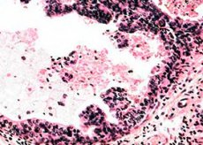
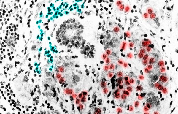
The degree of differentiation of the neoplasm (g) is assessed microscopically and can be determined by values from 1 to 3. The higher the g value, the lower the degree of differentiation of the tumor, and the more unfavorable the prognosis.
- g1 – high degree of differentiation.
- g2 – average degree of differentiation.
- g3 – low degree of differentiation.
- gx – there is no possibility to establish the degree of differentiation.
- g4 – undifferentiated tumor (invasive breast carcinoma of no special type).
Consequences and complications of invasive breast carcinoma
Invasive carcinoma is a very common pathology, and complications with this disease can occur both with and without treatment. The malignant tumor grows directly into the tissues of the mammary gland or milk ducts. It damages and presses on nearby tissues, nerve endings and blood vessels. The consequences of this situation can be bleeding, pain. An inflammatory reaction may join in if external damage to the skin occurs.
Mastitis can significantly worsen the course of carcinoma and accelerate the malignant process.
With distant metastasis, complications may also arise in the affected organs. The function of the respiratory or skeletal system, liver, brain (depending on the spread of metastases) is impaired. Constant headaches, loss of consciousness, problems with defecation and urination often appear.
Complications may also arise after surgery. For example, complete removal of the gland often provokes psychological problems, and surgical resection of the axillary lymph nodes can cause swelling and decreased range of motion in the upper limb.
Diagnosis of invasive breast carcinoma
External examination and palpation of the breast is the first and main examination if invasive carcinoma is suspected. It is advisable to palpate the gland in the first half of the monthly cycle - this will provide an opportunity to obtain sufficient information about the condition of the breast. Palpation helps to suspect carcinoma, but in the early stages of development with a small tumor size, this method may be ineffective.
Laboratory tests include tests for cancer markers, a poorly understood diagnostic method that demonstrates the body's tendency to develop cancerous tumors.
Instrumental diagnostics includes:
- mammography;
- ductography;
- pneumocystography;
- ultrasound examination of the mammary glands;
- magnetic resonance imaging and X-ray computed tomography.
Given the unpredictability of the malignant process, most specialists insist on a comprehensive examination of patients. It should include not only instrumental and laboratory diagnostic methods, but also an assessment of the function of the respiratory organs, liver, etc. This may require consultation with narrow specialists, such as a pulmonologist, orthopedist, gastroenterologist, gynecologist and surgeon.
Differential diagnostics are carried out with nodular mastopathy, adenoma, mastitis and erysipelas in the mammary gland.
Who to contact?
Treatment of invasive breast carcinoma
Treatment of carcinoma involves a comprehensive approach, using chemotherapy, hormonal therapy, radiation and, in most cases, surgery.
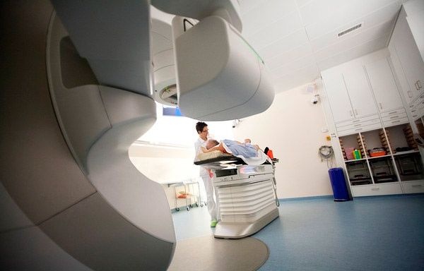
- Radiation therapy is always used in combination with other treatment procedures, and never on its own. Irradiation is prescribed after a course of medication, after surgery, etc. In this case, it affects not only the area of the affected breast, but also the sites of possible metastasis (for example, the area of the axillary lymph nodes). Sessions are carried out either immediately after resection or against the background of drug therapy, but no later than six months after surgery.

- Chemotherapy for the treatment of breast carcinoma is prescribed in the vast majority of cases, especially in the presence of metastases or in the late stages of the disease. The choice of drugs for this method of treatment is very wide. In the case of pronounced tumor progression, drugs such as cyclophosphamide, adriamycin, 5-fluorouracil are usually used, which help to prolong the life of patients even in the most advanced cases.
Often, chemotherapy is used in the preoperative period to reduce the volume of the tumor, which significantly improves the prognosis of the operation. And the simultaneous use of drugs such as trastuzumab or bevacizumab makes the treatment as effective as possible.
- Hormonal therapy is also rarely used independently - it is allowed only in old age to ensure long-term remission. Hormonal drugs are successfully used in combination with other treatment methods. In this case, drugs with an estrogen-like effect are prescribed, controlling tumor growth, or drugs that reduce estrogen synthesis. The first group of drugs includes tamoxifen, and the second group includes anastrozole or letrozole. The listed medications are considered first-choice drugs for invasive carcinoma. The scheme for the use of these drugs is prescribed strictly individually.
Surgical treatment can be carried out using several methods:
- the standard method of radical mastectomy involves the removal of the mammary gland (while preserving the chest muscles to allow for mammoplasty);
- Partial mastectomy, with the possibility of mammoplasty.
Subsequently, the shape and volume of the gland are restored using endoprosthetics or reconstruction with autogenous tissues.
In particularly severe advanced cases, operations are performed to alleviate the patient's condition and prolong his life. Such surgical interventions are called palliative.
Homeopathy for the treatment of invasive carcinoma is a rather controversial issue in medical circles. Most traditional medicine specialists allow the use of homeopathic remedies for prevention, but not for the treatment of malignant tumors. Of course, each patient decides for himself whether to trust homeopathy or not. The main thing is not to waste time and not to bring the disease to an advanced inoperable stage, when there can be no talk of successful treatment.
Among the most common homeopathic remedies for carcinoma of the gland are Conium, Thuja, Sulfuris, Kreosotum.
Traditional medicine can only be used in conjunction with traditional medicine, but not instead of it. Here are some of the most popular recipes that help slow down tumor growth.
- About 150 g of cherry pits are poured with 2 liters of goat milk and put in the oven on low heat for 6 hours. The resulting medicine is drunk 100 ml three times a day between meals. The duration of treatment is at least two months.
- Pure propolis is consumed 4-5 times a day, 6 g each, between meals.
- Potato flowers are collected, dried in the shade and an infusion is prepared: 1 teaspoon of raw material - 0.5 l of boiling water. Infuse for 3 hours. Take 100 ml three times a day 30 minutes before meals. Duration of intake is one month.
- The birch mushroom is grated and infused for 2 days in warm boiled water at a ratio of one to five. Then the infusion is filtered and drunk at least three times a day 30 minutes before meals. The medicine is stored in the refrigerator for no more than 4 days.
In addition, you can use gifts of nature - herbs, leaves, berries or fruits of plants. Herbal treatment involves the use of plants with the following properties:
- stimulate the immune system to fight malignant cells (euphorbia, astragalus, duckweed, red brush, etc.);
- damage tumor cells (natural cytostatics - periwinkle, colchicum, comfrey, meadowsweet, burdock, etc.);
- stabilize hormonal balance, compensate for the deficiency or excess of a particular hormone, for example, estrogen or prolactin (black cohosh, black cohosh, comfrey, comfrey, etc.);
- accelerate the removal of toxic substances and waste products from the body (milk thistle, dandelion, chicory, yarrow, etc.);
- relieve pain (comfrey, peony, willow, comfrey).
Prevention of invasive breast carcinoma
The danger of developing a cancerous tumor haunts almost every woman, especially those over 45. However, there is no need to be afraid, because there are preventive recommendations that can often help avoid the disease.
Of course, it is impossible to eliminate the existing hereditary predisposition. If it exists, the only way out is to regularly visit a gynecologist and mammologist, who will be able to monitor the health of the reproductive system in general and the mammary gland in particular.
What recommendations should all women without exception follow:
- do not smoke, do not abuse alcohol;
- treat infectious diseases and inflammatory processes in the genital area in a timely manner;
- avoid stress and excessive loads that can negatively affect hormonal levels;
- avoid X-ray exposure (only if absolutely necessary);
- eat properly and nutritiously;
- do not take hormonal drugs unnecessarily, and if using oral contraceptives for a long time, undergo periodic examinations and, if possible, take breaks or change contraceptives;
- avoid abortions, avoid injuries to the genitals and mammary glands;
- monitor your own weight and prevent the development of obesity.
Despite the fact that a person is not able to fully control his body and prevent all diseases, following the simple rules listed above will significantly reduce the risk of developing cancer.
Forecast
The prognosis for patients with invasive carcinoma depends on a number of factors:
- from the presence of metastases;
- from the size of the neoplasm;
- from the degree of penetration into surrounding tissues;
- from the rate of tumor growth.
Unfortunately, in recent years, the incidence of cancer worldwide has increased by more than 30%. For this reason, many countries have made preventive programs mandatory to help identify the disease at an early stage of development.
Invasive breast carcinoma diagnosed at stage one or two ends in recovery in more than 90% of cases. If the malignant pathology was detected much later, when the process of metastasis has already begun, the prognosis becomes much worse.

