Medical expert of the article

New publications
Hyposphagma
Last reviewed: 04.07.2025

All iLive content is medically reviewed or fact checked to ensure as much factual accuracy as possible.
We have strict sourcing guidelines and only link to reputable media sites, academic research institutions and, whenever possible, medically peer reviewed studies. Note that the numbers in parentheses ([1], [2], etc.) are clickable links to these studies.
If you feel that any of our content is inaccurate, out-of-date, or otherwise questionable, please select it and press Ctrl + Enter.
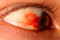
Subconjunctival or intraocular hemorrhage, otherwise known as hyposphagma, occurs when a small blood vessel is damaged, resulting in a small amount of blood leaking under the conjunctiva. Hyposphagma does not affect the quality of a person’s visual function and is only apparent externally. In the vast majority of cases, this phenomenon does not require special therapy, and in healthy people it passes without any intervention within a couple of weeks. Emergency medical care may be needed only if hyposphagma is caused by severe traumatic injury, a sharp increase in pressure (intraocular or arterial), and some other reasons. [ 1 ]
Epidemiology
Accurate statistics on the occurrence of hyposphagma are not kept because most people with relatively small subconjunctival hemorrhages simply do not seek medical attention. The incidence of hyposphagma was 2.9% in a study of 8,726 patients, and there was an increase with age, especially in those over 50 years of age. [ 2 ] It is also most common in young adults in their late teens and middle age;
The most common cause of the pathology is considered to be increased blood pressure (hypertension, physical or stress overload, lifting weights, vomiting, etc.), as well as injuries:
- industrial injuries;
- sports injuries (most often during football, hockey, tennis, baseball, boxing, paintball).
Less common are eye injuries that occur when an airbag deploys in an accident.
Hyposphagma is also common in children – its appearance is often caused by blows and touches received during active games.
Causes hypophagmas
One of the main causes of trauma to capillaries entering the blood supply system of the conjunctiva and conjunctival sac is high blood pressure. Blood with increased force affects fragile capillary walls, which break through, hemorrhage occurs in the subconjunctival space, and hyposphagma is formed.
Blood pressure in the capillaries can increase for many reasons, for example:
- direct trauma to the eyes, head, cervicothoracic spine;
- insufficient blood clotting function;
- leukemia; [ 3 ]
- chronic cardiovascular pathologies, such as hypertension, coronary heart disease, atherosclerosis, condition after a recent heart attack;
- Petechial hyposphagma may be seen in febrile systemic infections such as zoonoses (tsutsugamushi disease, typhus, leptospirosis), enteric fever, malaria, meningococcal septicemia, subacute bacterial endocarditis, scarlet fever, diphtheria, influenza, smallpox, and smallpox.[ 4 ],[ 5 ]
Acute hemorrhagic conjunctivitis caused by enterovirus type 70, coxsackievirus variant A24, and less commonly adenovirus types 8, 11, and 19, is characterized by sudden onset of follicular conjunctivitis with mucous discharge, epiphora, photophobia, eyelid edema, and conjunctival chemosis. It is often associated with multiple petechial hemorrhages in the superior palpebral and superior bulbar conjunctiva or widespread subconjunctival hemorrhage, especially localized to the temporal aspect.[ 6 ],[ 7 ]
Hyposphagma was found in 22.9% of 61 young immunocompetent men during a measles epidemic in addition to conjunctivitis, which is a well-known diagnostic feature of measles.[ 8 ] Patients with chickenpox and normal platelet counts were reported to develop unilateral hyposphagma after the onset of typical skin rashes without any other ocular complications.[ 9 ]
- chronic pathologies of the gastrointestinal tract, accompanied by bowel movements, frequent or prolonged constipation;
- respiratory diseases that are accompanied by coughing or sneezing attacks, such as asthmatic bronchitis, whooping cough, pneumonia, tuberculosis, etc.;
- enterovirus hemorrhagic conjunctivitis;
- infectious and inflammatory intestinal pathologies, poisoning accompanied by vomiting;
- any diseases or conditions in which asphyxia may develop.
- conjunctivochalasis. [ 10 ], [ 11 ]
- ocular amyloidosis. [ 12 ], [ 13 ]
Hyposphagma may appear after surgical procedures (in particular, after laser vision correction), after retro- and parabulbar administration of drugs, [ 14 ], [ 15 ] and in women – after childbirth (especially severe ones, associated with a long pushing period).
Risk factors
Conjunctival capillaries are more vulnerable and fragile compared to other vessels of the same caliber in the body. Their integrity can be affected by a variety of factors, both external and internal. Alcohol abuse, systematic smoking, lack of vitamins and microelements, and hypoxia play a special role. Under the influence of such causes, capillary fragility worsens, and periodic hyposphagma can become chronic with temporary visual impairment.
The most common provoking factors in the development of hyposphagma are considered to be professional activities or participation in certain sports, which increase the risk of injury to the head, organs of vision, neck, and spine. Other possible causes include circulatory disorders, cardiovascular pathologies, diabetes mellitus, atherosclerosis, and hypertension. [ 16 ] In these cases, treatment for hyposphagma is carried out in accordance with the underlying disease. It is believed that a significant increase in frequency depends on the increase in the prevalence of systemic hypertension after 50 years; diabetes mellitus, hyperlipidemia, and anticoagulant therapy also become more common with age.
With atherosclerosis and hypertension, absolutely all vessels in the body suffer: they lose elasticity and become brittle. Arteries narrow, while veins, on the contrary, expand. [ 17 ]
Patients with diabetes mellitus often develop angiopathy of the retinal vessels (diabetic retinopathy), which can also be complicated not only by hyposphagma, but also by retinal detachment with irreversible loss of visual function.
Other, less common factors that can lead to the development of hyposphagma include:
- tumor processes affecting the organs of vision, brain, spine; [ 18 ], [ 19 ]
- myopia, uveitis, iritis;
- vascular defects;
- physical and nervous overload.
- contact lens use. The incidence of hyposphagma associated with contact lenses has been reported to be 5.0%.[ 20 ]
- taking certain medications. In addition to anticoagulants and antiplatelet agents, some drugs related to hyposphagma (SCH) have been described in the literature. It should be borne in mind that interferon therapy in patients with chronic viral hepatitis may cause subconjunctival hemorrhage, and retinopathy and antiviral therapy, including polyethyleneglycolated interferon plus ribavirin, may cause hyposphagma in addition to vascular ophthalmologic side effects. [ 21 ], [ 22 ]
Pathogenesis
Hyposphagma is the release of blood (hemorrhagic fluid) from the vascular network of the conjunctival membrane with subsequent accumulation in the space between the sclera (white membrane of the eye) and the conjunctiva. The ocular conjunctiva is the outer fibrous membrane, which is localized on the inner side of the eyelids and the outer part of the eye. Visually, it is a thin transparent film through which any subconjunctival hemorrhage is clearly visible: against the background of the protein membrane, red spills, stripes or spots appear, which can change color to yellowish or dark.
The conjunctival membrane is very important for maintaining adequate functionality of the visual organs: the membrane structures produce lacrimal secretions, without which the hydrolipid state of the eyes will be disrupted. In addition, the membrane is saturated with numerous small capillaries - vessels with a small diameter. Conjunctival capillary walls are quite vulnerable and fragile. They are easily injured if blood pressure increases slightly - in particular, during a coughing fit, vomiting, strong vibration, etc. [ 23 ]
The blood flowing out of the injured capillary flows under the connective tissue, mixes with tear secretions, resulting in the formation of a hemorrhagic secretion, which is hyposphagma.
Symptoms hypophagmas
The symptoms of hyposphagma are logical and quite clear: blood comes out of a capillary vessel as a result of one reason or another (poor clotting, platelet abnormalities, endothelial membrane disorders, etc.), forming a blood clot, which appears as a peculiar scarlet spot. [ 24 ]
Most patients with hyposphagma do not voice any clear complaints related to deterioration of vision or with pronounced discomfort and pain. In addition to external manifestations, other symptoms are extremely rare and can be characteristic only of the third degree of hyposphagma, when the area of hematoma damage exceeds ¾ of the entire subconjunctival space. In such a situation, the following signs of hyposphagma are added:
- a slight feeling of discomfort that may bother you when blinking;
- a mild sensation of a foreign object in the eye, in the absence of stabbing or cutting sensations;
- the red spot is externally visible even from a great distance.
Since the conjunctival membrane does not have sensory light-perceiving neurons, the appearance of hyposphagma does not have any effect on the functioning of the visual analyzing system, therefore visual acuity (both central and peripheral) is not impaired.
The moment of hemorrhage and formation of hyposphagma usually goes unnoticed. A person notices the first signs after looking in the mirror. A red (bloody) spot of different sizes is found on the white part of the eye. There is no pain or deterioration of vision in the vast majority of cases.
Traumatic hyposphagma of the eye
Subconjunctival hemorrhage caused by trauma is easily visually determined. The hyposphagma spot may be small or quite large, occupying more than half or even the entire surface of the eyeball, and even extending beyond it.
A small hyposphagma is not dangerous, does not cause vision impairment and resolves without a trace in a short time. But it is important to understand that extensive traumatic hemorrhage may indicate a subconjunctival rupture of the sclera, which indicates an open eye injury. It is important for a medical specialist to exclude a through rupture of the sclera in widespread hyposphagma. This is taken into account when conducting diagnostics, which necessarily include diaphanoscopy and revision of the sclera, as well as determining the symptom of Pripechek - pain in the projection of the subconjunctival damage to the sclera in patients with massive hyposphagma when palpated with a glass rod. The symptom is assessed after preliminary anesthesia of the eyeball.
Stages
Hyposphagma is subdivided depending on the area of subconjunctival hemorrhage:
- At grade I hyposphagma, the subconjunctival space is filled less than ¼, while there is practically no discomfort for the patient.
- In grade II hyposphagma, the filling of the subconjunctival space is from ¼ to ½, and the symptoms are extremely weak.
- At stage III, more than ½ of the subconjunctival space is affected, patients may note slight discomfort when blinking. Pain and deterioration of vision are not typical.
If more than ¾ of the subconjunctival space is filled, then we speak of a pronounced third stage of hyposphagma. The condition may be accompanied by more severe discomfort, an unpleasant sensation of a foreign object in the eye. In such a situation, it is necessary to consult a doctor.
Complications and consequences
Hyposphagma is very rarely complicated by other pathologies. The hemorrhagic fluid that accumulates between the conjunctiva and sclera gradually dissolves, the spot disappears. How quickly this process occurs depends on several factors, the main one being the degree of hemorrhage. It can be determined by the color of the hyposphagma.
A red spot indicates that only a few capillaries are damaged. This problem usually disappears after a few days, the capillaries quickly recover without any consequences.
A burgundy-colored spot that covers approximately 50% of the white surface disappears within 2-3 weeks without complications.
A blood clot-like spot that extends over more than 50% of the ocular surface indicates damage to the visual tissues. In such a situation, complications with hyposphagma are possible, it is better to seek qualified medical advice.
In severe cases, the visual acuity and quality may decrease, sparks, flashes of light and flying spots may appear before the eyes. The possibility of infection with the development of infectious and inflammatory processes in the eye is not excluded.
Hyposphagma has a rather unpleasant appearance, but this phenomenon should not frighten: despite the external manifestations, the bloody spot does not affect the general health and does not affect the functionality of the visual organs. However, if the spot is large or if it recurs, it is necessary to consult an ophthalmologist.
Diagnostics hypophagmas
The initial stage of diagnosis for hyposphagma consists of an external examination, assessment of the visual condition of the eye, determination of the size of the spot and the scale of the affected subconjunctival space.
To exclude the possibility of infections and inflammatory processes in the conjunctiva, biomicroscopy is performed. To identify other possible hemorrhages and bleeding affecting the anterior chamber of the eye, gonioscopy is performed - a procedure during which the anterior chamber is examined using a slit lamp and special glasses - goniolenses.
During the examination, it is very important for the doctor to rule out damage to the integrity of the central venous vessel of the retina, as well as the retina itself and the optic nerve. For this purpose, ophthalmoscopy of the fundus is performed.
Laboratory tests for hyposphagma include a general blood test with a coagulogram. Such diagnostics are necessary for the possible identification of provoking factors that require systemic therapy. We are talking about hemostatic disorders, coagulo and hemoglobinopathies, etc.
Instrumental diagnostics are prescribed to patients with hyposphagma in the context of identifying ophthalmological pathologies, visual apparatus injuries, cardiovascular diseases, and hematopoietic organs. In some cases, the following diagnostic procedures are required:
- ultrasound examination of abdominal organs;
- ultrasound examination of the chest organs and heart;
- angiography;
- MRI of the brain;
- fluoroscopy.
Based on the results of the research, the doctor can create a complete clinical picture, discover the cause of hyposphagma and make a diagnosis.
Differential diagnosis
It is very important to distinguish common hyposphagma from other diseases with similar clinical manifestations, in particular, from hypophthalmos and hyphema.
With hyposphagma |
With hyphema |
In case of hemophthalmos |
|
Location of hemorrhage |
In the subconjunctival space |
In the anterior chamber of the eye in the iris area |
In the vitreous body |
Photophobia |
Absent |
Present |
Present |
The appearance of "fog" before the eyes |
Absent |
Present |
Present |
Dysfunction of the visual analyzing mechanism |
Only at stage III of pathology, when the blood clot fills more than ¾ of the subconjunctival space |
Present |
Present |
Neurological signs |
None |
Probable |
In most cases, they are present |
Who to contact?
Treatment hypophagmas
In the vast majority of patients with hyposphagma, the pathology disappears without any intervention within 1-3 weeks: there is no need for special treatment. The first treatment described in the literature was air therapy (AIR THERAPY). [ 25 ] Only sometimes is it necessary to carry out therapy that eliminates the underlying cause of bleeding - for example, the doctor prescribes medications to correct blood clotting, etc.
Depending on the indications, for hyposphagma the doctor may prescribe the following medications:
- Antimicrobial external agents – eye drops Levofloxacin, Levomycetin, Tobrex – are prescribed for proven infectious processes in the eye.
- Preparations for eliminating dry mucous membranes - Vizin, Taufon, Artificial tears - are prescribed to maintain adequate moisture and activate cellular restoration. Thanks to such preparations, the precorneal tear film is stabilized and thickened, and the absorption of hyposphagma is accelerated. These preparations are instilled into the eyes 5-6 times a day.
- Preparations with angioprotective and vasodilating properties - Diosmin, Pentoxifylline, Vincarmine - facilitate capillary blood circulation, strengthen vascular walls, make them elastic. In addition, angioprotectors prevent vascular congestion in hyposphagma.
Medicinal treatment is supplemented by taking multivitamin complex preparations. This is necessary to correct visual function and improve the condition of capillary walls. The complexes must necessarily contain ascorbic acid, vitamins A and E, B, as well as chromium and zinc. If hyposphagma has acquired a chronic relapsing course, then the dosage of vitamins is increased, vitamin P is added.
Patients with severe hyposphagma caused by acute hemorrhagic conjunctivitis are given nasal and temporal subconjunctival injections of tissue plasminogen activator.[ 26 ], [ 27 ], [ 28 ]
Patients with hypertension, diabetes, and atherosclerosis are treated for the corresponding diseases. If a patient with hyposphagma has taken antiplatelet or anticoagulant medications, [ 29 ] they are discontinued and a comprehensive examination of the body is performed with subsequent correction of prescriptions.
Prevention
There are no specific preventive measures to prevent hyposphagma. Doctors advise thinking in advance about preventing head injuries and, in particular, eye injuries, for which purpose using protective equipment when performing professional activities, during sports, etc. In addition, it is important to maintain your own health, monitor blood pressure and blood sugar levels.
Preventive measures can also be aimed at optimizing the functioning of the cardiovascular system, strengthening the vascular wall and ensuring its elasticity:
- Nutrition should be complete and varied, with the inclusion of plant products rich in vitamins and minerals in the diet. It is absolutely necessary to regularly consume sea fish, greens, vegetables, berries, legumes. These products will help strengthen the capillary network and prevent vascular fragility.
- To avoid tissue hypoxia, you should maintain physical activity and walk for at least 1-1.5 hours every day.
- In the presence of occupational hazards, it is important to protect the organs of vision with the help of special shields or glasses.
- Eye exercises should be performed daily, which include a set of exercises to maintain vascular tone and improve microcirculation. Typically, such exercises consist of repeated squinting, blinking, rotating the eyeballs, etc.
In order to prevent hyposphagma, it is necessary to visit an ophthalmologist at least annually. If there are somatic diseases - in particular, diabetes or hypertension - a mandatory medical examination every six months is important.
Forecast
Hyposphagma is a pathological condition characterized by the release of blood and hemorrhagic fluid into the space between the white of the eye and the conjunctiva. The condition is usually not accompanied by the development of complications and resolves on its own within a few days (sometimes weeks). Special treatment is not required in the vast majority of cases. The need for drug therapy appears with the development of infectious and inflammatory processes, or in the presence of primary diseases that served as an impetus for the occurrence of hyposphagma. [ 30 ]
In general, the prognosis for patients with hyposphagma is mostly favorable. Practicing ophthalmologists note that this disorder very rarely develops into serious complications.
If a patient develops chronic recurrent hyposphagma, he/she is recommended to see a doctor for a preventive examination at least every 6 months. Regular examinations will help to minimize the likelihood of relapse.