Medical expert of the article
New publications
Hepatitis C virus
Last reviewed: 08.07.2025

All iLive content is medically reviewed or fact checked to ensure as much factual accuracy as possible.
We have strict sourcing guidelines and only link to reputable media sites, academic research institutions and, whenever possible, medically peer reviewed studies. Note that the numbers in parentheses ([1], [2], etc.) are clickable links to these studies.
If you feel that any of our content is inaccurate, out-of-date, or otherwise questionable, please select it and press Ctrl + Enter.
Hepatitis C virus is a small RNA-containing virus with a shell of structural proteins that, together with a group of non-structural proteins, form the nucleocapsid of the virion.
Most researchers studying the biology of the hepatitis C virus believe that it belongs to the Flaviviridae family and is the only representative of the Hepacivirus gene (Dustin LB., Rice CM, 2007).

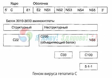
Hepatitis C virus (HCV) has a diameter of 30-60 nm, a buoyant density in a sucrose gradient of 1.0-1.14 g/cm, a sedimentation coefficient of 150 S, and a protein-lipid outer membrane. The HCV genome consists of a single-stranded positive RNA up to 10,000 nucleotide bases in size. The genome is represented by a single-stranded non-fragmented RNA of positive polarity 9,500-10,000 nucleotides long. The genome encodes one large polypeptide, which undergoes processing during maturation, in which two proteases participate: of viral origin and cellular. The HCV genome encodes 3 structural and 5 non-structural proteins of the virus. As shown in the figure, the main structural protein (C), which is part of the nucleocapsid, has a molecular weight of 21-33 kDa. Two other structural proteins E1 and E2 serve as viral envelope proteins and are glycoproteins with molecular weights of 31 and 70 kD, respectively. The remaining proteins are nonstructural polyproteins [NS2 (23 kD), NS3 (70 kD), NS4A (8 kD), NS4B (27 kD), NS5A (58 kD), NS5B].
When studying the molecular biology of HCV, a pronounced heterogeneity of the genomes of strains of this virus isolated in different countries, from different people, and even from the same person was established.
At present, there are up to 34 genotypes of the virus in 11 genetic groups. However, it is customary to distinguish 5 most common genotypes, numbered with Roman numerals I, Il, III, IV, V; they correspond to the designations of genotypes la, 1b, 2a, 2b and 3a. The genovariant of the virus determines the course of the infection, its transition to a chronic form and subsequently the development of cirrhosis and liver carcinoma. The most dangerous genovariants are lb and 4a. Genotypes lb, 2a, 2b and 3a circulate in Russia. The hepatitis C virus is widespread. According to WHO, about 1% of the world's population is infected with HCV.
Country |
Genotype, % |
|||
I (1a) 1 |
II (1b) |
III (2a) |
IV (2b) |
|
Japan |
74.0 |
24.0 |
1.0 |
- |
Italy |
51.0 |
35.0 |
5.0 |
1.0 |
USA |
75.0 |
16.0 |
5.0 |
1.0 |
England |
48.0 |
14.0 |
38.0 |
- |
Russia (Central European part) |
9.9 |
69.6 |
4.4 |
0.6 |
As can be seen from the table, the majority of people infected with the hepatitis C virus, regardless of continent and country, have genotype I (1a) or II (1b).
The distribution of genotypes is uneven across Russia. In the European part, genotype 1b is most often detected, while in Western Siberia and the Far East, genotypes 2a and 3a are most often detected.
The hepatitis C virus is found in the blood and liver in very low concentrations, in addition, it induces a weak immune response in the form of specific antibodies and has the ability to persist for a long time in the body of humans and experimental animals (monkeys). This often causes the occurrence of a chronic process in the liver in those infected with НСV.
The phenomenon of interference of НСV with hepatitis A and B viruses has been established; competitive infection with НСV leads to suppression of replication and expression of hepatitis A and B viruses in experimental animals (chimpanzees). This phenomenon may be of great clinical significance in case of coinfection of hepatitis C with hepatitis A and B.
The source of infection is only humans. The virus is detected in 100% of cases in the blood of patients and carriers (2/3 of all post-transfusion hepatitis is caused by HCV), in 50% - in saliva, in 25% - in sperm, in 5% - in urine. This determines the routes of infection.
The clinical course of hepatitis C is milder than that of hepatitis B. The hepatitis C virus is called a "soft killer". Jaundice is observed in 25% of cases; up to 70% of cases are latent. Regardless of the severity of the course, in 50-80% of cases hepatitis C becomes chronic, and in 20% of such patients cirrhosis and carcinoma subsequently develop. Experiments on mice have shown that the hepatitis C virus can affect nerve cells in addition to hepatocytes, causing severe consequences.
The hepatitis C virus reproduces poorly in cell culture, so its diagnosis is difficult. This is one of the few viruses for which RNA detection is the only method of identification. It is possible to detect the virus RNA using CPR in the reverse transcription variant, the ELISA method of antibodies to the virus using recombinant proteins and synthetic peptides.
Interferon, the production of which is impaired in chronic hepatitis, and the inducer of its endogenous synthesis, amixin, are the main pathogenetic agents for the treatment of all viral hepatitis.
 [ 1 ], [ 2 ], [ 3 ], [ 4 ], [ 5 ], [ 6 ], [ 7 ], [ 8 ], [ 9 ], [ 10 ]

