Medical expert of the article
New publications
Hairy leukoplakia of the mouth and tongue
Last reviewed: 04.07.2025

All iLive content is medically reviewed or fact checked to ensure as much factual accuracy as possible.
We have strict sourcing guidelines and only link to reputable media sites, academic research institutions and, whenever possible, medically peer reviewed studies. Note that the numbers in parentheses ([1], [2], etc.) are clickable links to these studies.
If you feel that any of our content is inaccurate, out-of-date, or otherwise questionable, please select it and press Ctrl + Enter.

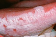
Hairy leukoplakia is not associated with hair growth on the superficial areas of the skin, but is a disease of the mucous membranes in which pathological areas are covered with filiform white villi, visible only during histological examination. Hairy leukoplakia of the oral cavity, first described in 1984, is a disease of the mucous membrane associated with the Epstein-Barr virus infection and occurs exclusively in people with immunosuppression. Visually, it looks like a plaque located symmetrically.
Epidemiology
The disease was first discovered and described in 1984 in America in a patient infected with AIDS. Scientists have traced the connection between the two pathologies. From a quarter to half of the cases of hairy leukoplakia were found in HIV-infected people.
The overall prevalence of oral leukoplakia in 2003 ranged from 1.7 to 2.7% in the general population.[ 1 ]
Hairy leukoplakia occurs more frequently in homosexual men with HIV infection (38%) than in heterosexual men with HIV infection (17%). [ 2 ] A cross-sectional study conducted in Brazil reported data collected from clinical examinations, interviews, and medical records of adult patients treated at the HIV/AIDS clinic at the University Hospital of the Federal University of Rio Grande. Three hundred individuals were followed (from April 2006 to January 2007). Of these patients, 51% were men and the mean age was 40 years. The most common lesion was candidiasis (59.1%), followed by hairy leukoplakia (19.5%).
Causes hairy leukoplakia
This pathology is one of the forms of leukoplakia - dystrophic changes in the mucosal epithelium, expressed in its keratinization. It occurs in 50% of patients with untreated HIV infection, especially in those with a CD4 count of less than 0.3 × 10 9 / l. [ 3 ] This pathology has a clear prognostic value for the subsequent development of AIDS and is classified as a clinical marker of HIV infection in the Centers for Disease Control and Prevention category B. [ 4 ] Hairy leukoplakia of the oral cavity also occurs in people with leukemia and organ and bone marrow transplantation, as well as in patients receiving systemic steroids.
Risk factors
In addition to HIV infection, AIDS, and other etiologies of immunodeficiency, risk factors include daily smoking of large quantities of cigarettes and promiscuous homosexual relations. Among the patients were people with ulcerative colitis, other gastrointestinal diseases, and Behcet's syndrome, which affects the mucous membranes of the oral cavity, genitals, and eyes. Hereditary predisposition is also important; diabetes mellitus and mechanical injuries (dentures, fillings, etc. in the mouth) contribute to the pathology.
Pathogenesis
The pathogenesis of oral hairy leukoplakia is complex and involves the interaction of persistent Epstein-Barr virus replication and virulence, systemic immunosuppression, and suppression of local host immunity. [ 5 ] The virus initially infects the basal epithelial cells in the pharynx, where it enters the replicative phase, is released, and remains in human saliva throughout life. It also penetrates B cells, where it can remain latent for an indefinite period until circumstances favorable for its reproduction occur, most often immune dysfunction.
Symptoms hairy leukoplakia
Hairy leukoplakia can develop asymptomatically for a long time. The first signs are expressed in the appearance of a white coating on the lateral surfaces of the tongue, in its upper and lower parts, less often on the inside of the cheeks, on the gums, soft palate. They are mainly symmetrical, can disappear for a while, and then appear. [ 6 ] Sometimes cracks form on the tongue, minor pain sensations appear, sensitivity is distorted, and taste changes. [ 7 ]
Gradually, the lesions merge into whitish stripes, alternating with healthy pink ones. Externally, it resembles a washboard. Hairy leukoplakia of the mouth and tongue slowly progresses, individual folds form plaques on the mucous membrane up to 3 mm in size, their borders are unclear and they are not removed by scraping.
In addition to the above-described localization, the pathology occurs much less frequently in women on the vulva, clitoris, cervix, and in men on the head of the penis, which is facilitated by mechanical and chemical factors (occurs in men aged 30 years and older).
Hairy leukoplakia in HIV is accompanied by weight loss, excessive sweating at night, unexplained diarrhea, and bouts of fever.
Stages
Hairy leukoplakia is a long-term chronic dystrophic process of the mucous membranes, which goes through several stages:
- proliferation, multiplication of cells;
- keratinization of squamous epithelium;
- sclerosis of cells (pathological regeneration, replacement with connective tissue).
Forms
There are several types of leukoplakia:
- flat - looks like a slightly rough film that cannot be removed with a spatula, with jagged outlines;
- warty - is represented by raised plaques with a diameter of 2-3 mm and a whitish color;
- erosive - appears in the foci of the first two leukoplakias in the form of erosions, sometimes cracks;
- smoker's leukoplakia or Tappeiner's leukoplakia - is formed on the areas of the hard and soft palate, where they become completely keratinized and acquire a gray-white color interspersed with reddish dots - the mouths of the salivary gland ducts;
- candidal - chronic candidal infection joins in;
- Hairy leukoplakia is a disease caused by the Epstein-Barr virus.
Complications and consequences
Unpleasant consequences and complications of hairy leukoplakia include changes in taste, inflammation of the oral mucosa due to infection with Candida fungi (candidal stomatitis), discomfort in the mouth: tingling, burning.
Diagnostics hairy leukoplakia
The diagnosis of the disease is based on the clinical picture and laboratory tests. Histology is performed, which reveals "shagginess" of the affected areas in the upper epithelial layer. A smear may show a superficial infection (candidiasis), keratinization of the mucous membrane, thickening and enlargement of the spinous and granular layers of the epithelium, and inflammation.
A mucosal biopsy reveals the Epstein-Barr virus. An HIV test is also used, and the number of T-helper lymphocytes is determined (in leukoplakia it is below normal). EBV can be detected by several methods, such as polymerase chain reaction (PCR), immunohistochemistry, electron microscopy and in situ hybridization (ISH), the latter being considered the gold standard for diagnosis. [ 8 ]
Additional methods include instrumental examination with a photodiagnostic scope (ultraviolet irradiation and observation of tissue glow), electron microscopy (by directing electron flows, the structure of tissues is studied at the subcellular and micromolecular level), and the use of optical coherence tomography.
Differential diagnosis
Differential diagnosis includes oral candidiasis, lichen planus, oral intraepithelial neoplasia caused by human papillomavirus, and oral squamous cell carcinoma. In most cases, oral hairy leukoplakia can be diagnosed clinically and does not require confirmatory biopsy.
Who to contact?
Treatment hairy leukoplakia
Hairy leukoplakia usually does not require special treatment and often resolves with HAART if it is associated with HIV infection. [ 9 ] Drug therapy is primarily aimed at suppressing the Epstein-Barr virus. There are also special dietary requirements: spicy, hot, salty, and sour foods are excluded from the diet.
Special care of the oral mucosa will be required, namely rinsing with antiseptics. Local preparations that improve tissue trophism are used, general tonics, biostimulants, and, if necessary, analgesics will be needed.
Treatment for hairy leukoplakia is aimed at restoring patient comfort, restoring the normal appearance of the tongue, and preventing other oral diseases. [ 10 ] Suggested treatments include surgery, systemic antiviral therapy, and topical treatment.
Medicines
Gentian violet is a triphenylmethane dye that was synthesized by Charles Laut in 1861 under the name "Violet de Paris". Churchman in 1912 demonstrated the bacteriostatic action of crystal violet against Gram-positive microorganisms in vitro and in animal models, as well as the antimycotic activity of this agent against several Candida species. [ 11 ] Since then, several studies have assessed the antibacterial and antifungal activity.
The antiviral properties of gentian violet were investigated based on the fact that EBV viral products induce reactive oxygen species formation and gentian violet is a potent inhibitor of reactive oxygen species.[ 12 ] Considering that crystal violet is well tolerated, approved for human use and is inexpensive, Bhandarkar et al.[ 13 ] conducted a study using gentian violet (2%) as a topical treatment for hairy leukoplakia in one HIV-infected man. Gentian violet was applied topically to the lesion three times for one month. Complete regression of the disease was observed after one month of follow-up and no relapse was observed after one year of treatment.
Podophyllin is a dry, alcoholic extract of the rhizomes and roots of Podophyllum peltatum. It is a fat-soluble substance that penetrates cell membranes and interferes with cell replication; it is commonly used as a topical chemotherapeutic agent. [ 14 ] It is inexpensive, easy to use, and effective over a long period of time.
The results of using 25% podophyllin alcoholic solution as a topical therapy for hairy leukoplakia are significant, especially in the first week after application. In a case series, nine patients were treated with 25% podophyllin sol in tincture of benzoin compound. The results showed complete regression of all lesions: five patients within one week and four after a second application one week later. These four patients had more extensive lesions. In another study, six male patients with hairy leukoplakia were treated with 25% podophyllin once daily and healing of all lesions was confirmed in three to five days. [ 15 ] Gowdy et al. evaluated ten HIV-infected patients with hairy leukoplakia on the tongue and treated one side with a single topical application of 25% podophyllin resin solution. The other side was used as a control. The patients were evaluated on the second, seventh and thirty days of the study. They described a slight change in taste, burning and pain of short duration. There was a regression of lesions, especially on the second day after application.
The dose commonly used in topical therapy for hairy leukoplakia ranged from 10 to 20 mg podophyllin.
Antiviral therapy includes drugs such as acyclovir, valacyclovir, famciclovir. After discontinuation of systemic antiviral drugs such as desciclovir, valacyclovir, acyclovir and ganciclovir, relapses of hairy leukoplakia were often observed. [ 16 ]
Acyclovir is a chemotherapeutic antiviral agent that is highly effective against herpes simplex virus types I and II, EBV, Varicella zoster virus, and cytomegalovirus. The only study using topical acyclovir cream was conducted by Ficarra et al. [ 17 ] The authors observed hairy leukoplakia in 23 of 120 HIV-positive patients (19%) and found complete resolution of the disease in two patients and partial regression in one patient after topical application of acyclovir cream.
Acyclovir - tablets, the recommended daily dose is 800 mg (one tablet contains 200 mg), divided into 5 doses. It is not prescribed to children under 2 years of age, pregnant and lactating women should use it with caution, taking into account the benefit-risk ratio. Side effects include nausea, diarrhea, fatigue, itching, rash, headache, dizziness. Anemia, jaundice, and hepatitis may develop. The drug is contraindicated in case of allergy to the components, patients with renal and hepatic insufficiency, and elderly people should reduce the dose.
If the disease occurs against the background of HIV infection, reverse transcriptase inhibitors are used: zidovudine, didanosine.
Candidal infection is treated with antifungal drugs: fluconazole, ketoconazole.
Fluconazole - capsules, 200-400 mg are taken on the first day of treatment, then 100-200 mg for 1-3 weeks until remission. Children in this form can be given the drug when they can swallow a capsule, usually after 5 years. The initial daily dose for them is 6 mg/kg, maintenance - 3 mg/kg.
Possible side effects are drowsiness, insomnia, anemia, diarrhea, nausea, headache, dry mouth, increased bilirubin levels, transaminases. There are contraindications for combined treatment with some medications (terfenadine, cisapride, astemizole, etc.).
In the treatment of hairy leukoplakia, local keratolytics and retinoic acid preparations are also used.
Vitamins
Vitamin therapy is appropriate for the treatment of leukoplakia. Oil solutions of tocopherol acetate and retinol are prescribed orally. Before swallowing, they are held in the mouth for some time.
Retinoids are dekeratinizing agents responsible for the modulation of Langerhans cells in hairy leukoplakia. Topical application of 0.1% vitamin A twice daily was performed in twelve cases of the disease and regression of lesions was observed after 10 days.[ 18 ] Daily application of tretinoin solution (Retin-A) for 15-20 days was performed in 22 patients and 37 patients were untreated. Healing of lesions was observed in 69% of treated patients and spontaneous regression in 10.8% of untreated patients.[ 19 ] Retin-A is an expensive drug and causes a burning sensation after prolonged use.[ 20 ]
Vitamins C, B group, including riboflavin, and others that strengthen the immune system are used.
Physiotherapy treatment
Physiotherapeutic methods are included in the hairy leukoplakia treatment protocol. These are diathermocoagulation and cryodestruction - procedures used to eliminate hyperkeratosis areas.
Folk remedies
Among folk methods, you can use mouthwash with decoctions of medicinal herbs that have an antiseptic effect: chamomile flowers, linden blossom, sage.
Surgical treatment
Excision is a surgical method used for hairy leukoplakia. The most modern is laser ablation, which uses a laser beam to remove the substance from the surface of the mucosa, it simply evaporates. Another method, cryotherapy, has not received widespread use.
No recurrence was observed for three months after surgical excision of hairy leukoplakia. However, most patients developed new lesions after three months of observation.[ 21 ]
Considering this and comparing surgery with systemic therapy, local treatment should be recommended to patients as it does not cause systemic side effects, is less invasive and is effective over a long period of time. [ 22 ]
Prevention
There are no preventive measures to prevent the disease.
Forecast
In half of the cases, the disease stabilizes after treatment. The same proportion is subject to complications (the appearance of new foci). The Epstein-Barr virus does not go away, the therapy only suppresses its productive replication.
Although hairy leukoplakia itself does not lead to death, its manifestation against the background of immunodeficiency is a very alarming signal, indicating an unfavorable prognosis for life expectancy (usually 1.5-2 years).

