Medical expert of the article

New publications
Fractures of the zygomatic bone and zygomatic arch: causes, symptoms, diagnosis, treatment
Last reviewed: 05.07.2025

All iLive content is medically reviewed or fact checked to ensure as much factual accuracy as possible.
We have strict sourcing guidelines and only link to reputable media sites, academic research institutions and, whenever possible, medically peer reviewed studies. Note that the numbers in parentheses ([1], [2], etc.) are clickable links to these studies.
If you feel that any of our content is inaccurate, out-of-date, or otherwise questionable, please select it and press Ctrl + Enter.
The zygomatic arch (arcus zygomaticus) is a complex formed by the temporal process of the zygomatic bone and the zygomatic process of the temporal bone.
Quite often, fractures of the zygomatic arch itself are observed that do not extend to the body of the zygomatic bone and its other processes.
 [
[ What causes a fracture of the zygomatic bone and zygomatic arch?
According to the literature, patients with fractures of the zygomatic bone and arch make up 6.5 to 19.4% of the total number of patients with facial bone injuries. They make up only 8.5%, since clinics receive not only emergency patients, but also a significant number of planned patients who need complex reconstructive surgeries after injury to other facial bones. They are often caused by domestic (fall, punch or hard object blow), industrial, transport or sports injuries.
According to the most common classification developed at the Central Research Institute of Surgery, fractures of the zygomatic bone and zygomatic arch are divided into the following groups:
- fresh closed or open isolated fractures without displacement or with slight displacement of fragments;
- fresh closed or open fractures with significant displacement of fragments;
- fresh closed or open combined fractures without displacement or with displacement of fragments;
- fresh closed or open combined fractures with simultaneous damage to other facial bones;
- old fractures and traumatic defects of the zygomatic bone and arch with facial deformation and impaired movement of the lower jaw.
Yu. E. Bragin classifies such fractures in approximately the same way.
In some cases, instead of the term "zygomatic bone", the term "anterior section of the zygomatic arch" is used, and instead of "zygomatic arch", the term "posterior section of the zygomatic arch" is used.
Non-gunshot injuries to the zygomatic bone and arch can be divided into three groups:
- zygomaticomaxillary fractures (closed or open, with or without displacement of fragments);
- fractures of the zygomatic arch (closed or open, with or without displacement of fragments);
- incorrectly fused zygomaticomaxillary fractures or fractures of the zygomatic arch (with facial deformation, persistent contracture of the lower jaw or signs of chronic inflammation of the maxillary sinus).
Taking into account the literature data and the experience of our clinic, all injuries to the zygomatic bone and arch, depending on the time elapsed since the injury, can be divided into three groups:
- fresh fractures - up to 10 days after injury;
- old fractures - 11-30 days;
- incorrectly fused and non-fused - over 30 days.
Direct contact of the facial bones with each other in general and with the zygomatic bone in particular, as well as the complexity and diversity of the vascular and nerve plexuses located here, determine! The occurrence of various injuries in this area, united under the name "Purcher syndrome", or traumatic retinopathy and angiopathy syndrome. This syndrome includes decreased visual acuity 1-2 days after the injury, cicatricial changes in the retina, pigmentation and atrophy of the optic nerve of varying degrees, up to retinal detachment several months after the injury.
Symptoms of a fracture of the zygomatic bone and zygomatic arch
Fractures of the zygomatic bones are usually combined with a closed craniocerebral injury: most often with a concussion, less often with a moderate or severe contusion.
In most cases, the zygomatic bone is displaced downward, inward, and backward; less often, the displacement is directed upward, inward, and backward, and even more rarely, outward and backward or forward. Any displacement of the zygomatic bone results in damage to the infraorbital nerve or its posterior superior alveolar branches, which manifests itself as a disturbance of the sensitivity of the skin of the infraorbital region, upper lip, wing of the nose, and a disturbance of the electrical excitability of the teeth of the upper jaw. Isolated fractures of the zygomatic bone, as a rule, do not occur. The frequently observed penetration of the zygomatic bone into the maxillary sinus leads to its filling with blood as a result of damage to the bone walls and mucous membrane of the sinus, which, in turn, contributes to the development of traumatic sinusitis. The size of the maxillary sinus decreases, but this remains unnoticed on the radiograph due to a sharp decrease in the pneumatization of the sinus. The veiled contours of the maxillary sinus can also be caused by the penetration of fatty tissue from the orbit into it.
Old fractures of the zygomatic bone. Cosmetic and functional disorders in old fractures depend on the location of the fracture, the degree of displacement of bone fragments, the decrease in bone substance, the duration of the injury, the nature of the treatment used, the extent of scar formations, the presence of chronic sinusitis or osteomyelitis of the zygomatic bone, upper jaw, the presence of a salivary fistula.
Diagnosis of fracture of the zygomatic bone and zygomatic arch
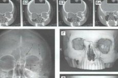
Diagnosis of fractures of the zygomatic bone and arch is based on anamnesis data, external examination, palpation of the damaged area, examination of the bite condition, anterior rhinoscopy, radiography in axial and sagittal (nasal-mental) projections. Table 4 presents subjective and objective symptoms of fracture of the zygomatic bone and zygomatic arch.
In the first hours after injury, before the appearance of edema, infiltrate or hematoma, palpation can provide so much valuable objective data that in some cases the need for radiographic examination disappears.
The displacement of fragments can be of varying degrees, and facial asymmetry and sunken eyeball, being a cosmetic defect, can be accompanied by functional disorders in the form of diplopia, limited mouth opening. Therefore, with each of the 8 listed classes of fresh fractures of the zygomatic bone, a combination of a number of symptoms of cosmetic and functional disorders, expressed to one degree or another, is noted.
Treatment of fractures of the zygomatic bone and arch
Treatment of fractures of the zygomatic bone and arch depends on the duration and location of the fracture, the direction and degree of displacement of the fragments, the presence of concomitant general disorders (concussion, brain contusion) and damage to the surrounding soft tissues.
In case of contusion syndrome, measures are taken that are necessary in such a case. Local interventions are determined primarily by the age of the fracture, the degree and direction of displacement of fragments, the presence or absence of damage to adjacent soft tissues and bones.
Treatment of fractures of the zygomatic bones and arches can be conservative and surgical. The latter, in turn, is divided into bloodless (non-operative) and bloody (operative).
All surgical treatment methods are also divided into intraoral and extraoral.
Non-operative surgical treatment of fractures of the zygomatic bone and zygomatic arch is indicated for easily reducible fresh closed fractures with varying degrees of displacement of the zygomatic bone, arch or fragments. There are two options for such treatment:
- the surgeon inserts the index finger or thumb of the hand into the posterior part of the superior vault of the vestibule of the mouth and repositions the zygomatic bone, monitoring the correctness and sufficiency of the reposition with the fingers of the other hand;
- a spatula or Buyalsky's scapula wrapped in gauze is inserted into the same area and the zygomatic bone, arch or their fragments are lifted with it. It is advisable not to rest the spatula on the zygomatic-alveolar ridge. The bloodless method may be effective for fresh fractures (in the first three days). If it is unsuccessful, one of the surgical methods is used.
Conservative treatment of fracture of the zygomatic bone and zygomatic arch
Conservative treatment is indicated for fresh fractures of the zygomatic arch or bone without significant displacement of fragments.
Keen intraoral method
This method is indicated for class III fractures and consists of making an incision in the upper posterior part of the vault of the vestibule of the mouth behind the zygomatic-alveolar ridge, through which a short and strong elevator is inserted, advanced under the displaced bone and with a vigorous upward and outward movement repositioned into the correct position.
Wielage Method
The method is a modification of the Keen method with the only difference being that it is used to realign both the zygomatic bone and the zygomatic arch.
For this purpose, it is also possible to use the retractor of A. G. Mamonov, A. A. Nesmeyanov, E. A. Glukina, which is bluntly passed through the wound into the area of the transitional fold at the level of the projection of the apices of the roots of the teeth, reaching the surface of the tubercle of the upper jaw (when reducing the zygomatic bone) or the squamous part of the temporal bone (when reducing the zygomatic arch). Pressing the branches of the retractor with the hand helps to shift the bone fragments and establish them in the correct position; with the free hand, the doctor controls the movement of the fragments. The therapeutic effect is determined by the results of the clinical and radiographic examination of the patient in the postoperative period.
Method of M. D. Dubov
The method involves extending the Keen-Wielage incision to the first incisor for simultaneous revision of the anterolateral wall of the maxilla and maxillary sinus. It is indicated in the treatment of zygomatic bone fractures combined with comminuted damage to the maxillary sinus. In these cases, the mucoperiosteal flap is peeled off, the soft tissues trapped between the fragments are released, the bone fragments are adjusted (using a spatula or Buyalsky spoon), and mucous membrane scraps and blood clots are removed. Then the fragments of the lower wall of the orbit are lifted with a finger and the cavity is tightly filled with an iodoform-gauze swab soaked in petroleum jelly (to hold the fragments in the correct position). The end of the swab is brought out through the junction with the inferior nasal passage formed (by the surgeon). In the vestibule of the mouth, the wound is tightly sutured. The tampon is removed after 14 days.
Duchange Method
The zygomatic bone is grasped and adjusted using special Duchange forceps, equipped with cheeks with sharp teeth. The zygomatic bone is repositioned in the same way using Sh. K. Cholariya forceps.
A. A. Limberg's method
The method is used when the fracture is relatively recent (up to 10 days). The displaced zygomatic arch or bone is grasped from the outside (through a puncture in the skin) with a special single-pronged hook with a transversely positioned handle and pulled into the correct position. However, in some patients with a V-shaped fracture of the zygomatic arch, the single-pronged hook of A. A. Limberg does not provide the same level of removal of fragments, since it can only be brought under one fragment, while the other either remains in place or is displaced (reset) with a lag from the first. To eliminate this drawback, Yu. E. Bragin proposed a two-pronged hook with a more convenient handle, made taking into account the anatomical features of the surgeon's hand, and a hole on each tooth. Ligatures are passed through these holes under the fragments of the zygomatic arch to fix them to the external splint.
Method of P. V. Khodorovich and V. I. Barinova
This method involves the use of improved forceps, which, if necessary, allow the displacement of bone fragments not only outward, but also in all other directions.
Method of Yu. E. Bragin
The method can be used even for very old fractures (more than 3 weeks old) due to the fact that the device is built on the screw principle, which allows, with minimal effort from the surgeon, to gradually increase the displacing (repositioning) force of action on the zygomatic bone, distributing and transmitting it to the bones of the skull through two support platforms. It is also important that the bone hooks of the device are applied to the edges of the zygomatic bone fragment without preliminary dissection of soft tissues.
Method of V. A. Malanchuk and P. V. Khodorovich
The specified method can be used for both fresh and old fractures. The advantage of the method is that only one support is required to install the apparatus (in the parietal bone area). The use of the apparatus of V. A. Malanchuk and P. V. Khodorovich allows to almost completely exclude more complex surgical methods of reduction of the zygomatic bone and arch with the imposition of bone sutures. Due to the use of this method in our clinic, good results were obtained in 95.2% of cases in the treatment of fresh fractures of the zygomatic complex, satisfactory results - in 4.8%, in the treatment of old (11-30 days) fractures - 90.9% and 9.1%, respectively, in the treatment of malunion fractures (over 30 days) - 57.2% and 35.7%, and unsatisfactory results - in 7.1% of cases. In case of a longer history of injury, open osteotomy and osteosynthesis of fragments are indicated.
Contour plastic surgery of the face in case of fractures of the zygomatic complex is indicated in case of normal function of the lower jaw and cosmetic defects of more than 1-2 years duration. Palliative surgeries - resection of the coronoid process of the lower jaw or osteotomy and reposition of the zygomatic arch - are indicated in case of dysfunction of the lower jaw.
If the surgeon does not have one of the above-described devices for reduction of old fractures with fragment displacement that occurred 10 or more days ago, it is often inappropriate to reduce the fragments using bloodless and operative methods. In such cases, one-stage refraction, reposition and fixation of the fragments of the zygomatic bone or slow reposition of the fragments by their elastic (rubber or spring) traction are performed.
If the listed methods have proven ineffective, various approaches can be used to perform one-stage surgical repositioning and fixation of the zygomatic bone, arch or their fragments: intraoral (subzygomatic and transsinus), temporal, subtemporal, orbital, zygomatic-arch.
Temporal method Gillis, Kilner, Stone (1927)
The hair in the temple area is shaved and an incision is made in the skin and subcutaneous tissue about 2 cm long, slightly back from the hairline border. A long wide elevator is inserted into the incision and advanced under the zygomatic arch. Controlling from the outside with the fingers of the other hand, the displaced bone is repositioned using the elevator.
Reposition of the zygomatic bone and the inferior wall of the orbit through the canine fossa and maxillary sinus according to Kazanjian-Converse
Having made an intraoral incision along the transitional fold within the canine fossa, it is exposed by lifting upward the mucoperiosteal flap, which is held with a curved hook. A window is made in the anterolateral wall of the intramaxillary sinus, through which blood clots are removed from it. The wall of the maxillary sinus is examined with a finger, the site of the fracture of the lower wall of the orbit is identified, and the degree of depression of the zygomatic bone into the maxillary sinus is specified. The bony walls of the maxillary sinus and the zygomatic bone are reduced by tamponade of the sinus cavity with a soft rubber tube filled with gauze strips (previously soaked in oil and antibiotic solution). The end of the rubber tube is inserted into the nasal cavity (as in Caldwell-Luc maxillary antrotomy). The wound along the transitional fold is tightly sutured; the tampon is removed after 2 weeks.
To simplify this method, an incision can be made in the mucous membrane along the entire length of the transitional fold on the side of the injury, which allows for lifting up the widely exfoliated soft tissues and examining the anterior and posterior surfaces of the maxilla, the zygomaticomaxillary suture area, and the lower parts of the zygomatic bone. After opening the maxillary sinus, the posterior and lower walls of the orbit are examined and palpated. This determines whether the zygomatic bone has penetrated the maxillary sinus, whether the lower wall of the orbit has fractured, whether the orbital or cheek fat has prolapsed into the maxillary sinus, or whether small bone fragments and blood clots have entered it. Then, using a narrow raspatory, the zygomatic bone and the walls of the maxillary sinus are adjusted, and then tightly tamponed with iodoform gauze, as recommended by Bonnet, A. I. Kosachev, A. V. Klementov, B. Ya. Kelman, and others. The tampon, the end of which is brought out into the lower nasal passage, is removed after 12-20 days (depending on the age of the fracture and the degree of difficulty in reducing bone fragments due to the formation of fibrous adhesions). Long-term tamponade of the maxillary sinus gives a good effect and does not cause complications, among which the development of diplopia is especially distressing for patients. Some authors recommend using inflatable rubber balloons instead of iodoform gauze.
Suturing the bone
Gill suggested that after repositioning the zygomatic bone with a raspatory, two additional incisions should be made in the area of the zygomatic-frontal and zygomatic-maxillary sutures through a temporal or intraoral incision, and then one hole should be made with a bur on either side of the fracture site. A steel wire (in our clinic, a polyamide thread is used) with a diameter of 0.4-0.6 mm is inserted into them. By pulling and tying the ends of the threaded wire or polyamide thread, the fragments are brought together and tightly contacted.
Suspension and traction of the zygomatic bone
Suspension and traction of the zygomatic bone is performed in cases where it is not possible to adjust it using the Wielage method through intraoral access. When suspending using the Kazanjian method, the zygomatic part of the infraorbital margin is exposed using an incision at the lower edge of the lower eyelid. A hole is drilled in the bone, through which a thin stainless steel wire is passed. Its end is brought out and bent in the form of a hook or loop, with the help of which elastic traction is performed to a rod-stand mounted in a plaster cap. The bone can also be approached through the Caldwell-Luc intraoral incision.
Zygomatic bone traction
The zygomatic bone is pulled outward and forward using a polyamide thread threaded through a hole in it. The zygomatic bone is exposed using an external incision at the point of its greatest depression. Experience shows that the polyamide thread irritates the soft tissues less than the wire and is easily removed after the traction is complete, which is done through a rod mounted on the side of the plaster cap.
Suspension of the zygomatic bone together with the upper jaw can be accomplished either by the dental-extraoral apparatus of Ya. M. Zbarzh, or by a custom-made plastic maxillary splint with extraoral rods, or by the surgical methods of Adams, Federspil or Adams-T. V. Chernyatina.
N. A. Shinbirev suggested fixing the zygomatic bone with a single-toothed hook of A. A. Limberg (with which he adjusted it) to the head plaster bandage.
Treatment methods for patients with isolated fractures of the zygomatic arch
In these cases there are usually two fragments, lying freely and with their approximal ends bent inwards. They are reduced by different methods.
Limberg-Bragin method
A. A. Limberg's single-pronged hook or Yu. E. Bragin's double-pronged hook are inserted through a 0.3-0.5 cm long puncture in the area of the projection of the lower edge of the zygomatic arch. The fragments are adjusted with an outward movement, placing the hook under their inwardly displaced ends. If the fragments do not shift in the correct position, the wound is sutured.
Suturing the bone
In this technique, the incision along the lower edge of the zygomatic bone is slightly enlarged (up to 1.5-2 cm). This is necessary in cases where, after the arch fragments have been reduced, they again take up an incorrect position with the formation of a diastasis between the ends of the fragments. If the arch is wide enough, holes are made in it with a small fissure bur, thin chromium catgut or polyamide thread is passed through them, the ends are pulled together, and thus the bone fragments are given the correct position.
Wire loop reduction using the Matas-Berini method
Using a large curved Bassini needle, a thin wire is passed into the thickness of the temporalis tendon, forming a grip loop. By pulling the wire loop, the fragments are fixed in the correct position.
Choice of method of reposition and fixation of fragments in fractures of the zygomatic bone and arch
Since the formation of bone tissue in zygomatic bone fractures occurs metaplastically and ends on average in two weeks, it is advisable to divide them into fresh (up to 10 days from the moment of injury) and old (more than 10 days) to select treatment tactics. All methods of reducing zygomatic bone fragments can be divided by the same principle.
In the period up to 10 days after the injury, treatment can be either conservative (non-operative) or surgical (radical-operative), and after 10 days - only surgical. In this case, the nature of the surgical intervention is determined by the features of the functional and cosmetic disorders caused by cicatricial fixation of bone fragments in an incorrect position, as well as the experience of the surgeon, the availability of the necessary instruments, equipment, etc. Of no less importance is the patient's attitude to the cosmetic defect that has arisen and the proposal to undergo surgical intervention.
The choice of surgical treatment method for fresh fractures of the zygomatic bone or arch depends primarily on the type (location) of the fracture, the number of fragments, the degree of their displacement and the presence of a tissue defect.
In old fractures (over 10 days old), it is usually impossible to reduce bone fragments using the simplest methods (finger method, through a Keen-Wielage incision, using a single-pronged hook by A. A. Limberg or a double-pronged hook by Yu. E. Bragin). In such cases, it is necessary to resort to more drastic surgical interventions: either to apply reduction using V. A. Malanchuk and P. V. Khodorovich, Yu. E. Bragin devices, or, having exposed the fracture site using intra- or extraoral access, to break the formed cicatricial adhesions, to fasten the reduced fragments with a suture or mini-plate. One of the methods of fixing the zygomatic bone and the lower wall of the orbit after reduction is the method of tight tamponade of the maxillary sinus with an iodoform-gauze tampon according to V. M. Gnevsheva, and O. D. Nemsadze and L. I. Khirseli (1989) use a rod of preserved allograft bone of the appropriate size as a support for the reduced zygomatic bone, inserted into the sinus: one end of it rests against the zygomatic bone from its inner side, the other - against the lateral wall of the nose.
Outcomes of zygomatic bone and zygomatic arch fractures
In cases of timely and correct repositioning and fixation of fragments in fresh fractures of the zygomatic bones and arches, complications are not observed.
If the reduction is not performed, complications such as facial deformation, persistent contracture of the lower jaw, visual impairment, chronic sinusitis, chronic osteomyelitis of the zygomatic bone and upper jaw, impaired sensitivity, mental disorders, etc. may occur.
The facial deformity is caused by a significant displacement or defect of the zygomatic bone (arch), which was not corrected during the treatment of the victim.
O. D. Nemsadze, M. N. Kiviladze, A. A. Bregadze (1993) suggest that after establishing the degree of displacement of the zygomatic bone in the lateral zone (in the case of an old or incorrectly healed fracture of the zygomatic bone), in order to reposition the bone fragments (after refraction of the fragments), resect the newly formed bone of the appropriate size in the area of the lateral wall of the orbit (in the area of the zygomatic-frontal suture).
Contracture of the lower jaw can be caused by two reasons:
- displacement of the zygomatic bone inward and backward with subsequent fusion of its fragments in an incorrect position;
- rough cicatricial degeneration of the soft tissues surrounding the coronoid process of the lower jaw.
Contracture develops especially often with injuries of classes 1, 3, 5-8.
Chronic traumatic sinusitis is quite common: for example, in so-called “zygomaticomaxillary fractures” it is observed in 15.6% of victims (V. M. Gnevsheva, 1968).
All of the listed complications, and especially chronic traumatic osteomyelitis, occur as a result of open infected fractures of the zygomatic bone, in the absence of timely and correct surgical treatment, reposition and fixation. In this regard, the infection can spread to the maxillary bone, the mucous membrane of the maxillary sinus, the conjunctiva, the ocular tissue, and the soft tissues of the face.